View Article

Abstract

Ethosomes are advanced phospholipid-based vesicular carriers widely used for enhanced transdermal drug delivery. They are mainly composed of phospholipids, ethanol, and water, where ethanol acts as an important penetration enhancer by disturbing the lipid arrangement of the stratum corneum. Due to their soft, flexible, and deformable nature, ethosomes can penetrate deeply into the skin and improve drug permeation and bioavailability. Compared to conventional liposomes, ethosomes show better skin penetration efficiency and improved therapeutic effectiveness. These vesicular systems are capable of carrying both hydrophilic and lipophilic drugs, making them suitable for a wide range of pharmaceutical and cosmetic applications. Ethosomes can be prepared by simple techniques such as hot and cold methods and are often incorporated into gels for better stability and patient compliance. Various evaluation parameters including vesicle size, zeta potential, entrapment efficiency, pH, viscosity, and in vitro drug release are used to characterize ethosomal formulations. Different types of ethosomal systems such as classical ethosomes, binary ethosomes, and transethosomes have been developed to further enhance drug delivery performance. Overall, ethosomes represent a promising, non-invasive, and effective approach for improving transdermal drug delivery with enhanced permeability, controlled drug release, and reduced side effects.

Keywords

Ethosomes, Phospholipid, Ethanol, Drug Penetration ,Transdermal drug delivery, Vesicle size.

Introduction

× Popup Image

Ethosomes are innovative lipid-based vesicular systems used in transdermal drug delivery. They have gained attention in recent years due to their ability to enhance the penetration of drugs through the skin. The skin acts as a strong barrier, mainly because of the outermost layer called the stratum corneum, which limits drug absorption. Ethosomes help to overcome this barrier effectively. Ethosomes are composed of phospholipids, a high concentration of ethanol, and water. The presence of ethanol makes these vesicles soft, flexible, and highly deformable, allowing them to penetrate deeply into the skin layers. Ethanol also disrupts the lipid structure of the skin, which further enhances drug permeation. Structurally, ethosomes consist of a phospholipid bilayernclosing an aqueous core. They can carry both hydrophilic and lipophilic drugs and improve their bioavailability. Compared to conventional systems like liposomes, ethosomes show better skin penetration and delivery efficiency. Ethosomes are non-invasive and offer several advantages such as improved drug delivery, reduced side effects, better patient compliance, and controlled drug release. Due to these benefits, they are widely used in pharmaceutical and cosmetic applications for topical and transdermal delivery systems.

Fig 1. Structure of Ethosomes [2] 

Ethosomes are soft, flexible lipid vesicles designed to enhance transdermal drug delivery. They are mainly composed of phospholipids, high concentrations of ethanol, and water, which make them more malleable and unique compared to conventional liposomes. The high ethanol content disturbs skin lipid organization and fluidizes both the ethosomal membrane and the stratum corneum lipids, allowing the vesicles to penetrate deeply into the skin layers. Because of their soft structure and strong penetration ability, ethosomes

significantly improve drug transport through the stratum corneum and provide efficiencydelivery to deeper skin tissues or systemic circulation. For Preparation of Ethosomes mostly preferred Hot Method and Cold Method. [1,2]

TYPES OF ETHOSOMAL SYSTEM:

1. Classical Ethosomes: Classical ethosomes are composed of phospholipids, water and high concentration of ethanol (40%). Drugs having molecular weight ranging from 130,077 Da to 24kDa can be entrapped in classical ethosomes.

2. Binary Ethosomes: Binary ethosomes can be prepared by adding another type of alcohol to the classical ethosomes. Propylene glycol (PG) and isopropyl alcohol (IPA) are most commonly used alcohols.

3. Transethosomes: It contains basic components from classical ethosomes and a penetrationEnhancer (surfactant). Developed to combine the advantages of classical ethosomes and transferosomes to produce transethosomes. Transethosomes are advanced type of ethosomes as they have the advantages of both i.e classical ethosome and transferosomes
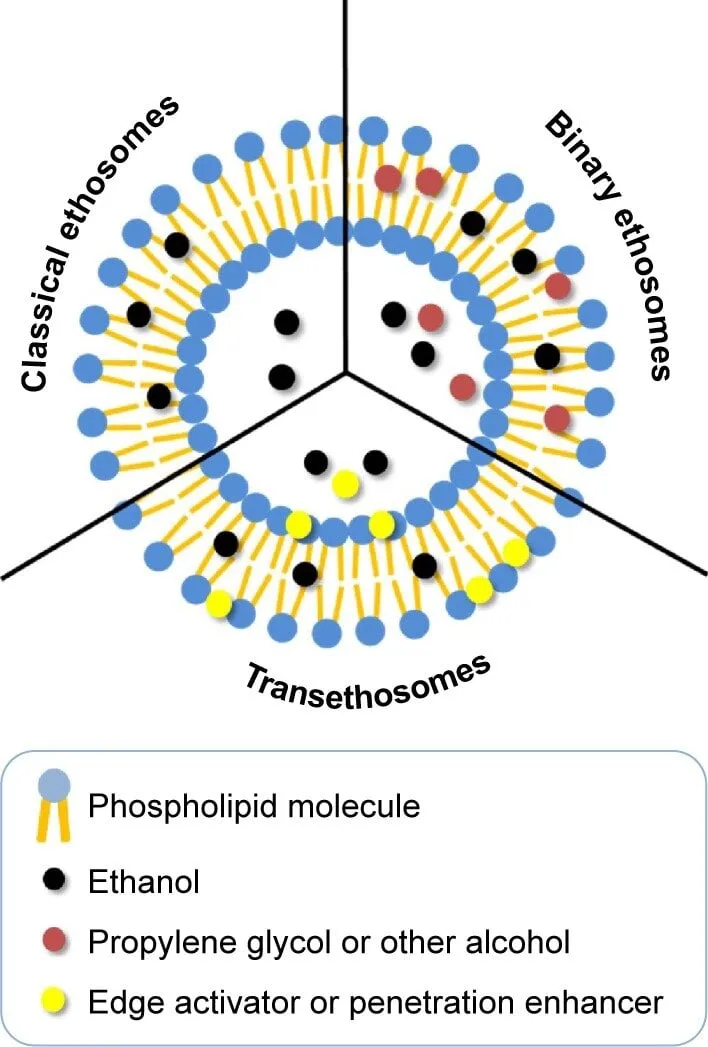
Fig 2. Types of Ethosomes [2]

METHOD:

1. Cold Method

2. Hot Method

3. Mechanical Dispersion Method 

4. Classic Method

Cold Method: Phospholipid, cholesterol, drug, ethanol, and PEG were mixed and heated to 40 °C. Distilled water was added slowly with continuous stirring at 700 rpm, and mixing was continued for 5 minutes. The dispersion was cooled for 30 minutes at room temperature. Finally, it was sonicated at 4 °C for five cycles (3 minutes each) using a probe sonicator. [3]
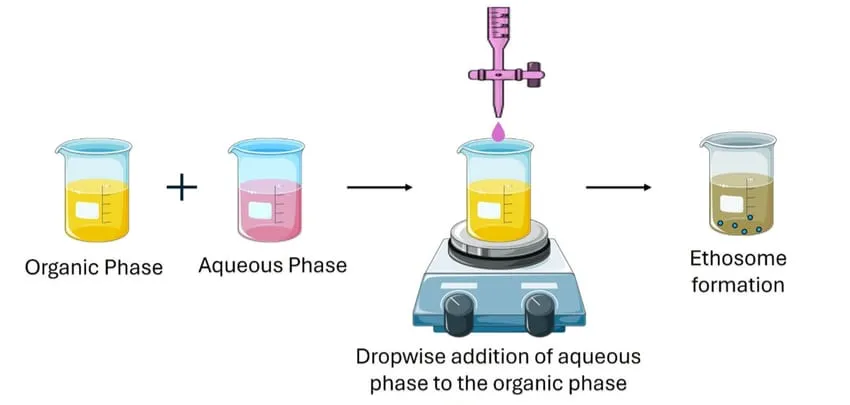
Fig 3. Cold Method [4]

Hot Method: In hot method, phospholipid was dispersed in water and dispersion was heated in a water bath or Hot plate at 40° C until a colloidal solution is obtained. In a separate vessel, ethanol, Drug and propylene glycol are mixed and heated to 40° C. Once both mixtures reach 40°C, the organic phase was added to the aqueous phase. The drug was dissolved in ethanol due to its hydrophobic property.  dispersion with continuous stirring on magnetic stirrer (1500 rpm) for 10 minutes. Finally, the prepared formulations were stored in refrigerator.[5]
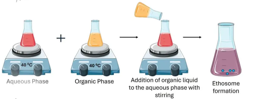
Fig 4. Hot Method [4]

Mechanical dispersion method: The phospholipid and drug are dissolved in ethanol and heated to 30 °C. Distilled water is then added slowly to this mixture with continuous stirring at 700 rpm. The formed vesicle suspension is collected in a closed vessel. Finally, the dispersion is homogenized by passing it through a polycarbonate membrane using a hand extruder for three cycles.[6]
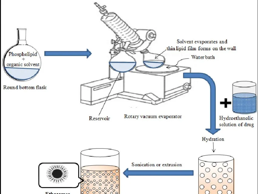
Fig 5. Mechanical Dispersion Method [7]

Classic Method: Soya phosphatidylcholine is dissolved in a chloroform:methanol (3:1) mixture in a round-bottom flask. The solvents are evaporated using a rotary vacuum evaporator above the lipid transition temperature to form a thin lipid film. Remaining solvent traces are removed by keeping the flask under vacuum overnight. The dry lipid film is then hydrated with a hydroethanolic drug solution by rotating the flask at an appropriate temperature.[6]
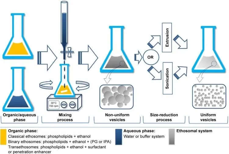
Fig 6. Classic Method [8]

Mechanism of drug penetration: 

Ethanol effect

 Classic Method Ethanol acts as a strong penetration enhancer by entering intercellular lipids of the stratum corneum, increasing lipid fluidity and decreasing lipid packing. This disruption makes the skin more permeable for drug diffusion. Ethosomes effect: In ethosomes, the high ethanol content fluidizes both vesicle lipids and skin lipids, allowing the vesicles to permeate deeply. Ethosomes then fuse with skin lipids and release the drug into deeper skin layers, enhancing drug delivery.[9,10]

Ethosomes effect:

In ethosomes, the high ethanol content fluidizes both vesicle lipids and skin lipids, allowing the vesicles to permeate deeply. Ethosomes then fuse with skin lipids and release the drug into deeper skin layers, enhancing drug delivery.[11]

Advantages:

1. Allows delivery of large molecules such as peptides and proteins.

2. Uses non-toxic and safe raw materials in the formulation.

3. Provides enhanced penetration of drugs through the skin for transdermal delivery.

 4. Can be widely applied in pharmaceutical, veterinary, and cosmetic fields.

5. High patient compliance because ethosomal formulations are usually given as gels or creams, which are easy to apply.[12,13]

Application of ethosomes :

 1. Ethosomes are effective in treating viral and microbial skin infections, as seen with bacitracin and erythromycin ethosomal systems.

2. Anti-inflammatory action is shown by ammonium glycyrrhizinate ethosomes on human volunteer skin.

3. Ethosomal patches help in treating androgen insufficiency in males and menopausal symptoms in women (animal studies).

4. Research suggests ethosomes may provide analgesic, antipyretic, and effects against erectile dysfunction.

5. Ethosomes can transport DNA molecules topically to help skin cells express specific genes.

6. Useful for delivering proteins and peptides effectively through skin

Skin :

 Skin is composed of multiple layers along with fine wrinkles, hair, and surface lipids. The layers form the main structure of the skin, while wrinkles, hair, and lipids are present on its outer surface. Together, these components show different physical and optical behaviours due to their unique structures.

Skin layers : The skin is an important and adaptable route for both systemic and topical drug delivery. Its outermost layer, the stratum corneum, acts as a strong protective barrier that restricts drug penetration and reduces the bioavailability of medications applied on the skin.
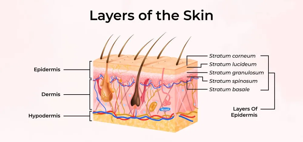
Fig.7 Layers of the skin [14]

Epidermis: The epidermis is the outermost layer of the skin, forming the first protective barrier of the body. Skin mainly has two layers—the epidermis and the dermis—and a clear wavy boundary separates them. The thickness of the epidermis varies depending on age, gender, individual differences, and body region. Its ability to hold water and maintain moisture also changes with age; for example, older skin retains less water due to reduced natural moisturizing factors (NMF). The transparency and condition of the stratum corneum, the outer sub-layer of the epidermis, depends on its water content.

Stratum Spinosum : The stratum spinosum, also known as the prickle cell layer, consists of 10 to 20 layers of cells located above the basal layer of the epidermis. During the natural skin turnover process, these cells become flatter and multi-sided, forming this layer. The cells are called prickle cells because they have small spine-like projections on their surface. The thickness of the stratum spinosum usually ranges from 50 to 150 µm, helping provide strength and support to the epidermis.

Stratum Granulosum (Granular Cell Layer):is a thin layer of about 2–4 rows of granular cells, typically around 3 µm thick. In this layer, keratinocytes begin keratinization, where their nuclei and mitochondria start to break down. The cells become filled with keratin fibers, lose moisture, and flatten as they move toward the skin surface.

Stratum Lucidum (Clear Layer): Stratum lucidum is a thin, clear layer present only on the palms and soles. The cells in this layer are flat, densely packed, and highly refractive. It provides extra protection in thick skin and forms during the normal process of cell turnover.

Stratum Corneum (Horny Cell Layer): Stratum corneum is the outermost layer of the epidermis, with a thickness of 8–15 µm. It is made of several layers of dead, flat keratinfilled cells called corneocytesp. This layer prevents water loss and protects the skin from dehydration. Corneocytes are surrounded by intercellular lipids and contain natural moisturizing factors (NMF) that help retain moisture.

Dermis: Dermis is the second, deeper layer of the skin located beneath the epidermis. It is much thicker than the epidermis (about 1–4 mm) and is mainly composed of collagen and elastin fibers, which provide strength, elasticity, and support. The dermis contains fewer cells but many fibers and also houses important structures like blood vessels, nerves, hair follicles, and glands.

Materials used for the Preparation of Ethosomes:[3]

Sr. No

Ingredients

Role

1

Drug

API

2

Phospholipid (soya lecithin)

Vesical forming agent

3

Ethanol

Penetration Enhancer

4

Propylene glycol

Stabilizer / Co-Solvent

5

Span 80

Surfactant (reduce vesical size)

6

Distilled Water

Vehicle

Table 1- Preparation of Ethosomes

Evaluation Parameter of Ethosomes:

  1. Particle Shape
  2. Zeta Potential & Particle Size
  3. pH
  4. Entrapment Efficiency
  5. Stability Study

Particle Shape:

The ethosomal particle shape study under a compound microscope is carried out by first preparing the sample, where a small amount of ethosomal suspension is taken and, if it appears too concentrated, it is diluted with distilled water. A drop of this prepared sample is then placed carefully on a clean glass slide. After that, a coverslip is gently placed over the drop to avoid the formation of air bubbles. The slide is then positioned under the compound microscope, and the observation is started at low magnification (10×), followed by higher magnification (40×) for better clarity. Finally, the fine focus knob is adjusted to obtain a clear view of the vesicles, allowing proper visualization of the shape and morphology of the ethosomal particles.

Zeta Potential & Particle Size :

The vesicle size, size distribution (polydispersity index), and zeta potential of the formulation were measured using a Zeta sizer based on dynamic light scattering (DLS) and electrophoretic light scattering techniques.

pH:

The pH of the ethosomal dispersion was measured using a calibrated digital pH meter at room temperature. The measurements were carried out directly on the dispersion, and readings were recorded in triplicate.

Entrapment Efficiency :

Entrapment efficiency was evaluated by estimating the amount of unentrapped (free) drug present in the aqueous phase of the ethosomal dispersion.Approximately 1 mL of the drug-loaded ethosomal formulation was transferred into centrifuge tubes and subjected to high-speed centrifugation for about 30 minutes. After centrifugation, the vesicles containing the entrapped drug formed a pellet at the bottom, while the unentrapped drug remained in the supernatant.The collected supernatant was then analyzed using a UV spectrophotometric method to determine the concentration of free drug.[3]

The entrapment efficiency was calculated using the formula:

% EE = Total drug content – Free drug content   x 100

                             Total drug content

Stability Study :

Stability study was carried out for drug loaded ethosomes at 3 different temperatures i.e. refrigeration temperature (4.0 ± 0.2°C) at room temperature (25-28 ± 2°C) and 45±1°C)  for 45days. The formulation subjected for stability study was stored in borosilicate container to avoid any interaction between the formulation and glass of container. The particle size of formulation was determined by optical microscopy using a calibrated ocular micrometer.[3]

CONCLUSION

Ethosomes represent an advanced and effective vesicular drug delivery system for enhancing transdermal drug penetration. Their unique composition, especially the high ethanol content, provides improved flexibility and permeability, allowing drugs to penetrate deeper into the skin layers compared to conventional systems. Ethosomes can encapsulate both hydrophilic and lipophilic drugs and offer advantages such as better bioavailability, controlled drug release, and reduced side effects.Various preparation methods like cold and hot methods make their formulation simple and reproducible. Evaluation parameters such as particle size, zeta potential, pH, and entrapment efficiency ensure the quality and stability of the system. Additionally, ethosomal formulations show wide applications in pharmaceutical, cosmetic, and therapeutic fields.Overall, ethosomes provide a promising, non-invasive, and efficient approach for transdermal drug delivery, improving patient compliance and therapeutic effectiveness. Future research can further enhance their potential in targeted and controlled drug delivery systems.

REFERENCES

  1. Dave V, Kumar D, Lewis S, & Paliwal S. Ethosome for enhanced transdermal drug Delivery of aceclofenac. International Journal of Drug Delivery, (2007) 2(1), 81–92.
  2. Review on Ethosomal Gel: For Effective Transdermal Delivery Hemanth SN, Lakshmi Radhika G, Venkanagouda Shridharagouda Agasimundin, Kavana Shree H. Review on ethosomal gel: for effective transdermal delivery. International Journal of Novel Research and Development. 2025.
  3. Khan S, Dubey B. K, Pandey G.K, Jain A, Dhakar S, & Jain P.K. Formulation and Evaluation of ethosomal gel of ciprofloxacin for bacterial infections. National Journal Of Advanced Research, (2019) 5(2), 21–25.
  4. Almuqbil RM, et al. Ethosome-Based Transdermal Drug Delivery: Its Structural Components, Preparation Techniques, and Therapeutic Applications Across Metabolic, Chronic, and Oncological Conditions. Pharmaceutics. 2025;17(5):583.
  5. Acharya A, Ahmed MG, Rao BD, Vinay CH. Development and evaluation of ethosomal gel of lornoxicam for transdermal delivery: In-vitro and in-vivo evaluation. Manipal J Pharm Sci. 2016;2(1):15-20.
  6. Touitou E, Dayan N, Bergelson L, Godin B, Eliaz M. Ethosomes – novel vesicular carriers for enhanced delivery: characterization and skin penetration properties. Journal of Controlled Release. 2000;65(3):403–418.
  7. Tyagi S, Chaurasia L, Islam M, Sagar B, Tyagi A. A review on novel approach ethosome as a nanocarrier for transdermal drug delivery. Biomedical Research. 2022:33(3)
  8. Moghassemi S, Hadjizadeh A. Nanoethosomal nanocarriers: the impact of constituents and formulation techniques on ethosomal properties, in vivo studies, and clinical trials. Journal of Controlled Release. 2014;185:12-22.
  9. Thomas A.P, Dubey R, & Jain P. Formulation and evaluation of ethosomal gel of Tazarotene for topical delivery. Asian Journal of Pharmaceutics, (2019) 13(1), 37–48.
  10. Satyan G, Shivani S, & Garima.G. Ethosomes: A novel tool for drug delivery through The skin. Journal of Pharmacy Research, (2010)3(4), 688–691.
  11. Giram, J, Mahabal, R, Bhosale N, & Jagtap V. Review on ethosomes incorporated Into gel: A novel approach for topical delivery of drug. International Journal of Pharmaceutical Sciences,( 2024)2(12),462-479.
  12. Ethosomal Gel Comprehensive Review Jain N, Sharma A, & Srivastava, A.K.Preparation and characterization of ethosomal gel drug delivery: A comprehensive review. Journal of Biomedical and Pharmaceutical Research, (2024). 13(6), 78–84.
  13. Kushkiwala A.M, Zankhwala F.M, Patel M.D, & Raval, A.M. Flurbiprofen loaded Ethosomal gel: Design, optimization, and anti-inflammatory activity. International Journal of Research and Analytical Reviews, (2024). 11(4), 709–720.
  14. Verma P, Pathak K. Therapeutic and cosmeceutical potential of ethosomes: An overview. Journal of Advanced Pharmaceutical Technology & Research. 2010;1(3):274-282. doi:10.4103/0110-5558.72415.
  15. Suprija R, & Sailaja A.K. Formulation of mefenamic acid loaded ethosomal gel by Hot and cold methods. Nano Biomedicine and Engineering, (2017) 9(1), 27–35.
  16. Chouksey A, Dubey B.K, Basediya D. K, & Mahajan V. Formulation of SDS aided And ethosomal fluconazole topical gel for improved antifungal effect. The Pharmaceutical and Chemical Journal, (2023)10(3), 59–72.
  17. Gadad A. P, Patil A.S, Singh Y, Dandagi P.M, Bolmal U.B, & Basu A.Development And evaluation of flurbiprofen loaded transethosomes to improve transdermal delivery. Indian Journal of Pharmaceutical Education and Research, (2020). 54(4), 953–963.
  18. Ghanbarzadeh, S.& Arami S. (2013). Enhanced transdermal delivery of diclofenac Sodium via conventional liposomes, ethosomes, and transfersomes. BioMed Research International, 2013, Article 616810.
  19. Kalva S, Rao P.S, Sundari B.T, & Diwan P.V. Preparation and evaluation of Mangifera Indica loaded ethosomal gel for anti-inflammatory activity in animal model. International Journal of Ayurvedic and Herbal Medicine, (2018)8(1), 3089–3098.
  20. Parmar P, Mishra A, & Pathak A. Preparation and evaluation of ethosomal gel of Clotrimazole for fungal infection by mechanical dispersion method. Current Research in Pharmaceutical Sciences, . (2016)6(2), 45–49.
  21. Astuti K. F, Surini S, & Bahtiar, A. Advances in ameliorating rheumatoid arthritis by Andrographolide ethosome-based gel:Pharmacokinetic and activity study in rats. International Journal of Applied Pharmaceutics, (2022)15(1), 79–86.
  22. Yadav N, & Prasad S. Formulation and characterization of anti-fungal ethosomal gel For effective topical fungal treatment. Panacea Journal of Pharmacy and Pharmaceutical Sciences,  (2024) 13(4), 1–10.
  23. Gharat S, Momin M, Panchal U, & Omri A. Novel ethosomal gel formulation for Enhanced transdermal delivery of curcumin and cyclosporine: A preclinical approach to Rheumatoid arthritis management. Drug Delivery, (2025).32(1), 1–20.
  24. Jain S, Tiwary A.K, Sapra B, & Jain N. K. Formulation and evaluation of ethosomes For transdermal delivery of lamivudine. AAPS PharmSciTech, (2007)8(4), Article 111.
  25. Arjun Gaire B.Pharma, Sangit Maharjan B.Pharm, Sumitra Shrestha M.Pharm, SonuPakhrin M.Pharmd, Naresh Thapa B.Pharma, Dr. Jeevan Raj Shrestha, Mrs. Bijaya Laxmi Giri M.Pharm Formulation & Evaluation of Fluconazole Gel for Topical Drug Delivery System American Scientific Research Journal for Engineering. Technology, and Sciences (ASRJETS).
  26. V. Sankar, V. Wilson, K. Siram, A. Karuppaiah, S. Hariharan, A. Justin (2019) Topical Delivery of Drugs Using Ethosomes: A review. Indian Drugs, 56(8), 7-14.
  27. Baghli, F., Khaqua, R., Guenifed, I., & Moussaoui-Khedam, N. (2024).Effect of formulation parameters on the characteristics of binary ethosomes loaded with Fluconazole. Magna Scientia Advanced Biology and Pharmacy, 13(02), 007-016
  28. Ethosomes: A Novel Tool for Transdermal Drug Delivery System Rita B. Ethosomes: A Novel Tool for Transdermal Drug Delivery System. International Journal of Pharma Research & Review. 2016;5(7):35-42.
  29. Ethosomes: Carrier for Enhanced Transdermal Drug Delivery System Ramakrishna GA, Manohar SD, Bhandas SR. Ethosomes: Carrier for Enhanced Transdermal Drug Delivery System. Journal of Advanced Pharmacy Education & Research. 2014;4(4):380-387.
  30. Ethosomes – A Modernistic Approach for Drug Delivery Ojha M, Chaudhary V, Tiwari P. Ethosomes – A Modernistic Approach for Drug Delivery. International Journal of Pharmaceutical Research and Applications. 2023;8(3):2566-2577.
  31. Sherin F, Saju F, Jagadeesh A, Sree S, Paul N, Venugopal A. Ethosome: A Novel Approach to Enhance Drug Permeation. International Journal of Pharmaceutical Sciences Review and Research. 2019;55(1):18-22.
  32. Simran Chaudhari, Prateeksha Pawar, Vaishnavi More, and Pranali Borse. “Ethosomes: A Novel Drug Delivery System.” In Futuristic Trends in Pharmacy & Nursing, Vol. 3, 2023, pp. 92–104.
  33. Formulation and Evaluation of Ethosomal Gel Containing Clarithromycin for the Treatment of Acne Vulgaris Ajikumar A, Dharan SS, Chandra SM, Soman M, Mathew LT. Formulation and evaluation of ethosomal gel containing clarithromycin for the treatment of acne vulgaris. Journal of Pharmaceutical Sciences and Research. 2022.
  34. Acharya A, Ahmed MG, Rao BD, Vinay CH. Development and evaluation of ethosomal gel of lornoxicam for transdermal delivery: In-vitro and in-vivo evaluation. Manipal J Pharm Sci. 2016;2(1):15-20.

Reference

  1. Dave V, Kumar D, Lewis S, & Paliwal S. Ethosome for enhanced transdermal drug Delivery of aceclofenac. International Journal of Drug Delivery, (2007) 2(1), 81–92.
  2. Review on Ethosomal Gel: For Effective Transdermal Delivery Hemanth SN, Lakshmi Radhika G, Venkanagouda Shridharagouda Agasimundin, Kavana Shree H. Review on ethosomal gel: for effective transdermal delivery. International Journal of Novel Research and Development. 2025.
  3. Khan S, Dubey B. K, Pandey G.K, Jain A, Dhakar S, & Jain P.K. Formulation and Evaluation of ethosomal gel of ciprofloxacin for bacterial infections. National Journal Of Advanced Research, (2019) 5(2), 21–25.
  4. Almuqbil RM, et al. Ethosome-Based Transdermal Drug Delivery: Its Structural Components, Preparation Techniques, and Therapeutic Applications Across Metabolic, Chronic, and Oncological Conditions. Pharmaceutics. 2025;17(5):583.
  5. Acharya A, Ahmed MG, Rao BD, Vinay CH. Development and evaluation of ethosomal gel of lornoxicam for transdermal delivery: In-vitro and in-vivo evaluation. Manipal J Pharm Sci. 2016;2(1):15-20.
  6. Touitou E, Dayan N, Bergelson L, Godin B, Eliaz M. Ethosomes – novel vesicular carriers for enhanced delivery: characterization and skin penetration properties. Journal of Controlled Release. 2000;65(3):403–418.
  7. Tyagi S, Chaurasia L, Islam M, Sagar B, Tyagi A. A review on novel approach ethosome as a nanocarrier for transdermal drug delivery. Biomedical Research. 2022:33(3)
  8. Moghassemi S, Hadjizadeh A. Nanoethosomal nanocarriers: the impact of constituents and formulation techniques on ethosomal properties, in vivo studies, and clinical trials. Journal of Controlled Release. 2014;185:12-22.
  9. Thomas A.P, Dubey R, & Jain P. Formulation and evaluation of ethosomal gel of Tazarotene for topical delivery. Asian Journal of Pharmaceutics, (2019) 13(1), 37–48.
  10. Satyan G, Shivani S, & Garima.G. Ethosomes: A novel tool for drug delivery through The skin. Journal of Pharmacy Research, (2010)3(4), 688–691.
  11. Giram, J, Mahabal, R, Bhosale N, & Jagtap V. Review on ethosomes incorporated Into gel: A novel approach for topical delivery of drug. International Journal of Pharmaceutical Sciences,( 2024)2(12),462-479.
  12. Ethosomal Gel Comprehensive Review Jain N, Sharma A, & Srivastava, A.K.Preparation and characterization of ethosomal gel drug delivery: A comprehensive review. Journal of Biomedical and Pharmaceutical Research, (2024). 13(6), 78–84.
  13. Kushkiwala A.M, Zankhwala F.M, Patel M.D, & Raval, A.M. Flurbiprofen loaded Ethosomal gel: Design, optimization, and anti-inflammatory activity. International Journal of Research and Analytical Reviews, (2024). 11(4), 709–720.
  14. Verma P, Pathak K. Therapeutic and cosmeceutical potential of ethosomes: An overview. Journal of Advanced Pharmaceutical Technology & Research. 2010;1(3):274-282. doi:10.4103/0110-5558.72415.
  15. Suprija R, & Sailaja A.K. Formulation of mefenamic acid loaded ethosomal gel by Hot and cold methods. Nano Biomedicine and Engineering, (2017) 9(1), 27–35.
  16. Chouksey A, Dubey B.K, Basediya D. K, & Mahajan V. Formulation of SDS aided And ethosomal fluconazole topical gel for improved antifungal effect. The Pharmaceutical and Chemical Journal, (2023)10(3), 59–72.
  17. Gadad A. P, Patil A.S, Singh Y, Dandagi P.M, Bolmal U.B, & Basu A.Development And evaluation of flurbiprofen loaded transethosomes to improve transdermal delivery. Indian Journal of Pharmaceutical Education and Research, (2020). 54(4), 953–963.
  18. Ghanbarzadeh, S.& Arami S. (2013). Enhanced transdermal delivery of diclofenac Sodium via conventional liposomes, ethosomes, and transfersomes. BioMed Research International, 2013, Article 616810.
  19. Kalva S, Rao P.S, Sundari B.T, & Diwan P.V. Preparation and evaluation of Mangifera Indica loaded ethosomal gel for anti-inflammatory activity in animal model. International Journal of Ayurvedic and Herbal Medicine, (2018)8(1), 3089–3098.
  20. Parmar P, Mishra A, & Pathak A. Preparation and evaluation of ethosomal gel of Clotrimazole for fungal infection by mechanical dispersion method. Current Research in Pharmaceutical Sciences, . (2016)6(2), 45–49.
  21. Astuti K. F, Surini S, & Bahtiar, A. Advances in ameliorating rheumatoid arthritis by Andrographolide ethosome-based gel:Pharmacokinetic and activity study in rats. International Journal of Applied Pharmaceutics, (2022)15(1), 79–86.
  22. Yadav N, & Prasad S. Formulation and characterization of anti-fungal ethosomal gel For effective topical fungal treatment. Panacea Journal of Pharmacy and Pharmaceutical Sciences,  (2024) 13(4), 1–10.
  23. Gharat S, Momin M, Panchal U, & Omri A. Novel ethosomal gel formulation for Enhanced transdermal delivery of curcumin and cyclosporine: A preclinical approach to Rheumatoid arthritis management. Drug Delivery, (2025).32(1), 1–20.
  24. Jain S, Tiwary A.K, Sapra B, & Jain N. K. Formulation and evaluation of ethosomes For transdermal delivery of lamivudine. AAPS PharmSciTech, (2007)8(4), Article 111.
  25. Arjun Gaire B.Pharma, Sangit Maharjan B.Pharm, Sumitra Shrestha M.Pharm, SonuPakhrin M.Pharmd, Naresh Thapa B.Pharma, Dr. Jeevan Raj Shrestha, Mrs. Bijaya Laxmi Giri M.Pharm Formulation & Evaluation of Fluconazole Gel for Topical Drug Delivery System American Scientific Research Journal for Engineering. Technology, and Sciences (ASRJETS).
  26. V. Sankar, V. Wilson, K. Siram, A. Karuppaiah, S. Hariharan, A. Justin (2019) Topical Delivery of Drugs Using Ethosomes: A review. Indian Drugs, 56(8), 7-14.
  27. Baghli, F., Khaqua, R., Guenifed, I., & Moussaoui-Khedam, N. (2024).Effect of formulation parameters on the characteristics of binary ethosomes loaded with Fluconazole. Magna Scientia Advanced Biology and Pharmacy, 13(02), 007-016
  28. Ethosomes: A Novel Tool for Transdermal Drug Delivery System Rita B. Ethosomes: A Novel Tool for Transdermal Drug Delivery System. International Journal of Pharma Research & Review. 2016;5(7):35-42.
  29. Ethosomes: Carrier for Enhanced Transdermal Drug Delivery System Ramakrishna GA, Manohar SD, Bhandas SR. Ethosomes: Carrier for Enhanced Transdermal Drug Delivery System. Journal of Advanced Pharmacy Education & Research. 2014;4(4):380-387.
  30. Ethosomes – A Modernistic Approach for Drug Delivery Ojha M, Chaudhary V, Tiwari P. Ethosomes – A Modernistic Approach for Drug Delivery. International Journal of Pharmaceutical Research and Applications. 2023;8(3):2566-2577.
  31. Sherin F, Saju F, Jagadeesh A, Sree S, Paul N, Venugopal A. Ethosome: A Novel Approach to Enhance Drug Permeation. International Journal of Pharmaceutical Sciences Review and Research. 2019;55(1):18-22.
  32. Simran Chaudhari, Prateeksha Pawar, Vaishnavi More, and Pranali Borse. “Ethosomes: A Novel Drug Delivery System.” In Futuristic Trends in Pharmacy & Nursing, Vol. 3, 2023, pp. 92–104.
  33. Formulation and Evaluation of Ethosomal Gel Containing Clarithromycin for the Treatment of Acne Vulgaris Ajikumar A, Dharan SS, Chandra SM, Soman M, Mathew LT. Formulation and evaluation of ethosomal gel containing clarithromycin for the treatment of acne vulgaris. Journal of Pharmaceutical Sciences and Research. 2022.
  34. Acharya A, Ahmed MG, Rao BD, Vinay CH. Development and evaluation of ethosomal gel of lornoxicam for transdermal delivery: In-vitro and in-vivo evaluation. Manipal J Pharm Sci. 2016;2(1):15-20.

Photo
Ghorpade Sakshi
Corresponding author

Rashtrasant Janardhan Swami College Of Pharmacy, Kokamthan, Kopargaon

Photo
Sawant Aparna
Co-author

Rashtrasant Janardhan Swami College Of Pharmacy, Kokamthan, Kopargaon

Photo
Gosavi Shruti
Co-author

Rashtrasant Janardhan Swami College Of Pharmacy, Kokamthan, Kopargaon

Photo
Pooja Lohakane
Co-author

Rashtrasant Janardhan Swami College Of Pharmacy, Kokamthan, Kopargaon

Photo
Sachin Chavan
Co-author

Rashtrasant Janardhan Swami College Of Pharmacy, Kokamthan, Kopargaon

Photo
Thorat Suvarna S.
Co-author

Rashtrasant Janardhan Swami College Of Pharmacy, Kokamthan, Kopargaon

Gosavi Shruti, Sawant Aparna, Ghorpade Sakshi*, Thorat Suvarna S., Sachin Chavan, Pooja Lohakane, Formulation And Evaluation Of Ethosomes For Improved Transdermal Drug Delivery: A Review, Int. J. Sci. R. Tech., 2026, 3 (5), 936-944. https://doi.org/10.5281/zenodo.20409744

Related Articles
Formulation and Evaluation of Ethosomes For Improved Transdermal Drug Delivery: ...
Shruti Gosavi, Thorat Suvarna.S, Ghorpade Sakshi, Sawant Aparna...
Ethosomes As Advanced Transdermal Drug Carriers: A Critical Review of Design, Sk...
Aishwarya Morkhade, M. A. Mapari , Vishvesh Bajad, Sakshi Sisodiya...
Formulation and Evaluation of Fluconazole Loaded Ethosomes for Topical Drug Deli...
Suvarna Sangale, Gursal Kanchan, Prerana Jamdhade, Tanvir Pathan, Snehal Gondkar, Vaishnavi Dhage...
Transethosomes: Novel Transdermal Drug Delivery Technology...
Diksha Mhatre, Rutuja Kokane, Dr. Ganesh Dama, Dr. Harshal Tare...