

We use cookies to ensure our website works properly and to personalise your experience. Cookies policy
Department of Physics, Zakia Afaque Islamia College, Siwan (Affiliated to Jai Prakash University), Bihar, India
Pure and L-tartaric acid (LTA) doped ammonium dihydrogen phosphate (ADP) single crystals were successfully grown by slow evaporation solution technique (SEST) and Sankaranarayanan–Ramasamy (SR) technique under ambient conditions. The influence of LTA incorporation and growth technique on the structural and optical characteristics of ADP crystals was systematically investigated. Powder X-ray diffraction (PXRD) analysis confirmed the crystalline structure, while UV–Vis–NIR spectroscopy revealed changes in optical transparency upon LTA doping. High-resolution X-ray diffraction (HRXRD) analysis provided insights into crystalline perfection and indicated noticeable modifications following LTA incorporation. The results demonstrate that LTA doping effectively influences the structural and optical properties of ADP single crystals.
Nonlinear optical (NLO) materials in single-crystal form are highly useful because of their wide applications in optoelectronics, telecommunication, laser technology, and optical storage devices [1]. The increasing demand for fast data retrieval, processing, transmission, and high data-storage capacity has stimulated research on new NLO materials with excellent optical and physical properties. These applications have encouraged the growth of bulk single crystals of NLO materials in both pure and doped forms for optical device applications. Therefore, considerable efforts have been devoted to the development and growth of new NLO materials in single-crystal form. The properties of NLO materials can be modified by employing suitable crystal-growth techniques and incorporating functional groups or dopants, including organic, inorganic, and semi-organic materials, into the parent compound [2, 3]. Several researchers have attempted to improve the quality of inorganic NLO crystals by incorporating organic materials [4–6]. These materials exhibit large optical susceptibilities, ultrafast response times, and high optical damage thresholds compared with inorganic materials [8–10]. ADP is a representative hydrogen-bonded material whose crystals exhibit excellent dielectric, piezoelectric, ferroelectric, electro-optic, and nonlinear optical properties [2, 11]. Owing to these properties and their wide applications, ADP crystals have attracted considerable research interest. They are used in integrated nonlinear optics and for second-, third-, and fourth-harmonic generation with Nd:YAG and Nd:YLF lasers. ADP crystals are also widely used as electro-optic Q-switches for Nd:YAG, Nd:YLF, Ti:sapphire, and Alexandrite lasers, as well as in acousto-optic applications [12–16]. ADP crystallizes in a body-centered tetragonal structure with the space group I4Ì…2d, and its unit-cell parameters are a = b = 7.510 Å and c = 7.564 Å [17, 18].
In the present work, the SR growth technique was employed to grow pure and 1% LTA-doped ADP single crystals. The effect of LTA doping on the various properties of ADP crystals was investigated, and the results are discussed in the subsequent sections.
EXPERIMENTAL
2.1. Crystal Growth
2.1.1. SEST
Commercially available ADP salt with a claimed purity of ~99% was further purified by repeated recrystallization and used for crystal growth by the SEST at room temperature. For the growth of pure and doped ADP single crystals, 500 mL of a saturated ADP solution was prepared in double-distilled water according to its solubility at approximately 300 K, with continuous stirring to obtain a homogeneous and transparent solution [20]. The solution was equally divided into two beakers using a measuring cylinder. One portion was used for pure ADP crystal growth, while 1% LTA was added to the other portion. The doped solution was thoroughly stirred, and a suitable amount of water was added to dissolve the remaining dopant particles. Both solutions were continuously stirred for more than 6 hours to ensure homogeneity and complete dissolution. The solutions were then filtered into separate beakers, covered with perforated plastic sheets, and placed in a constant-temperature bath (CTB) maintained at the same temperature with an accuracy of ±0.01 °C. After 20 days, well-faceted and transparent single crystals of pure and 1% LTA-doped ADP with distinct morphologies were harvested from their respective mother solutions.
2.1.2. SR Technique
Good-quality seed crystals of pure and 1% LTA-doped ADP required for SR growth were first obtained by the conventional SEST method. The seed crystals were harvested from the respective mother solutions after 10–12 days of growth, and a small, good-quality crystal was selected as the seed for SR growth.
For the SR method, hollow glass ampoules of approximately 22 mm diameter and 100 mm length, with a U-shaped top and tapered V-shaped bottom, were used. The selected seed crystal was mounted at the bottom of the ampoule, and the respective supersaturated solutions of pure and 1% LTA-doped ADP were carefully introduced. The ampoules were then placed in a constant-temperature bath (CTB) maintained at approximately 300 K.
Crystal growth was observed within 1–2 days, and after 20–50 days, good-quality single crystals of pure and 1% LTA-doped ADP was harvested. The crystals grown by SEST and SR methods are shown in Fig.1. LTA doping resulted in a change in crystal morphology and a comparatively higher growth rate. The morphological variation may be attributed to the interaction of LTA with different crystallographic surfaces of ADP.
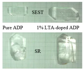
Fig.1. Pure and 1% LTA doped ADP crystal grown by SEST and SR
3. Characterizations
The powdered samples of pure and LTA-doped ADP crystals were subjected to powder X-ray diffraction (PXRD) analysis to determine their crystal structure and lattice parameters using a PW3710-based X-ray diffractometer equipped with Ni-filtered Cu Kα radiation (λ = 1.54056 Å). The diffraction patterns were recorded over a 2θ range of 10–70° at a scanning rate of 0.02° s⻹. The UV–Vis transmission spectra were recorded using a PerkinElmer Lambda 35 UV–Vis spectrophotometer in the wavelength range of 190–900 nm.
To investigate the influence of the growth technique and 1% LTA doping on the crystalline perfection of ADP crystals, high-resolution X-ray diffraction (HRXRD) analysis was performed. High-resolution rocking curves (diffraction curves, DCs) were recorded using a multicrystal X-ray diffractometer developed at the National Physical Laboratory (NPL), New Delhi [21].
RESULTS AND DISCUSSION
4.1. PXRD Studies
The PXRD patterns of the pure and LTA-doped ADP crystals are presented in Fig. 2. and compared with the corresponding JCPDS reference data. The diffraction peaks of the LTA-doped ADP crystal are consistent with those of pure ADP, and no additional diffraction peaks attributable to secondary crystalline phases are observed, indicating that LTA doping does not result in the formation of any detectable secondary crystalline phase. The sharp and well-defined diffraction peaks confirm the good crystallinity of the grown crystals.
Compared with pure ADP, the LTA-doped crystal exhibits a noticeable variation in the relative intensities of the diffraction peaks, with several reflections showing enhanced intensity. Such changes may be associated with modifications in crystal growth, crystallinity, or preferred orientation induced by the presence of LTA during crystal growth. These observations suggest that LTA doping influences the structural ordering and growth characteristics of ADP crystals. However, PXRD alone cannot conclusively establish the incorporation of LTA species into specific lattice sites.
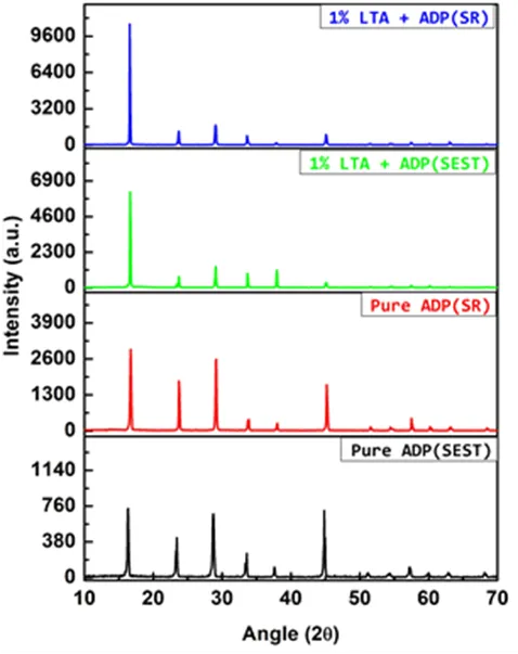
Figure 2: PXRD Patterns of Pure and 1% LTA Doped ADP Crystal Grown by SEST and SR
4.2. Optical Transmission Spectral Studies
Optical transparency is an important parameter for assessing the suitability of single crystals for optical and photonic applications. Pure and 1 mol% LTA-doped ADP single crystals of nearly identical thickness (~2 mm) were selected for optical characterization. The UV–Vis–NIR transmission spectra recorded for the crystals are presented in Fig. 3. All the investigated crystals exhibit good optical transparency over the measured wavelength range, indicating low optical absorption in the corresponding region.
The SEST-grown pure and 1 mol% LTA-doped ADP crystals exhibit transmittances of approximately 69% and 80%, respectively, whereas the corresponding SR-grown crystals show transmittances of approximately 73% and 85%, respectively. The higher transmittance of the SR-grown crystals compared with their SEST-grown counterparts indicates comparatively lower optical losses and improved optical quality. The enhancement in transmittance upon LTA doping may be associated with modifications in the crystal growth and defect characteristics induced by the dopant. Among the investigated samples, the SR-grown 1 mol% LTA-doped ADP crystal exhibits the highest optical transmittance, indicating its promising potential for optical, electro-optic, and nonlinear-optical applications.
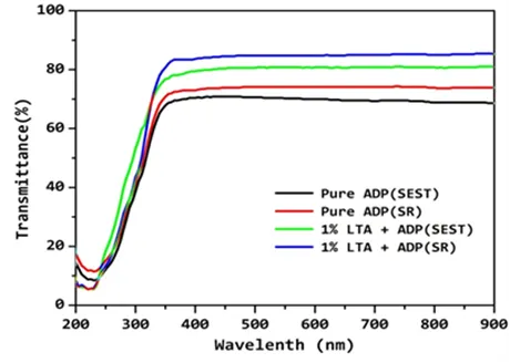
Figure 3: UV-VIS- NIR Transmission Spectra of Pure and 1% LTA Doped ADP Crystal Grown by SEST and SR
4.3. High resolution X-ray Diffraction Analysis
To evaluate the crystalline perfection of the specimen crystals, high-resolution X-ray diffraction (HRXRD) analysis was carried out. A multicrystal X-ray diffractometer developed at National Physical Laboratory (NPL), New Delhi [21] was used to record high-resolution rocking or diffraction curves (DCs). In this system, a fine focus (0.4 × 8 mm2; 2 kW Mo) X-ray source energized by a well-stabilized Philips X-ray generator (PW 1743) was employed. The well-collimated and monochromated MoKα1 beam obtained from the three monochromator Si crystals set in dispersive (+,-,-) configuration has been used as the exploring X-ray beam. This arrangement improves the spectral purity (Δλ/λ<< 10-5) of the MoKα1 beam. The divergence of the exploring beam in the horizontal plane (plane of diffraction) was estimated to be << 3 arc sec. The specimen crystal is aligned in the (+,-,-,+) configuration. Due to dispersive configuration of the third monochromator crystal with respect to the 2nd monochromator, the spectral quality of the diffracted beam emerged from the 3rd third monochromator is highly perfect (∆λ/λ~10-5; horizontal divergence >> 3 arc seconds) and hence though the lattice constant of the monochromator crystal and the specimen are different, the unwanted experimental dispersion broadening in the diffraction curve of the specimen crystal [∆FWHM=∆λ/λ(tanθM-tanθS); θM and θS are being the Bragg diffraction angles of monochromator and the specimen crystals] is insignificant. The advantage of dispersion configuration (+,−,−) over the non-dispersive configuration (+,−,+) of monochromator is well described in our recent article. The specimen can be rotated about a vertical axis, which is perpendicular to the plane of diffraction, with minimum angular interval of 0.4 arc sec. The diffracted intensity is measured by using a scintillation counter. The DCs were recorded by changing the glancing angle (angle between the incident X-ray beam and the surface of the specimen) around the Bragg diffraction peak position θB (taken zero as reference point) starting from a suitable arbitrary glancing angle (θ). The detector was kept at the same angular position 2θB with wide opening for its slit, the so-called ω scan [22]. The omega scan is very appropriate to record the short range order scattering caused by the defects or by the scattering from local Bragg diffractions from agglomerated point defects or due to low angle and very low angle structural grain boundaries [23]. Before recording the diffraction curve, to remove the non-crystallized solute atoms remained on the surface of the crystal and also to ensure the surface planarity, the specimens were first lapped and chemically etched in a non-preferential etchant of water and acetone mixture in 1:2 volume ratio.
(a)
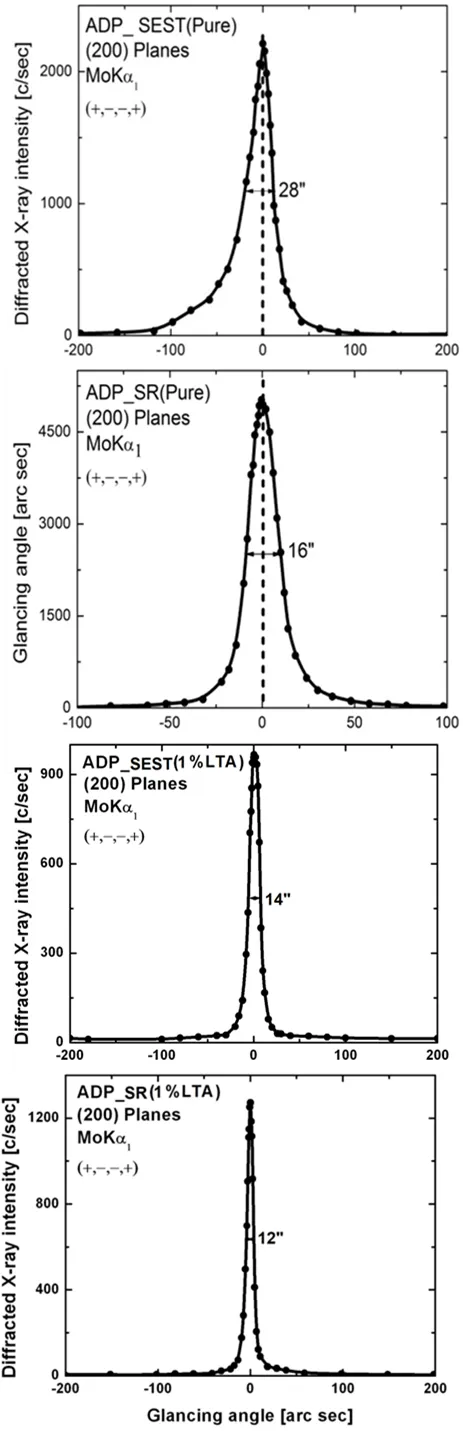
Figure 4: HR-XRD Diffraction Curves of Pure and 1% LTA Doped ADP Crystal Grown by SEST and SR
Fig. 2.4 (a) shows the high-resolution diffraction curve (DC) recorded for SEST grown pure ADP crystal using (200) diffracting planes in symmetrical Bragg geometry by employing the multicrystal X-ray diffractometer with MoKα1 radiation. As seen in the figure, the DC contains a single peak and indicates that the specimen is free from structural grain boundaries. The full width at half maximum (FWHM) of this curve is 28 arc sec which is somewhat higher than that expected from the plane wave theory of dynamical X-ray diffraction [24] and reveals the presence of point defects and their aggregates.
Fig. 4 (a) presents the HRXRD diffraction curve of the SEST-grown pure ADP single crystal. The curve consists of a single diffraction peak with an FWHM of approximately 28 arc sec. The absence of satellite or multiple diffraction peaks indicates that the crystal is free from detectable internal structural grain boundaries. However, the relatively broader peak suggests the presence of point defects and/or their aggregates, which contribute to the observed angular broadening of the diffraction curve.
Fig. 4 (b) shows the HRXRD diffraction curve obtained for the SR-grown pure ADP crystal. The curve exhibits a sharp and nearly symmetric single diffraction peak with an FWHM of approximately 16 arc sec. The significant reduction in FWHM compared with the SEST-grown pure ADP crystal indicates a substantial improvement in crystalline perfection. The sharp and symmetric nature of the peak, together with the absence of satellite reflections, confirms that the specimen does not possess significant internal structural grain boundaries. The observed narrow rocking curve is consistent with the characteristics expected for a highly perfect single crystal according to the dynamical theory of X-ray diffraction.
Fig. 4 (c) illustrates the HRXRD diffraction curve of a 1 mol% LTA-doped SEST-grown ADP single crystal. A single, sharp diffraction peak with an FWHM of approximately 14 arc sec is observed. No satellite peaks are detected, indicating the absence of significant internal structural grain boundaries or epitaxial layers. The considerably reduced FWHM compared with the SEST-grown pure ADP crystal demonstrates that LTA incorporation leads to an improvement in crystalline perfection and lattice uniformity.
Fig. 4 (d) represents the HRXRD diffraction curve of the 1 mol% LTA-doped SR-grown ADP single crystal. The curve exhibits a highly sharp and symmetric single diffraction peak with an FWHM of approximately 12 arc sec. The absence of satellite reflections indicates that the crystal is free from detectable internal structural grain boundaries and other significant extended defects. The lowest FWHM among the investigated samples signifies the minimum angular spread and superior crystalline perfection of the LTA-doped ADP crystal grown by the SR technique.
CONCLUSION
High-quality pure and 1 mol% L-tartaric acid (LTA)-doped ammonium dihydrogen phosphate (ADP) single crystals were successfully grown by the slow evaporation solution technique (SEST) and Sankaranarayanan–Ramasamy (SR) technique under ambient conditions. Powder X-ray diffraction analysis confirmed that LTA incorporation does not induce any structural phase transformation, although noticeable changes in the relative intensities of the diffraction peaks indicate modification of the crystallographic orientation. The grown crystals exhibited excellent optical transparency over the investigated spectral range, demonstrating their suitability for optical applications. HRXRD analysis further revealed good crystalline perfection, particularly for the 1 mol% LTA-doped crystal grown by the SR technique. Overall, the combined structural, optical, and crystalline-quality results establish 1 mol% LTA-doped ADP grown by the SR technique as a promising material with enhanced characteristics compared with pure ADP, making it attractive for potential nonlinear optical and optoelectronic device-fabrication applications.
REFERENCES

Mohd. Hasmuddin, A Comparative Study of Pure and L-Tartaric Acid Doped ADP Single Crystals Grown by SEST and SR Technique, Int. J. Sci. R. Tech., 2026, 3 (10), 713-718. https://doi.org/10.5281/zenodo.23311211
 10.5281/zenodo.23311211