

We use cookies to ensure our website works properly and to personalise your experience. Cookies policy
1Department of Medicine, King George’s Medical College
2Department of Radiodiagnosis, King George’s Medical College
Pial arteriovenous malformations (AVMs) are rare congenital vascular anomalies and an important cause of spontaneous intracranial haemorrhage in adolescents and young adults. We report the case of an 18-year-old female who presented with a one-week history of worsening headache and projectile vomiting without fever or focal neurological deficits. Magnetic resonance imaging (MRI) revealed a subacute intraparenchymal haemorrhage in the left parieto-occipital region extending into the splenium of the corpus callosum, with intraventricular extension into the left lateral ventricle. Digital subtraction angiography (DSA) demonstrated a 12 × 8 mm high-flow pial AVM supplied by the M3 branch of the left middle cerebral artery and draining into a cortical vein. The patient underwent successful endovascular Onyx embolisation with symptomatic improvement. This case underscores the importance of considering intracranial vascular malformations in young patients with persistent headache and vomiting, and highlights DSA and endovascular embolisation as key diagnostic and therapeutic modalities.
Cerebral arteriovenous malformations (AVMs) are congenital vascular anomalies characterised by direct shunting of blood from the arterial circulation to the venous system through a dysplastic nidus without an intervening capillary network. Although relatively uncommon, with an estimated annual incidence of approximately 1.1 per 100,000 person-years, AVMs are a major cause of spontaneous intracranial haemorrhage in children and young adults. [1,2]
Pial AVMs arise from pial vessels and are associated with high-flow haemodynamics, increasing the risk of rupture and haemorrhage. In adolescents and paediatric patients, haemorrhage is the most common mode of presentation, occurring in up to 80% of cases. [2,4] Clinical manifestations may range from seizures and focal neurological deficits to non-specific symptoms such as headache, nausea and vomiting.
Small AVMs, particularly those measuring less than 3 cm, paradoxically carry a higher risk of rupture because of elevated intranidal pressure. [3,4] Lesions involving deep structures, such as the corpus callosum, or those associated with intraventricular haemorrhage, are linked to increased morbidity and greater therapeutic complexity.
We report the case of a ruptured small pial AVM in an 18-year-old female who presented solely with headache and vomiting despite having a normal neurological examination.
Case Presentation
An 18-year-old female presented with a one-week history of progressively worsening headache associated with multiple episodes of projectile vomiting. There was no history of fever, seizures, trauma, loss of consciousness, visual disturbances, ear discharge or focal weakness.
On examination, the patient was haemodynamically stable and fully conscious with a Glasgow Coma Scale (GCS) score of 15/15. Meningeal signs were absent, and fundoscopy showed no evidence of papilloedema. Neurological examination revealed no focal deficits.
Given the persistence of her symptoms, neuroimaging was performed.
Imaging Findings
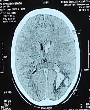
Non-contrast computed tomography (NCCT) of the head demonstrated an intraparenchymal haemorrhage in the left temporal region with surrounding oedema and intraventricular extension (Figure 1).
Magnetic resonance imaging (MRI) of the brain further characterised the lesion, demonstrating a subacute intraparenchymal haemorrhage extending into the splenium of the corpus callosum, with surrounding vasogenic oedema (Figures 2 and 3). Magnetic resonance angiography (MRA) suggested an underlying vascular malformation in the left temporal region (Figure 4).
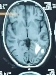
Figure 2
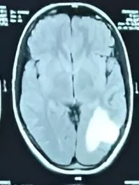
Figure 3
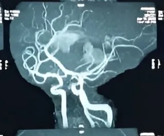
Figure 4
Subsequent digital subtraction angiography (DSA) confirmed a small high-flow pial AVM measuring approximately 12 × 8 mm, supplied by the M3 segment of the left middle cerebral artery and draining into a cortical vein (Figure 5). Post-embolisation angiographic imaging demonstrated successful obliteration of the nidus following Onyx embolisation (Figure 6).
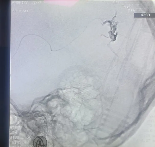
Figure 5
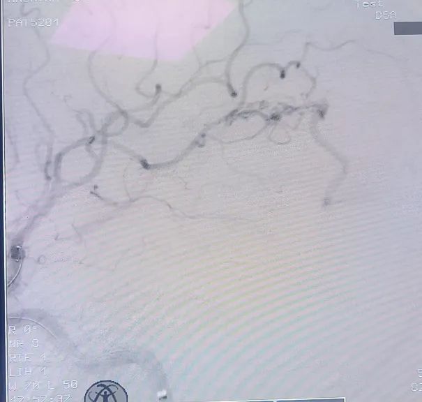
Figure 6
Management
Considering the small size of the nidus and its deep-seated location adjacent to the splenium of the corpus callosum, the patient underwent endovascular treatment using Onyx embolisation. Onyx, a non-adhesive liquid embolic agent, permits controlled penetration into the AVM nidus, facilitating effective obliteration while minimising the risk of re-bleeding.
Discussion
Brain AVMs are clinically significant vascular lesions because of their potential to cause catastrophic intracranial haemorrhage, particularly in younger individuals. Haemorrhage remains the most common presentation in paediatric and adolescent patients and is frequently associated with substantial neurological morbidity.
The present case is noteworthy because the patient presented with persistent headache and vomiting despite having no focal neurological deficits or papilloedema, even though imaging revealed a ruptured AVM with intraventricular extension. This highlights that a normal neurological examination does not exclude serious intracranial pathology in young patients presenting with 'red flag' symptoms. [2,4]
Small AVMs are recognised to have a greater propensity for rupture than larger lesions because of higher intranidal pressures and altered haemodynamics. [3,4] Furthermore, lesions involving deep cerebral structures and those associated with intraventricular haemorrhage are considered to carry a higher risk of adverse outcomes.
Digital subtraction angiography remains the gold standard investigation for cerebral AVMs because it provides detailed information regarding the angioarchitecture, including feeding arteries, nidus size and venous drainage.[5] In this case, DSA was essential for establishing the definitive diagnosis and guiding therapeutic planning.
Endovascular embolisation using Onyx has emerged as an effective minimally invasive treatment option for selected AVMs. Compared with older embolic agents such as n-butyl cyanoacrylate (n-BCA), Onyx offers slower solidification and greater controllability, allowing deeper penetration into the nidus and achieving higher rates of complete obliteration. [6–8]
CONCLUSION
This case highlights the importance of considering cerebral vascular malformations in adolescents presenting with persistent headache and vomiting, even in the absence of focal neurological deficits or papilloedema. Early neuroimaging plays a pivotal role in identifying underlying intracranial pathology.
Digital subtraction angiography remains indispensable for the definitive diagnosis and treatment planning of AVMs. Endovascular Onyx embolisation offers an effective and minimally invasive therapeutic option for small, deep-seated pial AVMs, with the aim of preventing recurrent haemorrhage and long-term neurological complications.
REFERENCES



Satyendra Kumar Sonkar1*, Pranjal Gulati1, Bhawana Lalwani1, Saurabh Kumar2, A Sinister Headache Without Focal Neurological Deficits: A Case Of Ruptured Pial Arteriovenous Malformation, Int. J. Sci. R. Tech., 2026, 3 (8), 1134-1137. https://doi.org/10.5281/zenodo.22207813
 10.5281/zenodo.22207813