

We use cookies to ensure our website works properly and to personalise your experience. Cookies policy
Department of Pharmacy Practice Santhiram College of Pharmacy Nandyal. 518112 Andhra Pradesh
BACKGROUND: A large lung mass in an older adult needs a careful and systematic evaluation. When malignancy is suspected, tissue confirmation is important. Superior sulcus (Pancoast-type) tumour is defined by its location at the lung apex, and an imaging impression alone cannot establish the diagnosis. CASE PRESENTATION: A 76-year old man who was a smoker and had coronary artery disease presented with gradually worsening breathlessness on exertion for 4 months and intermittent, non-radiating chest pain for 1 month. He also had reduced appetite and a recorded weight of 41kg. He did not report cough or fever, breath sounds were mildly reduced bilaterally. At the outpatient consultation, his blood pressure was 80/60mmHg and SPO2 was 97-98%. INVESTIGATIONS: Non-contrast HRCT of the chest showed a large irregular soft-tissue mass measuring 52×66×55mm in the apical anterior segment of the right upper lobe, with adjacent consolidation and pre-tracheal and bilateral para-tracheal lymph nodes. No pleural effusion was seen. The radiologist reported “Carcinoma of lung (Pancoast tumour)” and suggested a contrast study. Haemoglobin values were 12.4, 11.7 and 11.8 g/dl on 02, 05, and 06 July 2026, respectively. Renal and liver function tests were the laboratory’s printed reference ranges, Echocardiography showed an ejection fraction of 58% with grade 1 diastolic dysfunction, and ECG showed sinus tachycardia. MANAGEMENT: During admission, the patient received the treatment documented in the hospital record, including an injectable antibiotic, pantoprazole, nebulized medications and analgesics, while his cardiac medicines were continued. CT-guided biopsy of the right apical lesion was then performed under local anaesthesia. Five 2-cm tissue cores were obtained and placed in formalin. NO procedure related complication was recorded. OUTCOME: The patient was discharged on 07 July 2026. The histopathology report was not available in the supplied record, so the final diagnosis, stage, definitive treatment and subsequent follow-up could not be established. CONCLUSION: The HRCT findings in this older smoker raised for suspicion for a superior sulcus carcinoma, but the diagnosis could not be confirmed without tissue. The case highlights the importance of histopathological confirmation, appropriate contrast-enhanced staging, multi-disciplinary assessment and careful medication management in an older patient with cardiovascular disease.
Lung cancer is a major cause of cancer-related illness and death worldwide (1), and cigarette smoking remains an important risk-factor (2). A pulmonary lesion measuring more than 3cm is generally described as a mass rather than a nodule (3). Therefore, guidance intended for the management of small incidental nodules, such as the Fleischner Society recommendations, does not directly address the evaluation of a large symptomatic pulmonary mass (4).
The possible causes of a pulmonary mass in an older adult area broad. They include primary or metastatic malignancy, lymphoma, bacterial, mycobacterial or fungal infection, and inflammatory lesions. HRCT helps define the size and margins of a lesion and can show associated parenchymal changes, lymph nodes and possible chest-wall involvement. However, imaging findings alone cannot provide a histological diagnosis (5). When lung cancer is suspected, current guidance supports obtaining an adequate tissue diagnosis and staging information using the least invasive appropriate approach (5–7).
Superior sulcus, or Pancoast, tumours arise at the lung apex and may involve nearby structures such as the lower brachial plexus, first rib, vertebrae, subclavian vessels or stellate ganglion (8,9). The classical Pancoast-Tobis syndrome includes pain in the C8-T2 distribution, Horner syndrome and wasting of the intrinsic hand muscles (8). In older adults, coexisting coronary disease, multiple medicines and antiplatelet treatment can make diagnostic procedures more complex. These factors also make multidisciplinary care, including clinical pharmacy input, relevant (10–12).
This case describes an older man with a larger right apical lung mass documented in the hospital record. The imaging report raised the possibility of Pancoast-type carcinoma, but histopathological confirmation was not available. The case is presented to emphasise the difference between a radiological suspicion and a confirmed diagnosis, the steps involved in evaluating a suspected apical lung malignancy, and the medication-safety considerations in an older patient with coronary artery disease undergoing CT-guided biopsy.
CASE PRESENTATION:
A 76-year-old male was admitted in pulmonology department on 02 July 2026 for evaluation of respiratory and chest symptoms. The initial assessment documented breathlessness developing over approximately four months. The breathlessness was described as initially occurring with minimal activity, including indoor activity, and then becoming gradually progressive and more noticeable with exertion and distant walking, Intermittent chest pain had been present for approximately one month.
The history also documented cough, cold, and fever in the clinical course, The patient was described as a smoker and non-alcoholic, with reduced appetite and weight loss noted in the personal history. The record also contains a cardiovascular history, including coronary artery disease on medication and previous valvular repair. No significant past medical or family history was otherwise clearly documented.
On documented examination, oxygen saturation was approximately 97-98% on room air. Recorded blood pressure was around 110/70mmHg, with pulse values around 100-102 beats/min in some observations. The record did not document a classical Horner syndrome or a definite brachial-plexus neurological syndrome.
The initial working approach included evaluation for a possible pulmonary infective process, with CBC, renal and liver function testing, electrolytes, viral markers, ECG, echocardiography and HRCT chest planned/performed. The subsequent imaging findings shifted the diagnostic focus toward a structural right apical lung lesion.
CLINICAL TIMELINE:
| Stage | Documented clinical event |
|---|---|
| â“4 months before admission | progressive breathlessness, initially with minimal activity and later worsening on exertion/distant walking. |
| â“1 month before admission | Intermittent chest pain documented. |
| July 2026 admission | clinical assessment for dyspnea/chest pain; possible pulmonary infective process considered initially. |
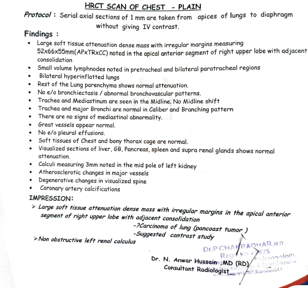
| During evaluation | HRCT chest demonstrated a 52×66×55mm irregular right upper-lobe apical/anterior mass with adjacent consolidation. |
| Radiological impression | Carcinoma of lung/possible Pancoast tumour suggested; contrast study recommended. |
| Subsequent procedure | CT-guided biopsy of the right apical lung mass performed; two core samples obtained and placed in formalin; no immediate complication documented. |
| Available endpoint | Histopathological report was not present |
DIAGNOSTIC INVESTIGATIONS:
HEMATOLOGY:
| Parameter | Result 1 | Result 2 |
|---|---|---|
| Haemoglobin | 11.8g/dl | 12.4g/dl |
| Total WBC count | 7,800/cmm | 12,800/cmm |
| Polymorphs | 17% | 69% |
| Lymphocytes | 19% | 22% |
| Eosinophils | 04% | 01% |
| Monocytes | 06% | 08% |
| Basophils | 00% | 00% |
| Platelets | 3.85 lakh/cmm | 3.17 lakh/cmm |
| Blood group | O positive | - |
RENAL FUNCTION, ELECTROLYTES AND LIVER FUNCTION:
| Test | Documented result |
|---|---|
| Random blood sugar | 109mg/dl |
| Sodium | 135mmol/L |
| Potassium | 4.3mmol/L |
| Chloride | 100mmol/L |
| Blood urea | 18mg/dl |
| Serum creatinine | 4.4mg/dl |
| Uric acid | - |
| Total bilirubin | 0.6mg/dl |
| Direct bilirubin | 0.3mg/dl |
| SGOT/AST | 26U/L |
| SGPT/ALT | 15U/L |
| Alkaline phosphatase | 49U/L |
| Total proteins | 6.4g/dl |
| Albumin | 3.6g/dl |
The creatinine value of 4.4mg/dl is clinically important and should be incorporated into medication-dose assessment and contrast-risk evaluation. The record does not provide a documented chronic kidney disease diagnosis, baseline creatinine or longitudinal renal trend, so CKD should not be inferred solely from this single value.
URINE ANALYSIS:
| Parameter | Result |
|---|---|
| Colour | Yellow |
| specific gravity | 1.020 |
| PH | Acidic |
| Protein | 1+ |
| Glucose | Nil |
| Urobilinogen | Nil |
| Ketone bodies | Negative |
| Bilirubin | Negative |
| Pus cells | 2-3/HPF |
| RBC | Nil |
| Epithelial cells | 3-4/HPF |
HRCT CHEST:
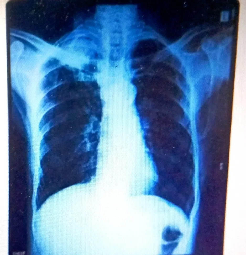
X-RAY:
ULTRASOUND OF ABDOMEN:
Abdominal ultrasonography reported no obvious sonological abnormality. The spleen was approximately 7.26 cm, and both kidneys were described as normal in size and echogenicity,
with right kidney approximately 8.5×3cm and left kidney approximately 8.3×4.6cm. the urinary bladder was minimally distended, the report did not identify an abdominal mass or obvious metastatic lesion.
ULTRASOUND OF CHEST:
Ultrasound of the chest documented no evidence of pleural effusion on either side. The liver, gallbladder and spleen were described without obvious sonological abnormality.
ECHOCARDIOGRAPHY:
Cardiac imaging showed normal mitral and aortic valve morphology, normal left atrial and right-sided chamber dimensions, TAPSE approximately 18mm, no regional wall-motion abnormality at rest, and preserved LV systolic function with an ejection fraction of approximately 58%. The report described trivial mitral, aortic and tricuspid regurgitation, without pulmonary hypertension or pericardial effusion.
DIAGNOSTIC CONSIDERATIONS:
| Possibility | Evidence in this case | Current status |
|---|---|---|
| Primary apical lung malignancy/superior sulcus tumour | Large irregular right apical mass; radiologist specifically suggested carcinoma/Pancoast tumour. | Strong radiological suspicion; histology not available in record. |
| Pulmonary infection with focal consolidation | Adjacent consolidation and initial provisional management for possible pneumonia. | Considered initially; mass morphology requires tissue diagnosis. |
| other primary or metastatic apical lesion. | An apical mass can have multiple etiologies. | Cannot be excluded without histopathology and complete staging. |
TISSUE DIAGNOSIS:
A subsequent progress note documents CT-guided biopsy of the right apical lung mass under local anesthesia. The record states that two cores were obtained and stored in formalin and that the procedure was uneventful, with no immediate complication documented.
This step is consistent with the recommended principle that a suspected superior sulcus tumour requires tissue diagnosis before definitive oncologic treatment. Consequently, this manuscript intentionally does not label the lesion as histologically confirmed lung cancer, adenocarcinoma, squamous-cell carcinoma or any other specific malignancy.
THERAPEUTIC MANAGEMENT:
The initial inpatient included supportive treatment for the presenting respiratory symptoms and possible infective component. Documented medications included antimicrobial therapy, gastric acid suppression, nebulized respiratory treatment and continuation of cardiac medications.
DISCUSSION:
This case highlights the importance of investigating persistent respiratory symptoms in older adult. HRCT revealed a 52×66×55mm right apical lung mass with adjacent consolidation and para-tracheal lymph nodes, raising suspicion of a superior sulcus (Pancoast) tumour. Although the patient lacked features such as Horner syndrome and severe shoulder or arm pain, their absence does not exclude the diagnosis. Overall, the case emphasizes early evaluation of persistent symptoms, appropriate imaging, tissue confirmation, and multidisciplinary management, particularly when significant renal and cardiovascular comorbidities are present.
CLINICAL PERSPECTIVE:
Persistent dyspnea and chest pain in an older adult should prompt further evaluation. Imaging revealed a large right apical lung mass suspicious for a superior sulcus tumour. Tissue confirmation and complete staging are essential before definitive treatment.
CONCLUSION:
This case highlights the importance of early evaluation of persistent respiratory symptoms. Imaging identified a large right apical lung mass suspicious for a superior sulcus tumour. Histopathological confirmation and complete staging are essential to establish the diagnosis the diagnosis and guide appropriate treatment.
REFERENCES




Syed Muskan, Dasari Madhusree, Venenimuddala Padma Priya, Kallapari Tharun, A Right Upper-Lobe Pulmonary Mass with Suspected Pancoast-type Carcinoma: A Case Report, Int. J. Sci. R. Tech., 2026, 3 (10), 896-901. https://doi.org/10.5281/zenodo.23392124
 10.5281/zenodo.23392124