

We use cookies to ensure our website works properly and to personalise your experience. Cookies policy
Genba Sopanrao Moze College of Pharmacy, Wagholi, Pune, Maharshtra, India.
Cancer continues to be a leading cause of morbidity and mortality worldwide, making it crucial to develop more efficient, specific, and durable therapeutic options. Conventional treatment options, including surgery, chemotherapy, and radiotherapy, have demonstrated significant clinical efficacy; however, tumor recurrence, innate and acquired resistance, toxic side effects, and damage to normal tissues limit their clinical implementation. The cancer-immunity cycle is a newly described series of events that involve the immune system and are necessary for an effective anti-tumor immune response. The process starts with the release of neoantigens from dying tumor cells via immunogenic cell death. Dendritic cells and other antigen-presenting cells (APCs) receive tumor-associated antigens, which are then presented to naïve T lymphocytes in the lymph nodes. Consequently, cancer immunotherapy has emerged as a promising strategy to fight tumors by stimulating and enhancing endogenous anti-cancer immune responses. The cancer immunotherapy area has undergone a paradigm shift from non-specific to tailored and precise treatments. Immune checkpoint inhibitors targeting programmed cell death protein-1 (PD-1) and programmed death-ligand 1 (PD-L1) and cytotoxic T-lymphocyte-associated protein 4 (CTLA-4) have significantly improved the overall survival of patients with various tumor types. Chimeric antigen receptor (CAR) T-cell therapy, which involves genetic modification of the patient’s T cells to bind to specific tumor-associated antigens, has also enhanced cancer treatment options. Cancer vaccines, monoclonal antibodies, bispecific antibodies, cytokines, and oncolytic viruses are other effective cancer immunotherapy options that have shown promising preclinical and clinical results. Current research is focused on combination immunotherapy, personalized cancer vaccines, biomarker-driven approaches, and novel cell-based cancer treatments, including updated versions of CAR T cells and T-cell receptor (TCR) therapies. Nevertheless, major therapeutic challenges, including immune-related toxicities, tumor heterogeneity and antigen variability, innate and acquired resistance, and cost, hinder the widespread application of immunotherapy. Further understanding of tumor immunology, genetic engineering, and precision medicine will improve cancer immunotherapy and patient outcomes. Combination immunotherapy is a powerful approach to increase therapeutic efficacy and overcome tumor resistance.
What is Cancer? and Cause of Cancer.
Cancer is a disease that has become the leading cause of death gradually, especially in countries where junk food is available and physical activity is scarce. Cancer cannot be transferred from one person to another, but it is growing rapidly. According to the World Health Organization (WHO), there were 20 million new cancer cases in 2020, and 10 million people died from the disease in that year [1]. These numbers keep rising every day in the world. According to the forecast, the number of
people who die from cancer could reach about 35 million per year by 2050 [2]. These numbers are some of the reasons most of us have lost friends and family members to this disease or know someone who has been diagnosed with some kind of cancer. The occurrence of cancer around the world is not distributed uniformly. As Fig. 1 shows, the rate in highly industrialized Western countries is high, with reports of approximately 450 cancer cases for every 100,000 inhabitants. Lung, colon, breast, and prostate cancers are prevalent in those countries. While, rates reported in Asian countries are low, with only about 97 cases per 100,000 individuals. In those nations, cancers of the stomach and liver are more common than other types of cancer [3].
By analyzing the genomic sequence of the tumor DNA, it is possible to determine which environmental or lifestyle factors caused the cancer. For example, it can determine whether it was tobacco smoke that caused lung cancer, UV radiation that caused skin cancer, or previous chemotherapy that suppressed the immune system and allowed secondary tumors to grow.[4]
It is not common that cancer can be transferred from one person to another;[5] the exceptions are cases associated with pregnancy and occasionally organ donors. However, there are infectious diseases that can be transmitted and cause cancer, such as hepatitis B, Epstein–Barr, HPV, or HIV infection.[6]
History of Cancer
Cancer is the second leading cause of death in the world after cardiovascular diseases. Half of men and one third of women in the United States will develop cancer during their lifetimes. Today, millions of cancer people extend their life due to early identification and treatment. Cancer is not a new disease and has afflicted people throughout the world. The word cancer came from a Greek words karkinos to describe carcinoma tumors by a physician Hippocrates (460-370 B.C), but he was not the first to discover this disease. Some of the earliest evidence of human bone cancer was found in mummies in ancient Egypt and in ancient manuscripts dates about 1600 B.C. The world’s oldest recorded case of breast cancer hails from ancient Egypt in 1500 BC and it was recorded that there was no treatment for the cancer, only palliative treatment. According to inscriptions, surface tumors were Surgically removed in a similar manner as they are removed today [7-8].
IMPORTANCE OF IMMUNOTHERAPY
ADVANCES IN IMMUNOTHERAPY OF CANCER
Immunotherapy : a type of cancer therapy that enhances the body's very own immune system so that it can target and destroy cancer cells, often times preserving healthy tissue better than traditional chemotherapy. Depending on the treatment and type of cancer, it is usually administered through IV infusions, injections, topical creams or pills [9].
How does it work..?
Immunotherapy modulates or boosts the patient's very own immune system so it can recognize and destroy tumors, unlike chemotherapy or radiation which destroy cancer cells directly. The main reason it is used, is because cancer cells often times exploit immunological checkpoints (natural brakes on T cells) so they can avoid being targeted by the immune system. Immunotherapy either directly empowers immune cells to fight the tumor or it takes those brakes away [10].
• Immune checkpoint inhibitors
• Adoptive cell therapy
• Cancer vaccines
• CAR-T cell therapy
• Monoclonal antibodies
• Bispecific antibodies
• Oncolytic viruses therapy
• Tumor microenvironment
1. Immune checkpoint inhibitors
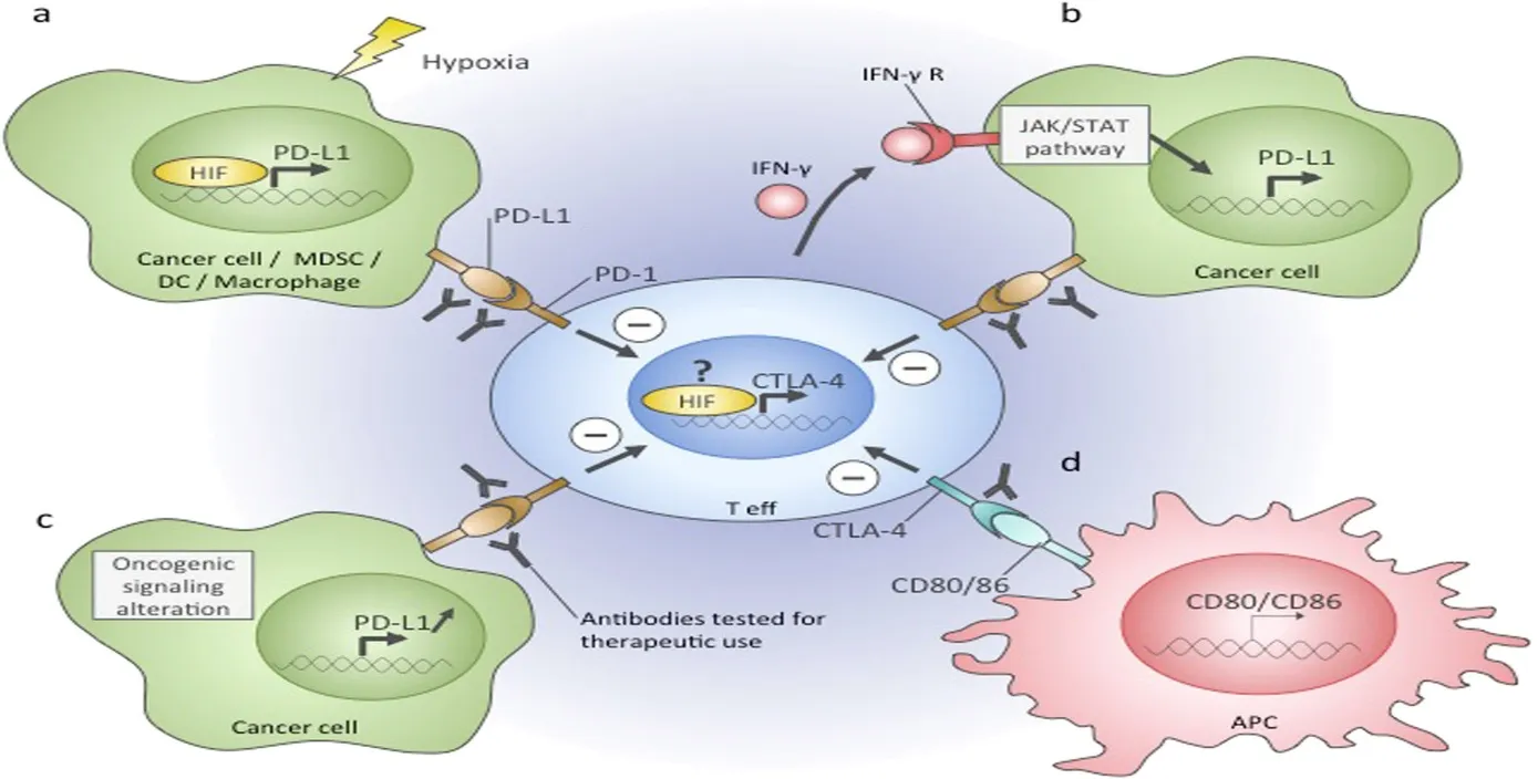
Figure 1: Mechanisms Regulating PD-L1 and CTLA-4 Immune Checkpoints in Cancer, Including Hypoxia, Ifn-Γ/Jak-Stat Signaling, Oncogenic Alterations, and APC Interactions[42].
Immunosuppressive molecules are referred to as immune checkpoints. Immune checkpoints are crucial for preserving immunological tolerance and controlling immune responses and avoiding tissue damage. However, by inhibiting immune cell function during tumor formation and activation, increased checkpoint expression can mediate tumor immune evasion. Fortunately, immune checkpoint inhibitors can improve or restore the body's antitumor immune response by preventing the transmission of immunosuppressive signals. Cytotoxic T lymphocyte antigen 4 (CTLA-4), programd cell death protein 1 (PD-1), and programd cell death ligand 1 (PD-L1) are the primary immunological checkpoint representatives. Activated CD8+ and CD4+ T lymphocytes express CTLA-4. CTLA-4 competes with the costimulatory receptor CD28 to bind to ligands B7-1 and B7-2 produced on antigen-presenting cells (APCs) during the early stages of T cell activation. T cell proliferation and IL-2 release are therefore suppressed as a result of the downstream negative regulation of immune response [11]. In the end, it prevents the body from responding appropriately to tumor cells. PD-1 is a transmembrane protein that is expressed on Tcells, just as CTLA-4. Tumor cells, APCs, and T cells themselves can all express PD-L1, one of its ligands. T cell responsiveness to T cell receptor (TCR) stimulation signals is decreased when PD-1 binds to PD-L1 via PI3K-AKT and as well as T lymphocytes. T cells' sensitivity to T cell receptor (TCR) stimulation signals is decreased when PD-1 binds to PD-L1 via the PI3K-AKT and JAK-STAT signaling pathways. The short anticancer mechanism of CTLA-4 and PD-1/PD-L1 blocking antibodies, which results in suppressive antitumor T cell responses. It is the first immune checkpoint inhibitor to receive clinical approval. Twenty percent of patients with metastatic melanoma who received ipilimumab lived for more than four years, and only a small fraction survived for ten years or more [12] . Additionally, ipilimumab is frequently used to treat cancers like prostate, renal, and lung cancer.
However, it is not as effective as metastatic melanoma. Efficiency levels below 10% are typically considered inadequate. In general, the antitumor therapy PD-1/PD-L1 blockers work better than CTLA-4 blockers. The clinical use of immune checkpoint inhibitors faces limitations due to their comparatively modest response rates along with a fairly elevated level of treatment-related toxicity. Certain biomarkers are capable of forecasting both the therapeutic outcomes and the adverse reactions associated with immune checkpoint blockade therapy. This offers hope for the broad ation of immune checkpoint blockade therapy. A greater mutational burden and higher neoantigen loads show a significant link to the clinical benefit derived from anti-CTLA-4 antibodies The expression of tumor PD-L1 was significantly linked to the overall response rate (ORR) achieved with PD-1/PD-L1 inhibitors. The ORR stood at 34.1% for the PD-L1 positive group, compared with 19.9% for the PD-L1 negative group [13] . Such biomarkers can prove useful in identifying cancer patients likely to respond well to treatment with immune checkpoint blockade. At the same time, the rise in CD177 and CEACAM1 expression is tightly linked to colitis following ipilimumab therapy. In other words, for individuals undergoing ipilimumab treatment, CD177 and CEACAM1 may assist in taking preventive measures ahead of time so as to lessen the harm brought about by immune-related colitis. Furthermore, numerous prospective biomarkers are valuable for forecasting adverse effects and therapeutic response; examples include mismatch repair (MMR) deficiency, interferon-γ 11 [14] .
2. Adoptive cell therapy
Adoptive cell therapy (ACT) is a kind of immunotherapy. In this therapy, the immune cells sampled from patients were activated or genetically modified in vitro to augment their therapeutic potential, expanded in bulk and readministered to recipient patients to elicit antitumor effect. Immune competent cells consist of lymphokine-activated killer cells (LAK), T cells transduced with chimeric antigen receptors (CAR-T), natural killer cells transduced with chimeric antigen receptors (CAR-NK), T cells transduced with T cell receptors (TCR-T), cytokine-induced killer cells (CIK) and tumor-infiltrating lymphocytes (TILs). Among various kinds of ACT therapies, TILs, TCR-T cells, CAR-T cells have been investigated most extensively. Since their continuous encounter with exogenous nucleic acids, bacteria and archaebacteria have also evolved an efficient adaptive defense system known as CRISPR-Cas [15] . ACT therapy using T cells, which have been genetically manipulated, can specifically recognize tumor antigens through tumor-related TCR or CAR. A phase I clinical trial including 20 patients with advanced myeloma demonstrated that NY-ESO1-specific TCR-T cells mediate durable antigen-specific antitumor responses. The combination of TCR-T therapy and ACT has partially demonstrated its efficacy in anti-tumor immunity, but it differs from person to person, which not only leads to different anti-tumor efficacies, but may even endanger the lives of patients. To reduce the toxicity, it is of great importance to find personalized targetable antigens, which are expressed specifically on tumor cells but not expressed on healthy tissues. Neoantigens, which originates from tumor-specific mutations, are bound to become a research focus in the future. Besides, MHC class I complex expression is usually down-regulated in tumor cells [16] .
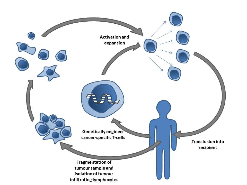
Figure 2: The Figure Illustrates, Adoptive Cell Therapy (Act): Tumor-Infiltrating Lymphocytes Are Isolated, Genetically Engineered, Activated and Expanded, Then Reinfused into the Patient to Target Cancer Cells [43] .
Because TILs and TCR-transduced T cells recognize the antigen in a restricted manner, this mechanism is likely to contribute to their limited efficacy. Moreover, although the ability of CAR-T cells to recognize tumor-associated antigens is not restricted by MHC, tumor-associated antigens are also expressed on normal tissues [17] . Therefore, ACT therapy faces the challenge of identifying tumor-specific targetable antigens. An MHC-dependent mechanism further restricts their clinical use. CAR-T cells rely on tumor surface proteins for antigen recognition, thereby removing the constraints imposed by the MHC to display antigens. CAR-T cell ACT relies on transgenic technology to drive CAR expression in T cells, and this receptor typically comprises an extracellular antigen-binding region (for instance, an antibody scFv) joined to an intracellular signaling domain that triggers T cell activation. Altering the signaling domain within the CAR architecture allows CAR to carry out diverse T cell roles, including amplification, cytokine secretion, and tissue selectivity. This can further broaden the reach and effectiveness of ACT. The extracellular glycoprotein CD19 serves as the most frequently used cellular target for CAR-T cell ACT, being a marker of B cell tumors. CD19 CAR-T has been applied successfully in treating patients suffering from refractory chronic lymphoma. For these three patients, the efficacy lasted over six months, and 2 of them achieved complete remission [18] . For adult and pediatric B cell malignancies, the CAR-T therapies (tisagenlecleucel and axicabtagene ciloleucel) received FDA approval in 2017. In addition, trials of CAR-T treatment aimed at CD22 and CD30 are likewise in progress. also underway. Moreover, the novel cellular targets for ACT involving CAR-T cells offer renewed optimism for turning relapse around. CAR-T cell therapy represents not just a promising approach for conquering blood cancers, but equally a modality possessing substantial therapeutic promise against solid tumors [19] .
3. Cancer Vaccines
Cancer vaccines are designed to expose the immune system to tumor-associated or tumor-specific anti-gens, thereby promoting antigen-specific immune activation and strengthening the response of T-cells against malignant cells. A wide variety of cancer vaccine platforms exist, including cell-based, RNA, DNA, and protein/peptide-based. Based on how cancer vac-cines are used clinically, they fall into two groups: preventive and therapeutic. A preventive cancer vaccine has the goal of stopping tumors from arising by provoking an immune response, whereas a therapeutic cancer vaccine is intended to eliminate tumor cells by triggering or strengthening tumor-specific immune reactions. Today, the preventive cancer vac-cines that have achieved relative success are those against cervical cancer, which work by targeting human papillomavirus (HPV). The approved cervical vaccines Gardasil and Cervarix brought about a de-cline in the prevalence of cervical intraepithelial neo-plasia in young women receiving the vaccine during adolescence [20,21] .
However, for the most part, they lose their validity once a person is battling cancer or a chronic HPV infection. Therefore, there is a need for therapeutic cancer vaccines, not just preventive ones. vaccines for cancer treatment are also required. It is heartening that sipuleucel-T (Provenge) is able to prolong the overall survival of patients with hormone-resistant prostate cancer by roughly 3 months .Although, following the use of cancer vaccines, numerous experiments have demonstrated that certain patients with advanced cancer experience favorable clinical outcomes, the rate of partial remission (PR) or complete remission (CR) is generally under 10%, or even under 5%. Up to now, in phase 3 randomized trials, no other therapeutic cancer vaccine has demon-strated notable clinical efficacy apart from sipuleucel-T. A cancer vaccine's effectiveness depends on immunogenicity, host immuno-suppression, preferential expression of tumor antigen, how the cancer vaccine is delivered . Because they are highly immunogenic, carry a lower risk of self-tolerance, are less commonly subject to tumor antigen deletions, and pose a lower risk of autoim-mune reactions, tumor-specific antigens—also called neoantigens—make ideal cancer vaccine targets [22] .
4. CAR-T cell therapy
Chimeric antigen receptors (CARs) are specialized receptors that are engineered to target a specific tumor antigen in order to functionally reprogram T lymphocytes. Since T lymphocytes are genetically modified to express these synthetic receptors that target cancer cells, this form of treatment can be called immunotherapy, gene therapy or cancer therapy. The concept of a chimeric T cell receptor, which merges antibody-derived variable regions (VH/VL) together with T cell receptor (TCR)-derived constant regions, was initially described in 1987 by a Japanese immunologist, Dr. Yoshikazu Kurosawa, and his team at the Institute for Comprehensive Medical Science in Aichi, Japan [23] .
Structure of CAR-T cells CARs consist of an
ectodomain, transmembrane domain and endodomain
Ectodomain:
ectodomain is the portion of a membrane protein that lies outside the cytoplasm and is exposed to the extracellular environment. In this instance, the ectodomain is made up of a signal peptide, an antigen recognition region, and a spacer. Correspondence: zhangxxi@sina.com 1Department of Hematology, Xinqiao Hospital, Third Military Medical University, Chongqing 400037, People’s Republic of China Full list of author information is available at the end of the article The function of a signal peptide is to guide the nascent protein into the endoplasmic reticulum. The scFv, which acts as the signal peptide of the ectodomain in a CAR, is composed of the variable regions of immunoglobulin heavy and light chains joined together by a flexible linker. An antigen recognition domain is typically an scFv with a simple ectodomain, along with more exotic recognition components, and it can be used to recognize any antigen provided that it can bind targets with high affinity. The spacer provided the connection between the antigen binding domain and the transmembrane domain. The hinge region of IgG1 is the simplest type of spacer, and it is sufficient for most scFv-based constructs [24] .
Transmembrane domain:
The transmembrane domain starts at the part of the endodomain closest to the membrane. It consists of a hydrophobic alpha-helix that crosses the membrane. How stable the receptor is depends on the transmembrane domain. If the natural CD3zeta transmembrane is present, the artificial TCR can be constructed into the natural TCR. Right now, the CD28 transmembrane domain gives the most stable receptor .
Endodomain:
The endodomain is the working end of the receptor. The most common part is CD3 ζ, which contains three immunoreceptor tyrosine-based activation motifs (ITAMs). When the antigen is recognized, the receptors cluster and signal was activated, then the sigal is transmitted to the T cell. Co-stimulatory signaling is needed during this profression [26] .
Evolution of CAR-T cells
A. first generation
B. Second generation
C Third generation
D Fourth generation
5. Monoclonal Antibodies
Monoclonal antibodies (mAbs) are artificially produced proteins made from the clone of a single B-cell that specifically recognizes and binds to one particular epitope of a given antigen. Unlike polyclonal antibodies, monoclonal ones are uniform and have mono-valent affinity, which makes them specifically targeted for diagnostic and therapeutic purposes. Hybridoma technology was developed by Georges Köhler and César Milstein in 1975, and it is considered the basic method for producing monoclonal antibodies. The process involves hybridization of antibody-producing B-lymphocytes obtained from an immunized animal with myeloma cells that have an immortal capability, resulting in hybridomas that can be cultured indefinitely to produce large quantities of a specific monoclonal antibody [27] .
Effector Mechanisms of Targeted Monoclonal Antibodies
1. Inhibition of Growth-Factor Signaling
A key direct impact of therapeutic antibodies is their ability to interrupt signaling on the growth factor receptor. Cancerous cells frequently rely on the existence of signaling routes in order to promote their growth, survival, progression and metastasis. By attaching to them, the mAb causes inhibition of ligand binding, receptor activation or dimerization, or modulation of receptor dimers internalization into the cell. The EGFR (epidermal growth factor receptor) is a well-known receptor that is frequently expressed in increased amounts on many tumour cells. Activated EGFR can promote tumour cell proliferation, migration, and invasion resulting in the formation of new cancers. Cetuximab, an EGFR monoclonal antibody, inhibits EGFR signaling pathways by blocking ligand binding and receptor dimerization, resulting in the induction of apoptosis in EGFR-sensitive tumor cells [28]
2. Complement-Dependent Cytotoxicity
Monoclonal antibodies have the capacity to cause tumour cell death indirectly via activation of innate immune responses. In particular, complement-dependent cytotoxicity (CDC) refers to the ability of many therapeutic antibodies to initiate and propagate the complement cascade following antibody-target engagement. Indeed, one of the mechanisms by which rituximab, an anti-CD20 monoclonal antibody, exerts its antitumor effect is CDC. Complement activation can be enhanced through various modifications during the engineering process of the antibody. In this regard, ofatumumab is a good example of an anti-CD20 antibody with improved complement-dependent cytotoxicity, which demonstrated better clinical efficacy in comparison with rituximab in patients with chronic lymphocytic leukaemia (CLL) [29] .
3. Antibody-Dependent Cellular Phagocytosis
Antibodies dependent cellular phagocytosis is a phenomenon in which phagocytic immune cells eliminate tumour cells that have been marked by an antibody through Fc receptors. For instance, FcγRI on macrophages, when cross-linked with monomeric or dimeric IgG1 or IgG3, stimulates phagocytosis of antibody-opsonized tumor cells. Despite being less well-understood, antibody-dependent cellular phagocytosis (ADCP) has been demonstrated to have a critical role in the clearance of circulating tumor cells following monoclonal antibody treatment [30] .
4. Antibody-Dependent Cellular Cytotoxicity
The activity of antibodies depends on the ability of their Fc regions to bind activating Fc receptors on immune cells, leading to the destruction of tumour cells. Thus, the process of antibody-dependent cellular cytotoxicity (ADCC) begins from the binding of Fab fragments of IgG1, IgG3, IgA, or IgE to the antigen on the surface of a target cell. After that, the Fc portions of the antibody bind to FcγRIII (CD16) on natural killer (NK) cells or other effector cells, leading to their activation and subsequent tumour cell lysis. Although antibodies of all classes are capable of engaging in ADCC, IgG1 plays a critical role in the context of cancer immunotherapy [81]. In order to engage in ADCC, effector cells must express Fc receptors specific for the type of antibody used. For instance, FcγR binds IgG, whereas FcαR binds IgA. On the other hand, Fcγ receptors play a particularly important role in the context of ADCC directed against tumour cells. There are activating and inhibitory Fcγ receptors that are implicated in the ADCC process. In particular, FcγRI (CD64), FcγRIIA (CD32A), and FcγRIIIA (CD16A) are activating receptors, whereas FcγRIIB (CD32B) is an inhibitory receptor [31] .
6. Biospecific Antibodies
The adaptive immune system uses antigen processing and presentation, which are achieved by means of MHC class I and class II antigens, to develop, survive, activate, and tolerate. On chromosome 6 there is the human MHC locus, which contains many polymorphic genes that present antigens, each with several haplotypes, leading to differences in HLA class I and II molecules. During tumorigenesis, mutations accumulate and give rise to new peptides called neo-peptides that are loaded onto HLAClass I molecules and presented on the outer membrane of tumour cells. Professional antigen-presenting cells can also cross-present these neoantigens. Although tumour antigens can be grouped into tumour-associated antigens (TAAs) and tumour-specific antigens (TSAs), TAAs are expressed abnormally by cancer cells but are also found in normal cells and are not specific to tumours. However, TSAs only appear on cancer cells as they arise from genetic somatic mutations [32] . An antibody consisting of one fragment that binds strongly to the mutant peptide-HLA complex on cancer cells but not the wild-type version expressed on normal tissue cells connected to a second antibody fragment that binds to the T cell receptor–CD3 complex on a T cell.
This agent stimulates T cells to secrete cytokines and efficiently kill the target cancer cells. This way successfully addresses the challenge to specifically target intracellular proteins such as p53 and RAS, which are among the genes most frequently mutated in cancers [33] and at the same time, exploits the patient's own antitumour response to eliminate the tumour. Indeed, p53 is a great target as a public neoantigen as it is by far the most frequently mutated gene and controls key factors in cancer biology, such as cell death, the cell cycle, cell metabolism, and commensal microbes. This is in line with how other members of the same family work, namely p63 and p73; and in line with the ability to predict overall survival of cancer patients like other prognostic factors in cancer progression do. TP53 has multiple hotspot mutations, the most frequent of which is Arg175His, which has been successfully targeted using bispecific antibodies [34] .
7. Oncolytic viruses therapy
Recently, oncolytic virus therapy has been recog-nized to be a promising new approach to cancer treatment. An oncolytic virus is a virus that is either genetically engineered or naturally occurring, and can selectively replicate inside cancer cells and destroy them without damaging normal tissues. Gene therapy only uses the virus as a carrier to deliver transgenes, but oncolytic virus therapy is different, and in this case, the virus acts as an agent itself. The oncolytic immunotherapy relies on the oncolytic vi-ruses (OVs). As OVs only replicate in cancer cells and leave healthy cells alone, they could destroy the tumor cells and trigger both innate and adaptive im-mune responses. In other words, oncolytic immune therapy could fight tumors both by directly breaking them and activating the body's immune system, thereby fighting cancer without relying on specific antigens for expression and being better than other immunotherapy approaches at that. In 2015, the United States FDA approved a genetical-ly modified oncolytic herpes simplex virus (HSV) called talimogene laherparepvec (T-VEC) for treating advanced melanoma. However, the immune response triggered by OVs is a double-edged sword, since it includes both the antitumor effect and the antiviral immune response [35] .
OVs, on the one hand, promote the immune system to recognize cancer cells and trigger antitumor immunity, mainly by inducing immunogenic cell death (ICD) in those cells. OVs, on the other hand, are recognized by the immune system as disease-causing agents.The immune system launches an antiviral response, which may impair the antitumor agent by eliminating the virus too early [36] . Intra-tumoral injection is chosen for its safety in oncolytic immunotherapy. It could prevent the humoral immunity from eliminating the viruses, while repeated intratumoral injection could trigger a powerful immune response against the tumor. As most cancers tend to metastasize to other parts of the body, intravenous injection is an ideal way for delivering tumors. How to prevent the antiviral response from eliminating OVs too early has become a matter of particular importance.In addition, it is impossible to simply eliminate the antiviral response as it could help reverse the tumor's suppression of immunity. Ideal oncolytic immunotherapy needs to achieve a balance between the antitumor and antiviral immune responses. To achieve this balance and maximize the therapeutic effect, a number of approaches have been explored, such as genetic modification, depleting antibodies, polymer coating, and carrier cells [37] .
Oncolytic Virus Types
1. Adenovirus
2. Herpes Simplex Virus
3. Reovirus
4. Vaccinia Virus
5. Measles Virus
6. Newcastle Disease Virus
8. Tumor microenvironment
The tumor microenvironment (TME) is the cellular environment in which tumors or cancer stem cells are found. Cancer stem cells are cells located inside a tumor that are capable of renewing themselves and tumor formation. A tumor is a highly complex tissue consisting of neoplastic cells and stromal cells. It is well known that stromal cells consist of numerous types of mesenchymal and non-mesenchymal cells, including fibroblasts, myofibroblasts, endothelial cells, pericytes, and inflammatory cells associated with the immune system. Accumulating evidence has demonstrated that tumor cells undergo a process of attracting and reprogramming normal neighboring cells required for their development, which are engaged in promoting tumor progression. Tumor cells and the normal cells attracting to them form a structure resembling an organ that is involved in rapid proliferation, local invasion, and metastases development. Normal cells which contribute to tumor progression are known as fibroblasts, immune cells, and vascular cells [44]. Previous studies have identified distinct cancer stem cells in patient samples of breast, blood, colon, lung, and brain cancers. These cells contribute to a better understanding of the TME, but also pose a number of major challenges in terms of identification and treatment of cancer. The TME consists of the local immune cells, blood vessels, extracellular matrix (ECM), fibroblasts, lymphocytes, bone marrow-derived inflammatory cells, and signaling molecules. The interaction of the malignant and nonmalignant cells leads to the development of a TME which dictates the processes of transformation and cancer growth. The nonmalignant cells in the TME tend to promote tumor growth at all stages of the carcinogenesis process by promoting cell replication without control. However, the malignant cells attack healthy tissues and spread into other parts of the body via the lymphatic or blood system [38,39] . The tumor microenvironment consists of the cellular, stromal, vascular, extracellular-matrix, and signaling parts surrounding the malignant cells, and can have a major influence on tumor growth, immune escape, and response to therapy. Tumor angiogenic blood vessels normally grow out from existing vessels or originate from endothelial progenitor cells. In this manner, these cells provide the nutrients required for the growth and development of the tumor. The immune cells, including granulocytes, lymphocytes, and macrophages, constitute the second component. These cells participate in a number of immune responses and activities, including the inflammatory reactions directed by the tumor to its advantage. Within the TME, the most common type of immune cell is the macrophage. Macrophages perform many functions related to the initiation and progression of cancer; they assist tumor cells from escaping into the circulatory system and that they are capable of suppressing immune mechanisms and responses directed against tumors [40] .
CONCLUSION
Cancer immunotherapy has proved to be a revolutionary approach in contemporary cancer treatment, which utilizes and enhances the body's immune system to identify and eliminate malignant cells. The development of immune checkpoint inhibitors targeting the cytotoxic T-lymphocyte-associated protein 4, programmed cell death protein 1 and its ligand 1 is capable of restoring antitumor T cell function and augmenting therapeutic efficacy. Adoptive cell therapy and chimeric antigen receptor (CAR) T-cell therapy have simultaneously provided a platform for personalized cancer treatment through the modification or expansion of the patient's immune cells to specifically target tumor-associated antigens. Moreover, cancer vaccines, monoclonal antibodies, bispecific antibodies and oncolytic viruses have contributed to a wider range of immunotherapeutic strategies that can stimulate tumor-specific immune response, promote immune-cell-mediated tumor destruction or directly trigger selective cancer cell destruction. The tumor microenvironment also plays a crucial role in determining the efficacy of cancer immunotherapy, as immune-suppressive cells, fibroblasts, blood vessels and extracellular matrix components can impact tumor progression and immune evasion. Although there has been remarkable progress in cancer immunotherapy, there are several challenges that remain, such as immune-related adverse effects, tumor resistance, antigen heterogeneity, limited response rate in certain patients and the high costs and complexities of advanced therapies. Cancer vaccines, for instance, still pose a challenge in the generation of strong and durable therapeutic response, and cellular therapies require further optimization to enable broader application.The future development of cancer immunotherapy will therefore focus on personalized and biomarker-guided treatment, combination therapies, improved cellular engineering, identification of suitable tumor-specific antigens and strategies to overcome the immunosuppressive tumor mi-croenvironment. The progress in cancer biology, genetic engineering and precision medicine will enhance the safety, efficacy and accessibility of these immunotherapeutic strategies. In conclusion, the continued development of cancer immunotherapy has the potential to achieve more effective, durable and personalized treatment outcomes for patients with cancer
REFERENCES




Prathamesh Saudagar, Rushikesh Rawate, Akshata Patil, Tushar Shelke, Advances in Immunotherapy of Cancer, Int. J. Sci. R. Tech., 2026, 3 (11), 341-352. https://doi.org/10.5281/zenodo.23975585
 10.5281/zenodo.23975585