
We use cookies to ensure our website works properly and to personalise your experience. Cookies policy
Department of Biochemistry, Bangalore University, Bangalore 560056, India
The current work reveals the manufacturing of silver nanoparticles (AgNps) mediated by caesalpinia crista seed coat extract and its impact on inflammation and diabetes caused by oxidative stress. Additionally, Aq CCSC AgNPs demonstrated a moderate level of anti-inflammatory properties when tested with PBMCS. Furthermore, the anti-diabetic properties of Aq CCSC AgNPs were demonstrated by their lack of action on ?-amylase and ?-glucosidase (20%) activity. To sum up, Aq CCSC AgNPs seem to be a promising option for controlling inflammation, diabetes, and oxidative stress.Although there are several methods for measuring oxidative DNA damage, the comet assay is among the most precise and sensitive due to its relative lack of artefacts.. The comet assay, also known as single cell gel electrophoresis, was developed as a sensitive technique for identifying breakage in DNA strands at the individual cell level. Additionally, it can be used to evaluate the antioxidant state of cells.The comet assay has evolved into a vital tool for genotoxicity assessment and human biomonitoring.An RBC (Red Blood Cell) ghost membrane stability test, which frequently uses hypotonic stress or mechanical force (shear), gauges how effectively a material prevents red blood cell membranes from rupturing (lysis) or changing in form. By measuring changes in membrane integrity, protein retention, or vesicle size, "ghosts" (hemoglobin-free membranes) are prepared and their resistance to disruption is tested. This process can be used as a model for anti-inflammatory effects, drug delivery system evaluation, or understanding cell mechanics.But ghost membrane and oxidative stress shows negative results against AqCCSC extract,calcined,uncalcined AgNps.
Nanoparticles, due to their small size range of 1 to 100 nm, have become versatile instruments in a variety of sectors, including electronics, optics, agriculture, and biomedicine [1]. They can be organic or inorganic, and have potential physical, chemical, and biological capabilities due to their higher surface area [2]. Dendrimers, micelles, liposomes, and protein/peptide-based nanoparticles are the primary organic nanoparticles that have been widely explored in medicine as drug carriers [3]. While inorganic nanoparticles often lack carbons, they do include metals and metal oxides [4]. Nanoparticles have been manufactured using a variety of ways, with green route approaches gaining popularity due to their eco-friendliness, nontoxicity, and high therapeutic index when compared to chemically synthesised ones [5]. Most importantly, they were discovered to have a wide range of pharmacological applications, including cell signalling, hyperthermia, magnetic resonance imaging (MRI), nanorobots, drug transport, anti-cancer activity, anti-fungi, anti-bacteria, biosensors, brain stimulation, and wound healing [6,7 ]. Biogenic nanoparticles have been created employing bacteria, fungi, seaweeds, microalgae, and plant extracts as essential precursors [8]. C. crista is often found in India, Burma, and Sri Lanka and is a member of the Fabaceae/Caesalpiniaceae family [18]. The seeds of C. crista are widely used in traditional medical systems, including Homoeopathy, Siddha, Ayurveda, and Unani [19]. According to multiple studies, C. crista seeds have antibacterial, antiviral, antifungal, antimalarial, antifilarial, antidiabetic, antihyperglycemic, antihypoglycemic, antilipidemic, immunomodulatory, protease inhibitory, proapoptotic, cox-2 inhibitory, anthelmintic, diuretic, adaptogenic, antiestrogenic, antipyretic, analgesic, antispermatogenic, and anti-inflammatory qualities [20]. Proteins, alkaloids, steroids, phenolic compounds, tannins, lignins, glycosides (bonducellin, bonducin, and saponin), and terpenoids (caesalpin, bcaesalpin, a-caesalpin, d-caesalpin, and g-caesalpin) may be the source of the discussed property of C.crista seed [21]. Plant parts have been extensively used for green synthesis because they contain a diverse variety of phytochemicals such as alkaloids, flavonoids, saponins, steroids, tannins, and other nutraceuticals [9]. Furthermore, the aforementioned phytochemicals stabilise the green-synthesized nanoparticles due to their reducing potential [10]. Oxidative stress, characterised by the production of reactive oxygen species, is linked to inflammation, tissue damage, diabetes, and cardiovascular disease (thrombosis). Chronic inflammation begins as a biological process that can damage vascular endothelial cells by producing large amounts of proinflammatory cytokines [11]. Endothelial dysfunction caused by oxidative stress is linked to diabetes and cardiovascular problems [12]. Thus, ROS-induced inflammatory-mediated diabetes and cardiovascular illnesses have been identified as a substantial public health burden because they increase worldwide morbidity and death [13, 14]. Anti-inflammatory (ibuprofen, naproxen, diclofenac, celecoxib, indomethacin, aspirin), antidiabetic (acarbose, miglitol, metformin, nateglinide, repaglinide), and antiplatelet drugs (aspirin, clopidogrel, ticagrelor, cilostazol, dipyridamole) are currently widely used to treat the aforementioned complications [15-17]. They do, however, cause inflammation, tissue damage, chromosomal abnormalities, miscarriage, and internal bleeding through the drug-induced ROS production mechanism [18]. As a result, developing a single molecule with several therapeutic applications aids in the better management of oxidative stress-induced pathogenesis. As a result, in the current study, we describe the green production of silver nanoparticles utilising caesalpinia crista seed coats and their anti-inflammatory, antidiabetic and DNA oxidative modification and RBC ghost membrane stability characteristics.
MATERIALS AND METHODS
Anti-Diabetic activity
A slightly modified version of the Sancheti et al. [19]method was used to determine the alpha-glucosidase inhibition assay. In short, 20 μl of 50 μg/ml alpha-glucosidase solution was applied to a well of a 96-well plate together with 5 μl of the plant extract (made at concentrations of 50 μg/ml, 100 μg/ml, and 200 μg/ml). After that, 60 μl of potassium phosphate buffer (pH 6.8) with a concentration of 67 mM was added. 10 μl of 10 mM ρ-nitrophenyl-α-D-glucoside solution (PNP-GLUC) was added after 5 minutes of incubation, and the mixture was then incubated for an additional 20 minutes at 37°C. Following incubation, 25 μl of a 100 mM sodium carbonate (Na2CO3) solution was added, and the absorbance at 405 nm was measured. The preparation of a blank and sample blank involved substituting 5 μl of deionised water for plant extract and 20 μl of deionised water for enzyme, respectively. 10 μg/ml of epigallocatechin gallate was employed as a positive control. The percentage inhibition was calculated using the following equation:

Statistical analysis
The results represent the means of at least 3 experiments ± standard deviation.
The distribution of data was tested by analysis of variance (ANOVA). As data
were normally distributed, the significance of differences between means was
evaluated by Student’s t-test at the significance level P < 0.05.
Anti inflammatory activity
In order to assess the samples' inhibitory effects on pro-inflammatory cytokines (TNF-α) in human PBMCs stimulated by LPS, PBMCs were first pre-treated for one hour with calcined, uncalcined, and Aq-CCSC extract at three different concentrations (50, 100, and 200 µg/mL). This was followed by the addition of 0.5 mg/mL LPS and a 24-hour incubation period. Sandwich ELISA was used to assess the cell culture supernatants and determine the amounts of TNF-α. In the absence of extracts, LPS stimulation, as anticipated, markedly increased TNFα production in the cultured human PBMCs. The induction of TNF-α by LPS stimulation was slightly and dose-dependently decreased by Aq-CCSC extract.
Impact of several extracts on human THP-1's pro-inflammatory cytokine TNF-α generated by LPS. A number of extracts were used to pre-treat the cells. Supernatants were extracted, TNF-α levels were assessed, and samples and their generated extracts were treated with LPS at the relevant dosages (50, 100, and 200 µg/mL) for 24 hours prior to treatment (0.5 µg/mL).[20]
Data represented as means ± SEM. Statistical analyses were carried out using one-way ANOVA followed by Tukey’s multiple comparison test of three independent experiments run in triplicates (n = 3). A p-value of <0.05 was considered significant. * p < 0.05, ** p < 0.001, *** p < 0.0001, and **** p < 0.00001.
DNA OXIDATIVE MODIFICATION ASSAY
The alkaline comet assay was used to quantify DNA damage. In short, 1% low melting point agarose in PBS was used to suspend pretreated, untreated, and treated cells. Pre-coated slides were filled with sixty microlitres of this suspension, or roughly 2 × 10⁴ cells. For one hour at 4°C, two slides per sample were submerged in a lysis solution containing 2.5M NaCl, 100mM Na2EDTA, 10mM Tris–HCl, pH 10, and 1% Triton X-100. Following lysis, the slides were either immediately transferred to an electrophoresis tank containing an electrophoresis solution (0.3M NaOH, 1mM Na2EDTA, pH > 13) for 20 minutes at 4°C before electrophoresis at 25V for 30 minutes, or they were incubated with hOGG1 (50 µl/gel, 30min, 37°C humidified atmosphere) or with a buffer. After neutralising the slides in PBS and ddH2O for 10 minutes at 4°C, 20 µl of Invitrogen's SYBR® Gold nucleic acid gel stain (0.1 µl of stock per ml of TE buffer—10 mM Tris–HCl, 1 mM EDTA, pH 7.5–8) was applied. Using image analysis (Comet Assay IV 4.2, Perceptive Instruments Ltd.), 100 comets per sample (50 per gel) were scored; the results were reported as a percentage of total DNA fluorescence in the tail. Positive controls were cells treated with hydrogen peroxide (250 µM, 5min, on ice) or photosensitiser Ro19-8022 + visible light (1 µM in PBS, 5min, on ice). There were three to six tests conducted using duplicate cell samples. By deducting the percentage of tail DNA following buffer incubation from the percentage of tail DNA following hOGG1 incubation, the net hOGG1-sensitive sites (representing 8-oxoG) were calculated.[21]
RBC Ghost membrane stability assay
The Jaja et al. approach was used[22]. This is based on the endosmosis-induced lysis of erythrocyte cells suspended in hypotonic saline solution. The process is as follows: 0.50 ml of AgNPs (1 g/5 ml) and 0.50 ml of suspended erythrocyte were added to duplicate centrifuge tubes that were properly labelled and contained 4.5 ml of various buffered saline concentrations. After 30 minutes of incubation at room temperature, the mixture was centrifuged for five minutes at 2000 rpm. Every tube's supernatant was gathered and loaded for SDS PAGE.
RESULTS AND DISCUSSIONS
Antidiabetic activity
The results of this investigation demonstrated that only uncalcined AgNps had a weakly significant influence on alpha-glucosidase at all tested concentrations, while calcined AgNps, uncalcined AgNps, and AqCCSC extracts had no discernible effect on alpha-amylase. The extract showed a notable 20.0% effect on alpha-glucosidase at the highest concentration (200 μg/ml) examined. But as positive controls, acarbose and EGCG performed significantly better in the corresponding tests than the extract and untreated control, showing percentage inhibitory activity of 94.7% and 57.5% against alpha-amylase and alpha-glucosidase, respectively.
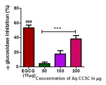
Fig 1- Antidiabetic assay
Anti inflammatory activity
Only Aq-CCSC extract inhibited LPS induced TNF-α in THP-1 monocytes at higher concentrations in a dose dependant manner with a percentage inhibition of 15% (p < 0.001), 10% (p > 0.05), and 05% (p < 0.05) at the 200 µg/mL, 100 µg/mL, and 50 µg/mL concentrations, respectively
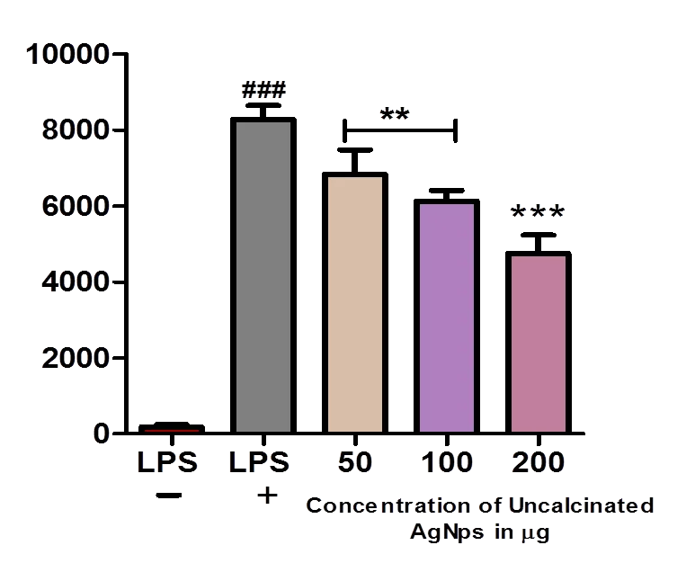
Fig 2- Anti inflammatory activity
Protective effect of calcined, uncalcined AgNps, Aq CCSC extracts against AgNP-induced DNA damage
The comet assay ± hOGG1 was used to assess the possible protective impact of calcined, uncalcined AgNps, Aq CCSC extracts against AgNP-induced DNA damage. For 24 hours, HEK 293 cells were pre-treated with non-cytotoxic and non-genotoxic doses of calcined, uncalcined AgNps, Aq CCSC extracts, then for 30 minutes, they were exposed to AgNPs. Positive controls were cells treated to either Ro19-8022 plus light (which induced 45% DNA in comet tails) or H2O2 (which induced 63% DNA in comet tails). Pre-treatment with 0.25 and 0.5 mg/ml of each extract shielded DNA from AgNP-induced SBs. This effect was also observed when cells were pretreated with 1 µg/ml AgNPs and the maximum concentration of extracts (2.5 mg/ml). Nevertheless, cells were not shielded from DNA damage by either 25 µg/ml or 100 µg/ml of AgNPs by the same concentration.
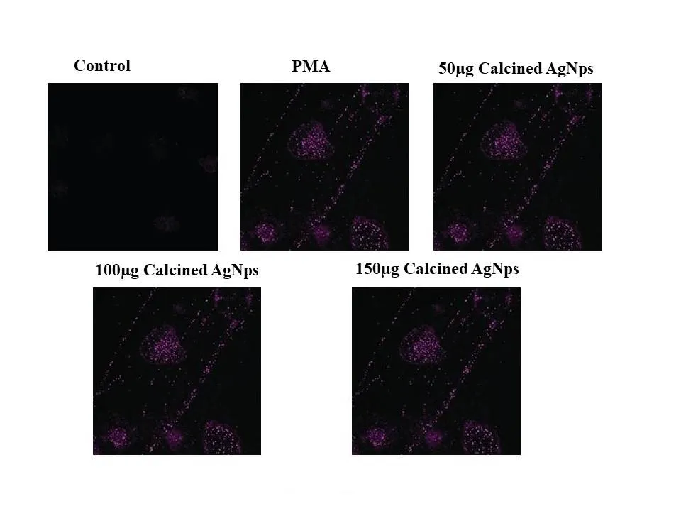
Fig 3- DNA OXIDATIVE MODIFICATION ASSAY of Calcined AgNps
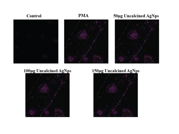
Fig 4- DNA OXIDATIVE MODIFICATION ASSAY of Uncalcined AgNps
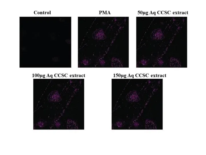
Fig 5- DNA OXIDATIVE MODIFICATION ASSAY of Aq CCSC extract
RBC Ghost membrane stability assay
Calcined , uncalcined AgNps,Aq CCSC extract samples are load along with ghost membrane unfortunately no reaction were found.
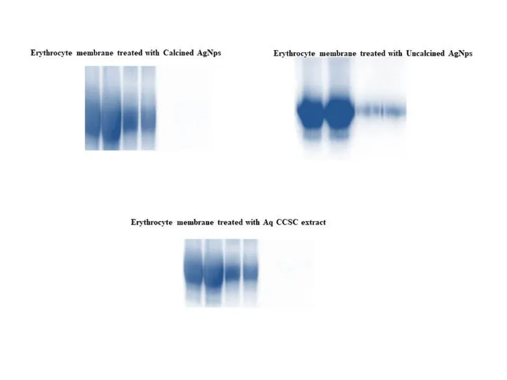
Fig 6- RBC Ghost membrane stability assay
CONCLUSION
Only uncalcined AgNps exhibited a weakly significant effect on alpha-glucosidase at all examined concentrations, whereas calcined AgNps, uncalcined AgNps, and AqCCSC extracts had no discernible effect on alpha-amylase. Aq-CCSC extract decreased LPS-induced TNF-α in THP-1 monocytes at increasing concentrations. AgNPs does not protected cells from DNA damage. Calcined and uncalcined AgNps, Aq CCSC extract samples were loaded, along with a ghost membrane, but no reaction was detected.
REFERENCES

Sowmya S., Chandramma Srinivasa, Manjunatha H.*, Antidiabetic, Anti Inflammatory, DNA Oxidative Modification And RBC Ghost Membrane Stability Of Caesalpinia Crista Seed Coats And Its Silver Nanoparticles, Int. J. Sci. R. Tech., 2026, 3 (6), 323-328. https://doi.org/10.5281/zenodo.20541623
 10.5281/zenodo.20541623