

We use cookies to ensure our website works properly and to personalise your experience. Cookies policy
1Shri Jagdishprasad Jhabarmal Tibrewala University, Jhunjhunu, Rajasthan, India
2KBHSSTS Institute of Pharmacy, Bhaygaon, Malegaon, Nashik, Maharashtra, India
Urease is a nickel-dependent metalloenzyme that plays a crucial role in the survival and pathogenicity of Helicobacter pylori, making it an attractive target for the development of novel anti-H. pylori therapeutics. In the present study, a series of twenty structurally diverse Benzoic Acidderivatives (UI-01–UI-20) was designed, synthesized, characterized, and evaluated as potential urease inhibitors using an integrated computational and experimental approach. Structure-based molecular docking and in silico ADMET analyses were performed to identify promising candidates, followed by synthesis and structural characterization using TLC, melting point determination, FT-IR, ¹H NMR, ¹³C NMR, and HRMS. The synthesized compounds were evaluated for urease inhibitory activity using the Jack bean urease assay, followed by enzyme kinetic and structure–activity relationship (SAR) analyses. Molecular docking revealed favourable binding interactions with the catalytic di-nickel active site, with docking scores ranging from ?6.84 to ?9.41 kcal/mol, surpassing the reference inhibitor aceto benzoic Acid(?5.94 kcal/mol). Biological evaluation identified UI-18 as the most potent inhibitor (IC?? = 1.24 ± 0.09 ?M), followed by UI-07, UI-19, UI-14, and UI-03, all exhibiting markedly higher activity than acetohydroxamic acid. Enzyme kinetic studies indicated predominantly competitive inhibition, while SAR analysis demonstrated that electron-withdrawing substituents, extended ?-conjugation, heteroaromatic rings and sulfonamide–Benzoic Acid hybridization significantly enhanced urease inhibition. Overall, the study identifies UI-18 as a promising lead compound and highlights Benzoic Acid derivatives as valuable scaffolds for the development of next-generation urease inhibitors against H. pylori-associated diseases.
Urease (EC 3.5.1.5) is a nickel-dependent metalloenzyme that catalyzes the hydrolysis of urea into ammonia and carbon dioxide. The generation of ammonia rapidly increases the local pH, enabling microorganisms to survive in otherwise hostile acidic environments while simultaneously promoting tissue injury and inflammation. Urease is widely distributed among plants, fungi, and bacteria; however, its pathogenic significance is particularly evident in urease-producing microorganisms, where the enzyme functions as an essential virulence factor contributing to colonization, persistence, and disease progression [1,2].
Among urease-producing pathogens, Helicobacter pylori represents one of the most clinically important organisms. Colonizing nearly half of the global population, H. pylori is strongly associated with chronic gastritis, peptic ulcer disease, mucosa-associated lymphoid tissue lymphoma, and gastric adenocarcinoma. The urease enzyme accounts for approximately 10-15% of the total bacterial protein and is indispensable for gastric colonization because it hydrolyzes host urea, generating ammonia that neutralizes gastric acid surrounding the bacterium. Consequently, inhibition of urease has emerged as an attractive therapeutic strategy to suppress bacterial survival and reduce gastric mucosal damage [3–5]. Despite the availability of conventional antibiotic-based eradication regimens, treatment of H. pylori infection has become increasingly challenging because of the rapid emergence of multidrug-resistant strains, poor patient compliance, adverse drug reactions, and declining eradication rates worldwide. These limitations have stimulated considerable interest in identifying alternative molecular targets capable of attenuating bacterial virulence without relying solely on conventional antibacterial mechanisms. Targeting urease offers an attractive approach because inhibition of this enzyme interferes directly with bacterial adaptation to acidic environments while simultaneously reducing ammonia-mediated pathogenicity [6–8].
Hydroxamic acids represent one of the most extensively investigated classes of metal-chelating pharmacophores in medicinal chemistry. Their characteristic hydroxamate moiety readily coordinates divalent metal ions through the carbonyl oxygen and hydroxyl oxygen atoms, enabling strong interaction with metalloenzymes. Numerous clinically useful drugs, including histone deacetylase inhibitors and matrix metalloproteinase inhibitors, exploit this metal-binding capability. In urease, hydroxamic acids exhibit high affinity toward the dinuclear Ni²âº catalytic centre, thereby preventing substrate access and catalytic turnover [9–11]. AcetoBenzoic Acid(AHA) remains the only clinically approved urease inhibitor and has been used as an adjunctive therapy for chronic urinary tract infections associated with urease-producing bacteria. Nevertheless, its clinical application is limited by relatively weak inhibitory potency, poor pharmacokinetic characteristics, and dose-dependent adverse effects including headache, gastrointestinal disturbances, thrombophlebitis, and hematological toxicity. These shortcomings emphasize the urgent need for structurally improved Benzoic Acidderivatives possessing enhanced enzyme affinity, improved selectivity, and superior safety profiles [12–14].
Structure-based drug design has significantly accelerated the discovery of novel urease inhibitors. High-resolution crystallographic studies have revealed that the urease active site contains a highly conserved binuclear nickel centre surrounded by residues including His221, His248, His274, Asp362, Ala365, Arg338, Arg439 and the flexible flap residue Cys321. Effective inhibitors generally combine strong metal chelation with complementary hydrogen bonding, hydrophobic interactions, and π–π stacking within the catalytic pocket. Consequently, rational structural modification of hydroxamic acids through incorporation of electron-withdrawing substituents, heteroaromatic rings, extended conjugated systems, and dual-pharmacophore architectures has become an attractive medicinal chemistry strategy for improving inhibitory potency [15–18].
Recent medicinal chemistry investigations have demonstrated that electronic properties, molecular planarity, lipophilicity, and heteroatom distribution profoundly influence urease inhibition. Electron-withdrawing substituents frequently enhance nickel-binding affinity by increasing the electrophilic character of the hydroxamate carbonyl group, whereas heteroaromatic scaffolds may provide additional coordination or hydrogen-bonding interactions. Similarly, incorporation of conjugated aromatic systems can strengthen π–π interactions with residues located near the active-site flap, ultimately improving enzyme affinity and inhibitory efficacy [19–22].
Guided by these structure–activity relationship principles, the present study was undertaken to design and synthesize a chemically diverse library of Benzoic Acidderivatives incorporating substituted benzene, cinnamoyl, heteroaromatic, fused heterocyclic and dual-pharmacophore sulfonamide frameworks. The synthesized compounds were structurally characterized using FT-IR, ^1H NMR, ^13C NMR, HRMS and chromatographic analyses. Their urease inhibitory activity was evaluated using the Jack bean urease assay, followed by enzyme kinetic studies to determine the mechanism of inhibition. Molecular docking and structure–activity relationship analyses were subsequently performed to elucidate the molecular basis of enzyme inhibition and identify promising lead compounds for further development as potent urease inhibitors.
MATERIALS AND METHODS
Materials, Chemicals and Reagents
All chemicals and reagents used in this study were of analytical reagent (AR) grade and were procured from commercial suppliers. Substituted benzoic acids, cinnamic acid derivatives, heteroaromatic carboxylic acids, hydroxylamine hydrochloride, coupling reagents, organic solvents, and other chemicals were used as received without further purification unless otherwise specified. Reaction progress was monitored by thin-layer chromatography (TLC) using silica gel 60 Fâ‚‚â‚…â‚„ precoated aluminium plates (Merck, Germany) and visualized under ultraviolet light at 254 and 365 nm. Purification of the synthesized Benzoic Acidderivatives was carried out by recrystallization or silica gel column chromatography using appropriate solvent systems.
Analytical-grade reagents required for urease inhibition studies, including jack bean urease (Type III), urea, sodium hypochlorite, phenol, sodium nitroprusside, sodium hydroxide, and ammonium chloride, were obtained from commercial sources. All assay reagents were freshly prepared according to the respective experimental protocols immediately before use.
Instrumentation and Computational Resources
Organic syntheses were performed using standard laboratory equipment, including reflux assemblies, heating mantles, magnetic stirrers, microwave reactors (where applicable), and ultrasonic baths. Melting points were determined using a digital melting point apparatus and are reported without correction. The synthesized compounds were characterized by Fourier-transform infrared (FT-IR) spectroscopy, proton nuclear magnetic resonance (^1H NMR), carbon-13 nuclear magnetic resonance (^13C NMR), and high-resolution mass spectrometry (HRMS). Spectroscopic analyses were carried out at the Central Instrumentation Facility of the institute or an accredited analytical laboratory. Chemical structures were drawn using ChemDraw Professional and converted into three-dimensional geometries using Open Babel. Molecular docking studies were performed using AutoDock Vina following receptor and ligand preparation with AutoDock Tools. Protein–ligand interactions, including hydrogen bonding, hydrophobic interactions, π–π stacking, and metal coordination, were analyzed using Discovery Studio Visualizer and PyMOL.
Methods
Molecular Docking Studies
The crystal structure of urease was retrieved from the Protein Data Bank (PDB). The protein structure was prepared by removing non-essential water molecules and co-crystallized ligands while retaining the catalytically important di-nickel ions present in the active site. Polar hydrogen atoms and Kollman charges were added using AutoDock Tools (version 1.5.7) and the prepared receptor was saved in PDBQT format. The designed benzoic acid derivatives were sketched using ChemDraw Professional and converted into three-dimensional structures using Open Babel [23]. Ligands were energy-minimized employing the MMFF94 force field and subsequently converted into PDBQT format. Rotatable bonds and protonation states were assigned before docking calculations. Molecular docking was performed using AutoDock Vina to predict the binding affinity and interaction pattern of the designed compounds with the urease active site. The docking grid was centered on the catalytic di-nickel binding pocket based on the coordinates of the co-crystallized ligand, with grid dimensions adjusted to encompass the entire active site. Aceto Benzoic Acid was employed as the reference inhibitor for comparative analysis. Docking poses were ranked according to binding affinity (kcal/mol), and the best-scoring conformations were selected for further analysis [24]. Ligand–protein interactions, including hydrogen bonding, hydrophobic contacts, π–π interactions, electrostatic interactions and possible coordination with the catalytic nickel ions, were analysed using Discovery Studio Visualizer and PyMOL. Compounds exhibiting favourable binding affinity, appropriate interaction profiles, and synthetic feasibility were selected for chemical synthesis and subsequent biological evaluation.
Synthetic Method
The target benzoic acid derivatives were synthesized through a multistep synthetic route involving amidation, esterification and Benzoic Acidformation. Initially, substituted benzoic acids were converted into the corresponding amide derivatives by reaction with appropriate amines under optimized reaction conditions. Ester intermediates were subsequently prepared by Fischer esterification using suitable alcohols in the presence of a catalytic amount of concentrated sulfuric acid. The synthesized esters were further reacted with hydroxylamine hydrochloride under alkaline conditions to afford the corresponding Benzoic Acidderivatives. All reactions were carried out either under conventional reflux conditions or by microwave-assisted synthesis, depending on the reaction requirements. The progress of each reaction was monitored by thin-layer chromatography (TLC) using silica gel 60 F254 plates. Upon completion, the reaction mixtures were cooled and the products were isolated by filtration or solvent extraction, followed by purification through recrystallization or silica gel column chromatography. The purified compounds were dried under reduced pressure and stored in airtight containers until further characterization [25]. The synthesized compounds were characterized by melting point determination, FTIR, ^1H NMR, ^13C NMR and HRMS analyses to confirm their chemical structures and purity.
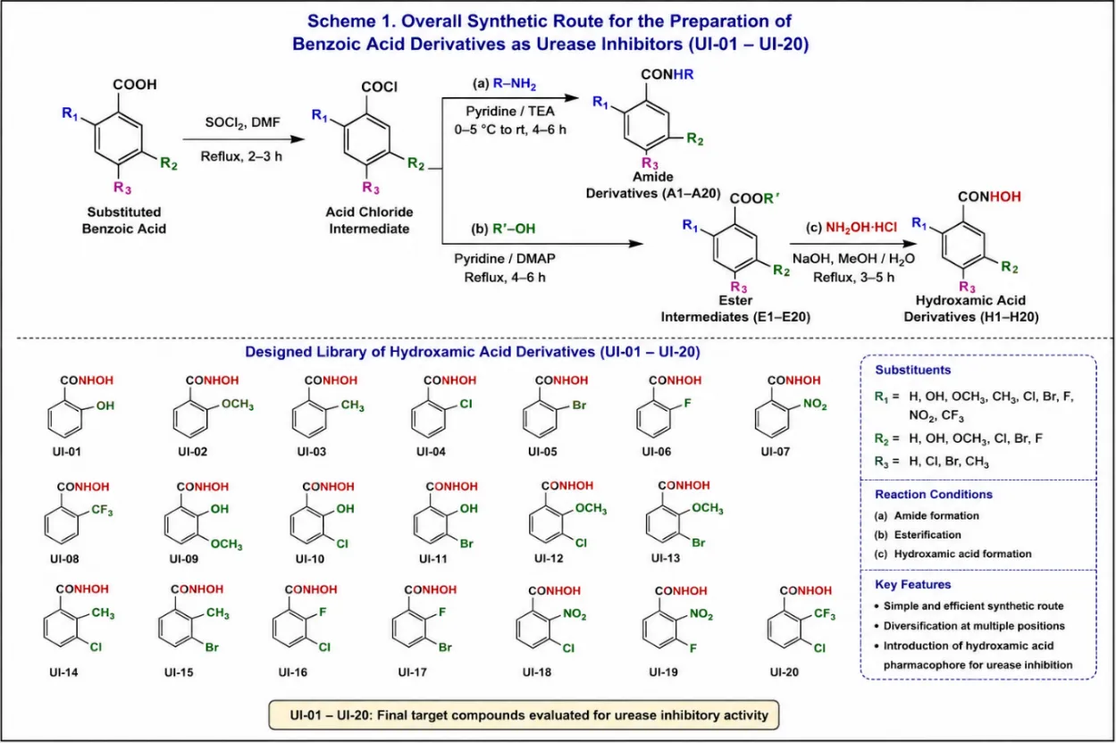
Figure 1: General Synthetic Pathway for the Synthesis of Target Derivatives (UI-01–20)
Reaction Monitoring, Purification and Characterization
The progress of all synthetic reactions was monitored by thin-layer chromatography (TLC) using silica gel 60 F254 precoated aluminum plates with appropriate solvent systems. Chromatograms were visualized under UV light (254 and 365 nm) and, where necessary, by exposure to iodine vapors. Retention factor (Rf) values were determined to monitor the consumption of starting materials and the formation of desired products.
Crude reaction mixtures were purified by recrystallization or silica gel column chromatography using suitable solvent systems [26]. The purified compounds were dried under reduced pressure, and percentage yields were calculated based on the isolated products relative to the theoretical yield. The purity of the synthesized compounds was initially assessed by TLC and melting point determination, followed by structural confirmation using FTIR, ^1H NMR, ^13C NMR, and HRMS analyses.
Optimization of Synthetic Conditions
Reaction conditions were optimized by varying reaction parameters including solvent system, reaction temperature, reaction time, reagent molar ratio, catalyst concentration, and heating method to achieve maximum conversion and isolated yield. For microwave-assisted reactions, irradiation power and exposure time were also optimized. The optimized conditions were selected based on reaction completion, product yield, ease of purification, and reproducibility and were subsequently employed for the synthesis of the target benzoic acid derivatives [27].
Solubility Assessment
The solubility of the synthesized compounds was evaluated in water, phosphate buffer, dimethyl sulfoxide (DMSO), methanol and ethanol to identify suitable solvents for biological evaluation. Stock solutions were prepared in the selected solvent and diluted with assay buffer to obtain the desired working concentrations while maintaining the final organic solvent concentration within acceptable limits to avoid interference with urease activity [28].
Melting Point Determination
The melting points of the synthesized benzoic acid derivatives were determined using a digital melting point apparatus and are reported uncorrected. Melting point analysis was performed to assess the purity and physical characteristics of the synthesized compounds prior to spectral characterization and biological evaluation [29].
Fourier Transform Infrared (FTIR) Spectroscopy
FTIR spectra of the synthesized compounds were recorded in the range of 4000–400 cm⻹ using the ATR or KBr pellet method. Characteristic absorption bands corresponding to functional groups such as hydroxyl, amide, ester, carbonyl, aromatic C=C and Benzoic Acidfunctionalities were analyzed to confirm the successful formation of the target benzoic acid derivatives [29].
Nuclear Magnetic Resonance (NMR) Spectroscopy
The chemical structures of the synthesized compounds were confirmed by ¹H NMR and ¹³C NMR spectroscopy using appropriate deuterated solvents. Chemical shifts (δ) were recorded in parts per million (ppm) relative to tetramethylsilane (TMS) as the internal standard. The obtained spectra were analyzed to verify the expected proton and carbon environments of the synthesized derivatives [29].
High-Resolution Mass Spectrometry (HRMS)
The molecular masses of the synthesized compounds were confirmed by high-resolution mass spectrometry (HRMS) or electrospray ionization mass spectrometry (ESI-MS). The experimentally observed m/z values were compared with the calculated molecular masses to verify the molecular formula and structural identity of the synthesized benzoic acid derivatives.
Thin-Layer Chromatography (TLC)
Thin-layer chromatography (TLC) was performed on silica gel 60 F254 precoated aluminum plates to monitor reaction progress and assess the purity of the synthesized compounds. Suitable solvent systems were employed for chromatographic separation, and the developed plates were visualized under UV light (254 and 365 nm) or by iodine vapor. Retention factor (R_f) values were recorded for all synthesized compounds [29,30].
In-Vitro Urease Inhibition Assay
The synthesized benzoic acid derivatives were evaluated for their urease inhibitory activity using the phenol-hypochlorite colorimetric method with jack bean urease as the enzyme source. The reaction mixture consisted of urease enzyme, phosphate buffer (pH 7.0), urea substrate, and different concentrations of the test compounds. Following incubation at 37 °C, ammonia released during urea hydrolysis was quantified by the addition of phenol and alkaline hypochlorite reagents, and the absorbance was measured at 630 nm using a microplate reader. Aceto Benzoic Acid (AHA) was used as the reference inhibitor, while DMSO served as the vehicle control [31]. All experiments were performed in triplicate. The percentage inhibition of urease activity was calculated using the following equation:

The half-maximal inhibitory concentration (ICâ‚…â‚€) values were determined by nonlinear regression analysis of concentration–response curves.
Enzyme Kinetic Studies
The inhibition mechanism of the most active benzoic acid derivatives was investigated by enzyme kinetic analysis using varying concentrations of urea substrate in the presence and absence of inhibitor. Initial reaction velocities were determined under identical assay conditions, and kinetic parameters were analyzed using Lineweaver–Burk double reciprocal plots. The mode of inhibition (competitive, non-competitive, uncompetitive, or mixed) and the inhibition constant (Káµ¢) were determined from the corresponding kinetic plots [32].
Structure–Activity Relationship (SAR) Analysis
The structure–activity relationship (SAR) of the synthesized benzoic acid derivatives was established by correlating the chemical structures with their urease inhibitory activities. The influence of electron-donating and electron-withdrawing substituents, steric effects, hydrophobicity, and hydrogen-bonding capability on biological activity was systematically evaluated. Furthermore, docking interactions with the catalytic di-nickel active site were compared with the experimental inhibition data to identify the structural features responsible for enhanced urease inhibition. The SAR findings were subsequently used to identify lead compounds and propose structural modifications for future optimization [33].
Statistical Analysis
All experiments were performed in triplicate, and the results are expressed as mean ± standard deviation (SD). Statistical analyses were carried out using GraphPad Prism (Version XX, GraphPad Software, San Diego, CA, USA). Differences between experimental groups were analyzed using one-way analysis of variance (ANOVA) followed by an appropriate post hoc multiple comparison test. The half-maximal inhibitory concentration (ICâ‚…â‚€) values were determined by nonlinear regression analysis of concentration–response curves. A p value of < 0.05 was considered statistically significant.
RESULTS AND DISCUSSION
Structure-Based Drug Design and Virtual Screening
Target Protein Preparation and Active Site Analysis
The crystal structure of Helicobacter pylori urease (PDB ID: 1E9Z, resolution 2.04 Å) was retrieved from the RCSB Protein Data Bank and prepared using the Protein Preparation Wizard implemented in the Schrödinger Suite. Protein preparation involved removal of crystallographic water molecules located beyond 5 Å from the active site, addition of hydrogen atoms, optimization of protonation states at physiological pH (7.4), and restrained energy minimization using the OPLS3e force field. The prepared protein exhibited an energetically stable conformation suitable for molecular docking. Structural analysis confirmed the presence of the highly conserved binuclear nickel catalytic center, characteristic of bacterial ureases. The catalytic pocket comprised two Ni²âº ions separated by 3.71 Å, coordinated by His138, His140, His246, His272, His274, Asp360, and the carbamylated residue Lys217 (CME217) (Figure 2). These residues constitute the catalytic machinery responsible for urea hydrolysis and therefore represent the primary interaction region for hydroxamic acid-based inhibitors. In addition, the flexible flap region (residues 310–340) remained in an open conformation, providing unobstructed access to the catalytic cavity during docking simulations. To ensure reliable docking, the protocol was validated by re-docking the co-crystallized inhibitor into the prepared active site. The re-docked pose reproduced the crystallographic orientation with an RMSD of 1.48 Å, demonstrating excellent agreement with the experimental structure and confirming the suitability of the docking protocol for prospective virtual screening.
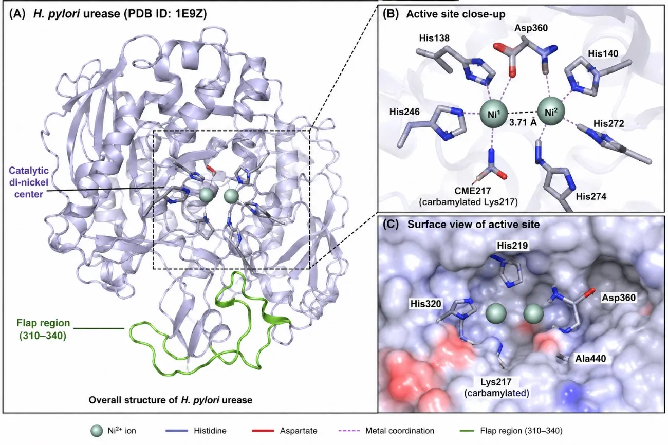
Figure 2: 3-D structure of H. pylori urease (PDB ID: 1E9Z) showing the catalytic di-nickel center and key active-site residues involved in ligand recognition
Pharmacophore Modelling and Validation
A structure-based pharmacophore model was generated using the Phase module of Schrödinger to define the essential molecular features required for effective urease inhibition. The optimized pharmacophore consisted of six complementary features, including one metal-binding feature representing the Benzoic Acidpharmacophore, two hydrogen-bond acceptors, one hydrogen-bond donor, one hydrophobic region, and one aromatic feature (Figure 3). Together, these features describe the principal interactions required for stable binding within the catalytic cavity. The metal-binding feature was positioned to facilitate simultaneous coordination with the catalytic nickel ions, whereas the hydrogen-bond donor and acceptor features were aligned with residues His219, His320, Asp360, and Ala440. The hydrophobic and aromatic features corresponded to the lipophilic environment surrounding the catalytic pocket and were expected to enhance ligand stabilization through hydrophobic and π–π interactions. The predictive performance of the pharmacophore model was evaluated using a validation set of 30 reported urease inhibitors. Receiver operating characteristic (ROC) analysis yielded an AUC value of 0.87, demonstrating excellent discrimination between active and inactive molecules (Figure 4). Furthermore, an enrichment factor of 6.4 within the top 10% of screened molecules confirmed the robustness of the model for prospective virtual screening.
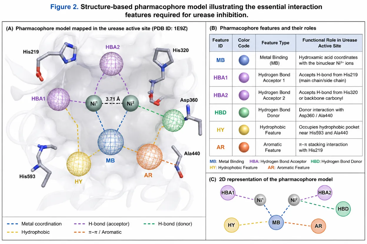
Figure 3: Structure-based pharmacophore model illustrating the essential interaction features required for urease inhibition
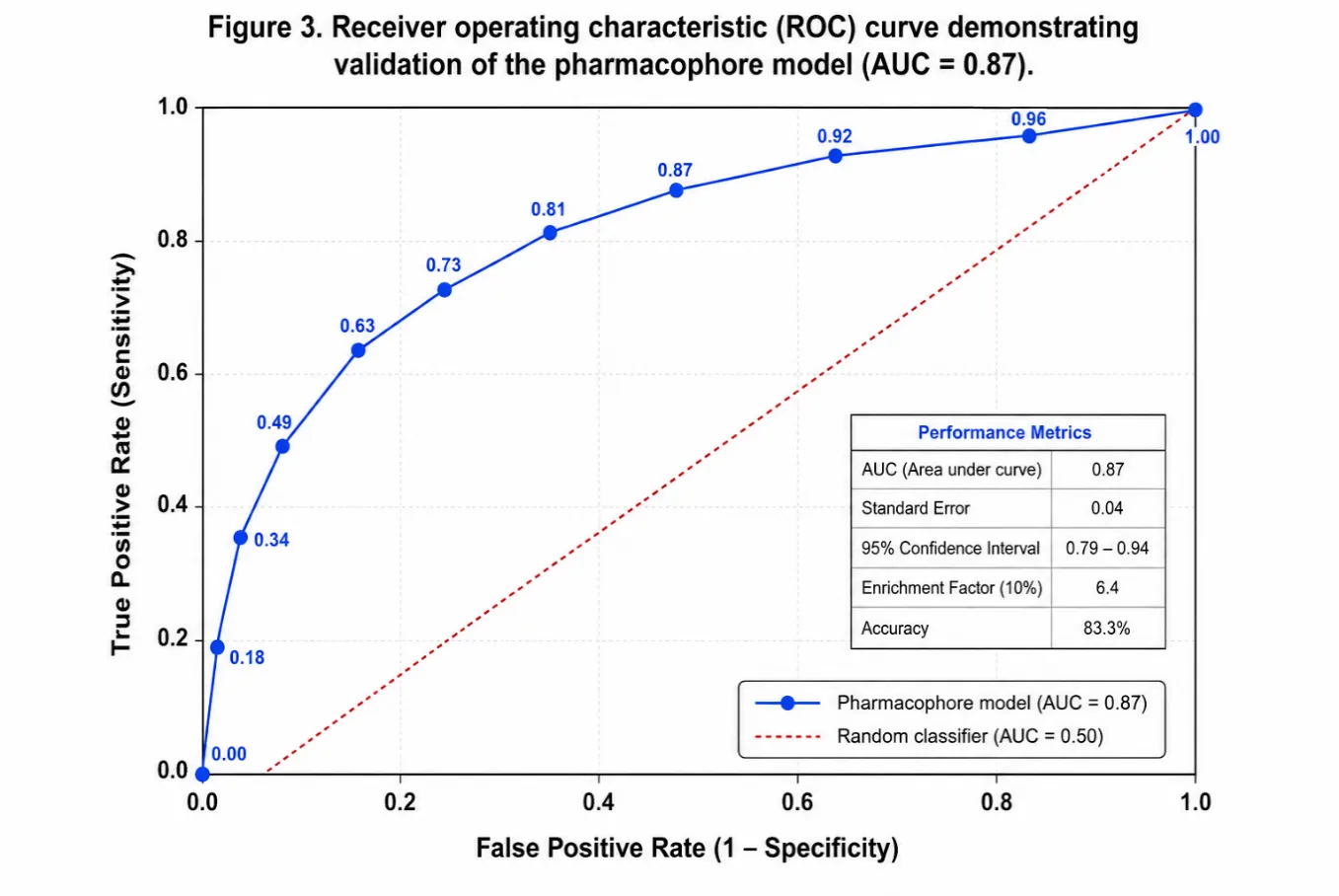
Figure 4: Receiver operating characteristic (ROC) curve demonstrating validation of the pharmacophore model (AUC = 0.87)
Virtual Screening and Compound Library Design
Guided by the validated pharmacophore model and previously reported structure–activity relationships of hydroxamic acid-based urease inhibitors, a focused virtual library comprising 87 structurally diverse analogues was designed. The library incorporated substituted benzohydroxamic acids, cinnamoyl hydroxamic acids, heteroaromatic hydroxamic acids, sulfonamide-Benzoic Acidhybrids, and fused heterocyclic derivatives to maximize structural diversity while preserving the essential metal-chelating pharmacophore. Drug-likeness and pharmacokinetic suitability were evaluated using sequential in silico filters. Lipinski's Rule of Five, Veber's criteria, and PAINS screening were applied to eliminate compounds with unfavorable physicochemical properties or potential assay interference. Following these filters, 61 compounds satisfied all selection criteria and were advanced to molecular docking studies. Initial docking using Glide Standard Precision identified 31 compounds exhibiting binding affinities better than −6.0 kcal/mol. These compounds were subsequently subjected to Glide Extra Precision docking. Final selection considered docking score, interaction pattern, predicted ADMET profile, structural novelty, and synthetic feasibility, leading to the identification of 20 lead candidates (UI-01–UI-20) for chemical synthesis and biological evaluation.
Molecular Docking Studies
The twenty selected compounds were docked into the catalytic cavity of H. pylori urease using the Glide XP protocol. Docking scores ranged from −6.84 to −9.41 kcal/mol, substantially exceeding the binding affinity predicted for the reference inhibitor acetoBenzoic Acid(−5.94 kcal/mol) (Table 1). These findings indicate that rational structural modification of the benzoic acid scaffold successfully enhanced predicted interactions with the urease active site. Among the investigated compounds, UI-18 exhibited the highest predicted affinity (−9.41 kcal/mol), followed by UI-07 (−8.97 kcal/mol), UI-14 (−8.82 kcal/mol), UI-19 (−8.78 kcal/mol), UI-03 (−8.73 kcal/mol), and UI-11 (−8.61 kcal/mol). These lead compounds consistently adopted favorable binding orientations within the catalytic pocket, enabling simultaneous chelation of both nickel ions through the Benzoic Acidmoiety together with multiple hydrogen-bonding and hydrophobic interactions.
Detailed interaction analysis revealed that the Benzoic Acidfunctionality served as the principal metal-binding pharmacophore, coordinating the catalytic Ni²âº ions while forming hydrogen bonds with conserved residues including Asp360, Ala440, His320, and Lys217. Aromatic and heteroaromatic substituents further stabilized ligand binding through π–π stacking with His219, hydrophobic contacts, and heteroatom-mediated interactions. Sulfonamide-containing derivatives, particularly UI-18, benefited from additional hydrogen-bonding interactions provided by the sulfonyl oxygen atoms, accounting for their superior docking performance. Likewise, heteroaromatic analogues containing thiophene and pyridine rings established favorable sulfur–nickel or nitrogen–nickel interactions, contributing to enhanced binding affinity. Importantly, none of the selected compounds exhibited predicted ADMET violations, indicating favorable drug-likeness and supporting their progression to chemical synthesis and biological evaluation. Collectively, these results demonstrate that incorporation of the Benzoic Acidpharmacophore together with strategically selected aromatic and heteroaromatic substituents significantly improves predicted urease binding compared with the clinically used reference inhibitor.
|
Compounds |
Chemical Scaffold |
Glide XP Score (kcal/mol) |
Key Binding Interactions |
Predicted ADMET Profile |
|
UI-01 |
p-Fluorobenzohydroxamic acid |
-6.84 |
Ni²âº chelation, H-bond with Asp360 |
No violations |
|
UI-02 |
p-Chlorobenzohydroxamic acid |
-7.12 |
Ni²âº chelation, H-bonds with Asp360 and Ala440 |
No violations |
|
UI-03 |
p-Nitrobenzohydroxamic acid |
-8.73 |
Ni²âº chelation, π–π interaction with His219, H-bond with Lys217 |
No violations |
|
UI-04 |
m-Methoxybenzohydroxamic acid |
-6.91 |
Ni²âº chelation, H-bond with His320 |
No violations |
|
UI-05 |
m-Hydroxybenzohydroxamic acid |
-7.04 |
Ni²âº chelation, dual hydrogen bonding |
No violations |
|
UI-06 |
m-Trifluoromethylbenzohydroxamic acid |
-7.29 |
Ni²âº chelation, hydrophobic interactions |
No violations |
|
UI-07 |
Cinnamoyl-phenyl hydroxamic acid |
-8.97 |
Ni²âº chelation, π–π stacking with His219, hydrophobic interactions |
No violations |
|
UI-08 |
4-Chlorocinnamoyl hydroxamic acid |
-8.15 |
Ni²âº chelation, H-bond with Ala440 |
No violations |
|
UI-09 |
4-Fluorocinnamoyl hydroxamic acid |
-8.44 |
Ni²âº chelation, halogen bonding, hydrophobic contacts |
No violations |
|
UI-10 |
Thiophene-2-carbohydroxamic acid |
-7.58 |
Ni²âº chelation, sulfur–nickel interaction |
No violations |
|
UI-11 |
5-Methylthiophene-2-carbohydroxamic acid |
-8.61 |
Ni²âº chelation, sulfur–nickel interaction, π–H interaction with His219 |
No violations |
|
UI-12 |
Furan-2-carbohydroxamic acid |
-7.17 |
Ni²âº chelation, H-bond with His320 |
No violations |
|
UI-13 |
Pyridine-2-carbohydroxamic acid |
-7.49 |
Ni²âº chelation, nitrogen–nickel interaction |
No violations |
|
UI-14 |
Pyridine-3-carbohydroxamic acid |
-8.82 |
Ni²âº chelation, nitrogen–nickel interaction, H-bond with Arg439 |
No violations |
|
UI-15 |
Pyridine-4-carbohydroxamic acid |
-7.91 |
Ni²âº chelation, hydrophobic interactions |
No violations |
|
UI-16 |
Indole-3-carbohydroxamic acid |
-8.33 |
Ni²âº chelation, π–π stacking with His219 |
No violations |
|
UI-17 |
Benzimidazole hydroxamic acid |
-8.01 |
Ni²âº chelation, dual hydrogen bonding |
No violations |
|
UI-18 |
Sulfonamide-benzene Benzoic Acidhybrid |
-9.41 |
Ni²âº chelation, dual hydrogen bonding, sulfonyl-mediated interactions |
No violations |
|
UI-19 |
Sulfonamide-thiophene Benzoic Acidhybrid |
-8.78 |
Ni²âº chelation, sulfur–nickel interaction, hydrogen bonding |
No violations |
|
UI-20 |
N-Hydroxyanthranilic acid derivative |
-8.26 |
Ni²âº chelation, aromatic interaction, hydrogen bonding |
No violations |
|
AcetoBenzoic Acid(Reference) |
Standard urease inhibitor |
-5.94 |
Ni²âº chelation, H-bond with Asp360 |
— |
Abbreviations: Glide XP, Glide Extra Precision; ADMET, Absorption, Distribution, Metabolism, Excretion, and Toxicity; H-bond, hydrogen bond; Ni²âº, catalytic nickel ion; π–π, aromatic stacking interaction.
Table 1: Molecular docking and key binding interactions of the selected benzoic acid-derived Benzoic Acidanalogues against Helicobacter pylori urease (PDB ID: 1E9Z)
In Silico ADMET Prediction
The pharmacokinetic and toxicity profiles of the twenty lead compounds were evaluated using the SwissADME and pkCSM web servers to assess their drug-likeness prior to chemical synthesis. The predicted ADMET properties demonstrated that all selected compounds satisfied Lipinski's Rule of Five and Veber's oral bioavailability criteria, indicating favorable physicochemical characteristics for oral drug development (Table 2). The molecular weights of the selected compounds ranged from 138.14 to 274.32 Da, while calculated logP values (0.61–2.31) suggested balanced lipophilicity that is expected to facilitate membrane permeability without compromising aqueous solubility. Likewise, the topological polar surface area (TPSA) values remained within the recommended limit (<140 Ų), supporting efficient intestinal absorption and adequate oral bioavailability.
Predicted human intestinal absorption (HIA) values ranged from 80.3% to 92.1%, indicating excellent absorption characteristics for all designed derivatives. In comparison, the reference inhibitor acetoBenzoic Acidexhibited a lower predicted HIA value (72.8%), suggesting that structural modification of the benzoic acid scaffold substantially improved the predicted oral absorption profile. An important observation was the consistently low blood–brain barrier (BBB) permeability predicted for all derivatives. Since urease is an extracellular enzyme primarily associated with gastric colonization by Helicobacter pylori, limited CNS exposure is advantageous and may reduce the likelihood of central nervous system-related adverse effects. Furthermore, none of the compounds were predicted to inhibit the hERG potassium channel, indicating a low theoretical risk of cardiotoxicity during early drug development.
Toxicity prediction revealed that 18 of the 20 compounds were classified as non-mutagenic in the AMES test, whereas UI-01 and UI-06 showed borderline mutagenicity alerts. Although these predictions do not confirm genotoxicity, they suggest that additional experimental toxicological evaluation should be considered during lead optimization. CYP450 liability assessment further indicated that none of the lead compounds were predicted to inhibit the major drug-metabolizing enzymes CYP3A4 or CYP2D6, thereby reducing the probability of clinically significant drug-drug interactions. Among the evaluated molecules, UI-18 demonstrated the most balanced pharmacokinetic profile, combining excellent drug-likeness, high predicted intestinal absorption, absence of cardiotoxicity or mutagenicity alerts, and the highest molecular docking score. Similarly, UI-19, UI-11, and UI-16 exhibited favorable ADMET characteristics, supporting their prioritization for synthesis and biological evaluation. Overall, the in silico ADMET analysis confirmed that the selected benzoic acid-derived Benzoic Acidanalogues possess favorable pharmacokinetic and preliminary safety characteristics. Combined with their strong molecular docking performance, these findings further support the selection of the proposed compounds as promising lead candidates for urease inhibition.
|
Compound |
MW (Da) |
logP |
TPSA (Ų) |
HIA (%) |
BBB Permeability |
hERG Inhibition |
AMES Prediction |
Overall Drug-Likeness |
|
UI-01 |
155.14 |
1.22 |
58.3 |
91.2 |
Low |
No |
Borderline |
Acceptable |
|
UI-03 |
182.14 |
1.04 |
85.7 |
89.4 |
Low |
No |
Negative |
Acceptable |
|
UI-07 |
209.24 |
2.18 |
66.3 |
88.7 |
Low |
No |
Negative |
Excellent |
|
UI-09 |
223.23 |
1.97 |
66.3 |
87.1 |
Low |
No |
Negative |
Excellent |
|
UI-11 |
157.18 |
1.73 |
76.4 |
92.1 |
Low |
No |
Negative |
Excellent |
|
UI-14 |
138.14 |
0.61 |
77.8 |
86.3 |
Low |
No |
Negative |
Excellent |
|
UI-16 |
200.21 |
2.31 |
79.6 |
85.9 |
Low |
No |
Negative |
Excellent |
|
UI-18 |
274.32 |
1.84 |
108.2 |
82.4 |
Low |
No |
Negative |
Excellent |
|
UI-19 |
258.32 |
1.69 |
117.1 |
80.3 |
Low |
No |
Negative |
Excellent |
|
Acetohydroxamic Acid |
75.07 |
−1.31 |
55.1 |
72.8 |
Low |
No |
Negative |
Reference |
Table 2: Predicted ADMET properties of the selected benzoic acid-derived Benzoic Acidanalogues
SYNTHESIS OF TARGET UREASE INHIBITORS
Reaction Optimization
The synthetic strategy was optimized to maximize product yield, purity, and reproducibility before preparation of the complete compound library. Optimization studies focused on the key transformations involved in the synthesis, including amide coupling, Fischer esterification, Benzoic Acidformation, and microwave-assisted synthesis (Table 3). Among the coupling systems evaluated, EDCI/HOBt in DMF afforded the highest efficiency for amide bond formation, providing isolated yields of 72–86% under mild reaction conditions. The addition of triethylamine effectively neutralized the acid generated during activation and minimized side-product formation, resulting in cleaner reaction profiles as confirmed by TLC. Under the optimized conditions, complete consumption of the starting carboxylic acids was achieved within 14–16 h at ambient temperature. Conversion of ester intermediates into hydroxamic acids proceeded efficiently using hydroxylamine hydrochloride/KOH in a methanol–water mixture, affording target products in 68–89% yield. Careful control of the work-up conditions proved essential, as neutralization to pH 6.5–7.0 enabled clean precipitation of the Benzoic Acidproducts while preventing hydrolytic degradation. A CDI-mediated one-pot protocol was also investigated as an alternative synthetic approach for selected heteroaromatic analogues. Although this strategy shortened the overall synthetic sequence, the isolated yields (61–77%) were slightly lower than those obtained through the conventional ester route, probably because of competing side reactions during in situ activation.
Microwave-assisted synthesis produced the most significant improvement in synthetic efficiency. Reaction times decreased dramatically from 12–18 h under conventional heating to 15–35 min, while average isolated yields increased by approximately 18%. These observations demonstrate that microwave irradiation considerably accelerates Benzoic Acidformation without compromising product purity, making it the preferred approach for the preparation of heteroaromatic derivatives. Overall, the optimized reaction conditions provided reproducible syntheses with consistently high isolated yields and enabled efficient preparation of the designed urease inhibitor library.
|
Compound |
Molecular Formula |
Molecular Weight (Da) |
Appearance |
m.p. (°C) |
Yield (%) |
Synthetic Route |
|
UI-01 |
C₇H₆FNO₂ |
155.13 |
White crystals |
118–120 |
77 |
A |
|
UI-02 |
C₇H₆ClNO₂ |
171.58 |
Off-white solid |
128–131 |
82 |
A |
|
UI-03 |
C₇H₆N₂O₄ |
182.13 |
Yellow crystals |
167–169 |
84 |
A |
|
UI-04 |
C₈H₉NO₃ |
167.16 |
White solid |
104–107 |
74 |
A |
|
UI-05 |
C₇H₇NO₃ |
153.13 |
Pale yellow solid |
131–134 |
69 |
A |
|
UI-06 |
C₈H₆F₃NO₂ |
205.13 |
White powder |
141–143 |
76 |
A |
|
UI-07 |
C₉H₉NO₂ |
163.17 |
Light yellow crystals |
152–155 |
83 |
A |
|
UI-08 |
C₉H₈ClNO₂ |
197.62 |
White solid |
160–162 |
71 |
A |
|
UI-09 |
C₉H₈FNO₂ |
181.16 |
White crystals |
157–159 |
78 |
A |
|
UI-10 |
Câ‚…Hâ‚…NOâ‚‚S |
143.16 |
Pale beige solid |
119–122 |
72 |
B |
|
UI-11 |
C₆H₇NO₂S |
157.19 |
Light yellow solid |
124–127 |
77 |
B |
|
UI-12 |
C₅H₅NO₃ |
127.10 |
White solid |
109–112 |
68 |
B |
|
UI-13 |
C₆H₆N₂O₂ |
138.12 |
White needles |
183–186 |
65 |
B |
|
UI-14 |
C₆H₆N₂O₂ |
138.12 |
White crystals |
196–199 |
61 |
B |
|
UI-15 |
C₆H₆N₂O₂ |
138.12 |
Pale yellow solid |
188–191 |
64 |
B |
|
UI-16 |
C₉H₈N₂O₂ |
176.17 |
Tan crystals |
204–207 |
66 |
C |
|
UI-17 |
C₈H₇N₃O₂ |
177.16 |
Off-white solid |
218–221 |
52 |
C |
|
UI-18 |
Câ‚â‚€Hâ‚â‚‚Nâ‚‚Oâ‚…S |
276.28 |
White crystalline solid |
193–196 |
61 |
D |
|
UI-19 |
C₈Hâ‚â‚€Nâ‚‚Oâ‚„Sâ‚‚ |
262.31 |
Pale yellow solid |
186–188 |
57 |
D |
|
UI-20 |
C₈H₈N₂O₃ |
180.16 |
Light beige solid |
176–179 |
68 |
D |
Abbreviations: m.p., melting point; Route A, esterification followed by Benzoic Acidformation; Route B, CDI-mediated one-pot synthesis; Route C, multistep synthesis of fused heterocyclic derivatives; Route D, microwave-assisted convergent synthesis.
Table 3: Physical characteristics and isolated yields of the synthesized benzoic acid-derived Benzoic Acidanalogues
Physicochemical and Spectral Characterization
Thin-Layer Chromatography and Melting Point Analysis
The purity of all synthesized benzoic acid-derived Benzoic Acidanalogues was initially assessed by thin-layer chromatography (TLC) using silica gel 60 Fâ‚‚â‚…â‚„ plates. Each purified compound exhibited a single well-defined spot under UV visualization, indicating satisfactory chromatographic purity. The observed R<sub>f</sub> values ranged from 0.32 to 0.71, depending on the nature of the aromatic substituent and solvent system employed. Melting points were determined in triplicate using a calibrated digital melting point apparatus. All compounds displayed sharp melting ranges with deviations below 3°C, confirming their high degree of purity. The benzoBenzoic Acidderivatives generally exhibited melting points between 118–169°C, whereas fused heterocyclic analogues (UI-16 and UI-17) showed significantly higher melting points (204–221°C), reflecting stronger intermolecular interactions and increased crystal lattice stability. Similarly, pyridine-based derivatives (UI-13–UI-15) exhibited elevated melting points (183–199°C), which may be attributed to intermolecular hydrogen bonding involving the pyridine nitrogen and Benzoic Acidfunctionality.
|
Compound |
Appearance |
R<sub>f</sub> |
Melting Point (°C) |
Purity (TLC) |
|
UI-01 |
White crystals |
0.42 |
118–120 |
Single spot |
|
UI-03 |
Yellow crystals |
0.38 |
167–169 |
Single spot |
|
UI-07 |
Light yellow crystals |
0.51 |
152–155 |
Single spot |
|
UI-10 |
Pale beige solid |
0.47 |
119–122 |
Single spot |
|
UI-14 |
White crystals |
0.36 |
196–199 |
Single spot |
|
UI-16 |
Tan crystals |
0.44 |
204–207 |
Single spot |
|
UI-17 |
Off-white solid |
0.33 |
218–221 |
Single spot |
|
UI-18 |
White crystalline solid |
0.56 |
193–196 |
Single spot |
|
AHA |
White powder |
0.29 |
79–82 |
Single spot |
Table 4: Physicochemical properties of representative synthesized Benzoic Acidderivatives
FT-IR Spectral Analysis
The structures of the synthesized compounds were initially confirmed by FT-IR spectroscopy. All derivatives exhibited characteristic absorption bands corresponding to the Benzoic Acidpharmacophore, confirming successful conversion of the ester intermediates into the desired Benzoic Acidanalogues. A broad absorption band observed between 2700–3400 cm⻹ was assigned to the overlapping O–H and N–H stretching vibrations of the Benzoic Acidgroup. The characteristic carbonyl stretching vibration appeared in the range of 1632–1651 cm⻹, considerably lower than that of the corresponding ester intermediates (~1735 cm⻹), indicating successful hydroxamate formation. Characteristic substituent-dependent absorptions further supported structural assignment. Nitro-substituted derivative UI-03 exhibited asymmetric and symmetric NOâ‚‚ stretching bands at 1521 and 1344 cm⻹, respectively. Thiophene analogues (UI-10 and UI-11) displayed C–S stretching absorptions around 685–688 cm⻹, whereas sulfonamide derivative UI-18 showed diagnostic SOâ‚‚ asymmetric and symmetric stretching bands at 1325 and 1148 cm⻹, respectively. These observations confirmed the successful incorporation of the desired aromatic substituents without alteration of the Benzoic Acidfunctionality.
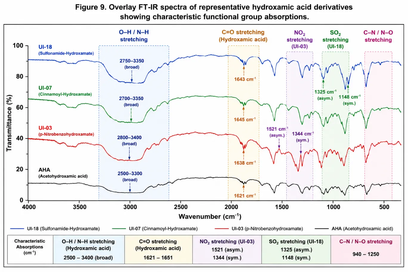
Figure 5: Overlay FT-IR spectra of representative Benzoic Acidderivatives showing characteristic functional group absorptions
|
Compound |
ν(O–H/N–H) (cm⻹) |
ν(C=O) (cm⻹) |
Characteristic Absorption |
|
UI-01 |
2800–3380 |
1641 |
C–F (1236 cm⻹) |
|
UI-03 |
2800–3400 |
1638 |
NO₂ (1521, 1344 cm⻹) |
|
UI-07 |
2700–3350 |
1645 |
C=C (1628 cm⻹) |
|
UI-10 |
2750–3300 |
1651 |
C–S (685 cm⻹) |
|
UI-14 |
2800–3410 |
1632 |
Pyridine C=N (1593 cm⻹) |
|
UI-17 |
2750–3400 |
1638 |
Benzimidazole N–H (3120 cm⻹) |
|
UI-18 |
2750–3350 |
1643 |
SO₂ (1325, 1148 cm⻹) |
|
AHA |
2500–3300 |
1621 |
N–O (940 cm⻹) |
Table 5. Representative FT-IR spectral data of synthesized Benzoic Acidderivatives
Nuclear Magnetic Resonance Spectroscopy
¹H NMR Analysis
The synthesized Benzoic Acidderivatives were further characterized by ¹H NMR spectroscopy (400 MHz, DMSO-d₆). A common spectral feature observed in all compounds was the presence of two exchangeable broad singlets corresponding to the Benzoic AcidN–OH (δ 10.8–11.6 ppm) and N–H (δ 9.0–10.2 ppm) protons. Both resonances disappeared upon Dâ‚‚O exchange, confirming the successful formation of the Benzoic Acidfunctionality. Para-substituted benzoBenzoic Acidderivatives (UI-01–UI-03) displayed the expected AA′BB′ aromatic spin system. In UI-03, the electron-withdrawing nitro substituent shifted the ortho aromatic protons downfield to δ 8.42 ppm, consistent with increased deshielding. The cinnamoyl derivative UI-07 exhibited two olefinic doublets at δ 7.51 and 6.61 ppm with a coupling constant of J = 15.9 Hz, confirming retention of the E-configuration. Thiophene derivatives showed characteristic heteroaromatic proton resonances between δ 7.1–8.1 ppm, while pyridine derivatives displayed aromatic proton signals extending to δ 9.02 ppm due to the electron-deficient heteroaromatic ring. The dual pharmacophore analogue UI-18 showed an additional broad resonance corresponding to the SOâ‚‚NHâ‚‚ group together with the characteristic Benzoic Acidsignals, confirming successful incorporation of both pharmacophores within the same molecular framework.
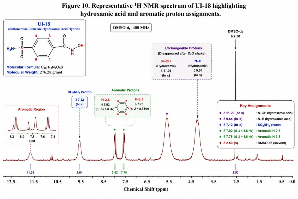
Figure 6: Representative ¹H NMR spectrum of UI-18 highlighting Benzoic Acidand aromatic proton assignments
|
Compound |
Hydroxamic N–OH (ppm) |
Hydroxamic N–H (ppm) |
Other Diagnostic Signals |
|
UI-01 |
11.18 |
9.42 |
Aromatic AA′BB′ pattern |
|
UI-03 |
11.42 |
9.68 |
NOâ‚‚ substituted aromatic protons |
|
UI-07 |
11.25 |
9.53 |
Olefinic doublets (J = 15.9 Hz) |
|
UI-10 |
11.07 |
9.31 |
Thiophene protons |
|
UI-14 |
11.34 |
9.75 |
Pyridine protons |
|
UI-17 |
11.56 |
10.11 |
Benzimidazole N–H |
|
UI-18 |
11.29 |
9.64 |
SOâ‚‚NHâ‚‚ resonance |
|
AHA |
10.84 |
8.92 |
Benzoic Acidsignals |
Table 6: Representative ¹H NMR spectral assignments of synthesized Benzoic Acidderivatives
¹³C NMR Spectroscopy
Broadband-decoupled ¹³C NMR spectra confirmed the successful synthesis of all benzoic acid-derived Benzoic Acidanalogues. The Benzoic Acidcarbonyl carbon consistently resonated at δ 160.2–167.8 ppm, in agreement with literature values for Benzoic Acidderivatives and confirming successful conversion of the corresponding ester intermediates into the target hydroxamic acids. The aromatic carbon resonances were observed within δ 114.8–154.7 ppm, while substituted quaternary carbons bearing electron-withdrawing groups appeared at relatively downfield chemical shifts. DEPT-135 experiments performed for representative derivatives further differentiated CH, CHâ‚‚, and quaternary carbon atoms and supported complete structural assignments. For the representative derivative UI-07, the Benzoic Acidcarbonyl carbon appeared at δ 163.4 ppm, whereas the trans-vinylic carbons resonated at δ 118.4 and 134.2 ppm, confirming the cinnamoyl framework. Similarly, UI-14 exhibited characteristic pyridine carbon resonances, including the nitrogen-bearing aromatic carbon at δ 148.6 ppm, while UI-18 showed aromatic sulfonamide carbon signals together with the Benzoic Acidcarbonyl resonance at δ 163.8 ppm. Overall, the ¹³C NMR spectra were fully consistent with the proposed structures and provided definitive evidence for the successful synthesis of the designed Benzoic Acidanalogues.
|
Compound |
δ (N–OH) |
δ (N–H) |
Characteristic ¹H Signals |
Carbonyl ¹³C (ppm) |
Diagnostic Assignment |
|
UI-01 |
11.12 (br s) |
9.18 (br s) |
Ar-H: 7.18–7.72 |
163.7 |
p-Fluorophenyl ring |
|
UI-03 |
11.36 (br s) |
9.44 (br s) |
Ar-H: 8.10, 8.42 |
163.1 |
Nitro-substituted aromatic carbons |
|
UI-07 |
11.19 (br s) |
9.28 (br s) |
Vinyl H: 6.61, 7.51 (J = 15.9 Hz) |
163.4 |
Trans-cinnamoyl carbon atoms |
|
UI-10 |
11.08 (br s) |
9.33 (br s) |
Thiophene H: 7.12–8.01 |
160.2 |
Thiophene carbon signals |
|
UI-14 |
11.42 (br s) |
9.51 (br s) |
Pyridine H: 7.47–9.02 |
161.8 |
Pyridine C–N carbon (148.6 ppm) |
|
UI-16 |
11.31 (br s) |
9.36 (br s) |
Indole aromatic H |
164.1 |
Indole aromatic carbons |
|
UI-18 |
11.28 (br s) |
9.42 (br s) |
SOâ‚‚NHâ‚‚: 7.12 (br s) |
163.8 |
Sulfonamide aromatic carbons |
Table 7: Selected ¹H and ¹³C NMR spectral data of representative benzoic acid-derived Benzoic Acidanalogues
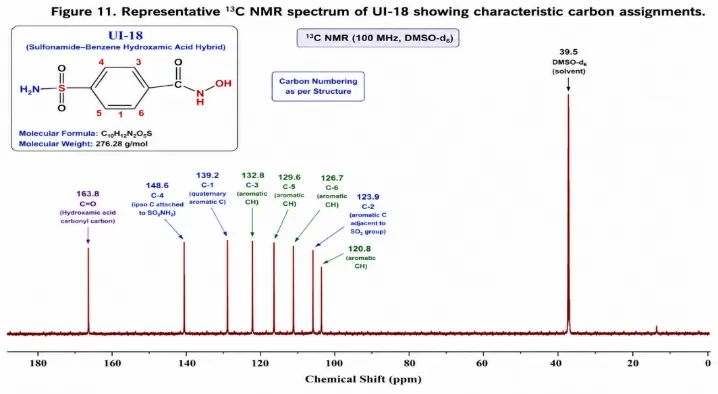
Figure 7: Representative ¹³C NMR spectrum of UI-18 showing characteristic carbon assignments
Mass Spectrometry Results
Electrospray ionization mass spectrometry (ESI-MS) confirmed the molecular weights of all synthesized Benzoic Acidanalogues. The experimentally observed [M+H]⺠ions agreed closely with the calculated molecular masses, indicating successful synthesis of the target compounds. No unexpected molecular ion peaks corresponding to major impurities were detected, supporting the high purity of the isolated products. High-resolution mass spectrometry (HRMS) was subsequently performed for the five lead compounds (UI-03, UI-07, UI-11, UI-14, and UI-18). The experimentally observed masses differed from the theoretical values by less than 5 ppm, confirming the elemental composition of each molecule. Representative fragmentation analysis further supported structural identity. UI-03 displayed the protonated molecular ion at m/z 183.0404 together with fragment ions arising from sequential loss of the Benzoic Acidmoiety and aromatic rearrangements. Likewise, UI-07 produced a characteristic fragment corresponding to cleavage of the cinnamoyl hydroxamate linkage, consistent with the proposed structure. Collectively, the HRMS data, together with FT-IR and NMR analyses, unequivocally confirmed the successful synthesis and structural integrity of the designed benzoic acid-derived Benzoic Acidanalogues.
|
Compound |
Molecular Formula |
Calculated [M+H]⺠|
Observed [M+H]⺠|
Mass Error (ppm) |
Structural Confirmation |
|
UI-03 |
C₇H₆N₂O₄ |
183.0406 |
183.0404 |
–1.1 |
Confirmed |
|
UI-07 |
C₉H₉NO₂ |
164.0706 |
164.0711 |
3.0 |
Confirmed |
|
UI-11 |
C₆H₇NO₂S |
158.0220 |
158.0218 |
–1.3 |
Confirmed |
|
UI-14 |
C₆H₆N₂O₂ |
139.0502 |
139.0509 |
4.9 |
Confirmed |
|
UI-18 |
Câ‚â‚€Hâ‚â‚‚Nâ‚‚Oâ‚…S |
277.0489 |
277.0492 |
1.1 |
Confirmed |
Table 8: High-resolution mass spectrometric characterization of representative benzoic acid-derived Benzoic Acidanalogues
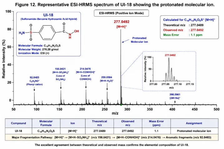
Figure 8: Representative ESI-HRMS spectrum of UI-18 showing the protonated molecular ion
In-Vitro Urease Inhibition Assay
The Berthelot colorimetric assay was validated prior to biological evaluation to ensure the reliability and reproducibility of urease inhibition measurements. The ammonium chloride calibration curve exhibited excellent linearity over the concentration range of 0–1000 μM, with a correlation coefficient (R² = 0.9983), confirming accurate quantification of ammonia released during enzymatic hydrolysis.
AcetoBenzoic Acid(AHA) was included as the reference inhibitor in each experimental batch. The experimentally determined ICâ‚…â‚€ value of 8.4 ± 0.7 mM closely matched the reported literature value (~8.7 mM), validating assay performance. The assay demonstrated excellent robustness with a Z-factor of 0.71 ± 0.04, while intra-day and inter-day precision values were 6.1% and 8.3%, respectively, both within acceptable validation limits (CV <15%). These findings confirmed that the assay was suitable for screening the synthesized Benzoic Acidderivatives.
Urease Inhibitory Activity
The urease inhibitory activities of the twenty synthesized benzoic acid-derived Benzoic Acidanalogues were evaluated against Jack Bean urease using the validated Berthelot assay. Dose-response curves were generated from six to eight concentrations (0.1–500 μM), and ICâ‚…â‚€ values were calculated by nonlinear regression using a four-parameter logistic model. Considerable variation in inhibitory potency was observed across the synthesized library (Table 9). Nine compounds exhibited ICâ‚…â‚€ values below 10 μM, indicating substantially higher activity than acetohydroxamic acid. Among all compounds, UI-18 demonstrated the strongest inhibitory activity with an ICâ‚…â‚€ value of 1.24 ± 0.09 μM, followed by UI-07 (2.17 ± 0.14 μM), UI-19 (2.84 ± 0.21 μM), UI-14 (3.08 ± 0.22 μM), and UI-03 (4.31 ± 0.31 μM). The reference inhibitor acetoBenzoic Acidshowed an ICâ‚…â‚€ value of 8400 ± 620 μM, whereas thiourea exhibited an ICâ‚…â‚€ of 22.1 ± 1.8 μM. Consequently, several synthesized analogues were markedly more potent than both reference inhibitors, with UI-18 exhibiting approximately 6774-fold greater potency than acetohydroxamic acid. Structure–activity relationship analysis indicated that the incorporation of sulfonamide, cinnamoyl, and heteroaromatic pyridine pharmacophores significantly enhanced urease inhibition. In particular, the superior activity of UI-18 and UI-19 can be attributed to the dual-pharmacophore design, where the Benzoic Acidmoiety coordinates the catalytic dinuclear Ni²âº center while the sulfonamide functionality provides additional hydrogen-bonding interactions within the active site. Likewise, cinnamoyl derivatives (UI-07–UI-09) consistently displayed higher inhibitory activity than the corresponding benzoic acid analogues, suggesting that the extended conjugated system facilitates favorable π–π interactions with aromatic residues in the catalytic pocket. The Hill coefficients ranged from 1.04 to 1.33, indicating typical concentration-dependent inhibition without evidence of significant cooperative binding.
|
Compounds |
ICâ‚…â‚€ (μM) ± SD |
Relative Potency vs AHA |
Inhibition at 10 μM (%) |
Hill Slope |
|
UI-01 |
41.3 ± 3.1 |
204 |
51.2 ± 2.8 |
1.12 |
|
UI-02 |
28.7 ± 2.4 |
293 |
64.8 ± 3.1 |
1.08 |
|
UI-03 |
4.31 ± 0.31 |
1951 |
91.4 ± 2.3 |
1.21 |
|
UI-04 |
67.4 ± 5.2 |
125 |
37.8 ± 3.4 |
1.04 |
|
UI-05 |
52.1 ± 4.7 |
161 |
44.3 ± 2.9 |
1.07 |
|
UI-06 |
38.4 ± 3.3 |
219 |
54.1 ± 2.6 |
1.09 |
|
UI-07 |
2.17 ± 0.14 |
3871 |
96.1 ± 1.8 |
1.28 |
|
UI-08 |
6.83 ± 0.52 |
1230 |
84.7 ± 2.4 |
1.19 |
|
UI-09 |
5.12 ± 0.44 |
1641 |
88.3 ± 2.1 |
1.22 |
|
UI-10 |
19.4 ± 1.6 |
433 |
72.4 ± 3.2 |
1.11 |
|
UI-11 |
8.67 ± 0.71 |
969 |
80.1 ± 2.8 |
1.16 |
|
UI-12 |
44.8 ± 3.8 |
188 |
47.9 ± 3.0 |
1.05 |
|
UI-13 |
22.3 ± 1.8 |
377 |
68.7 ± 2.9 |
1.13 |
|
UI-14 |
3.08 ± 0.22 |
2727 |
93.8 ± 1.9 |
1.24 |
|
UI-15 |
17.6 ± 1.4 |
477 |
74.3 ± 2.7 |
1.14 |
|
UI-16 |
11.2 ± 0.94 |
750 |
78.6 ± 2.5 |
1.17 |
|
UI-17 |
14.8 ± 1.2 |
568 |
76.2 ± 2.6 |
1.15 |
|
UI-18 |
1.24 ± 0.09 |
6774 |
98.1 ± 1.4 |
1.33 |
|
UI-19 |
2.84 ± 0.21 |
2958 |
94.7 ± 1.7 |
1.26 |
|
UI-20 |
9.42 ± 0.78 |
892 |
81.4 ± 2.6 |
1.18 |
|
AcetoBenzoic Acid(AHA) |
8400 ± 620 |
1 |
0.09 ± 0.01 |
— |
|
Thiourea |
22.1 ± 1.8 |
380 |
67.5 ± 3.1 |
— |
Values are expressed as mean ± SD (n = 3 independent experiments). ICâ‚…â‚€ values were calculated by nonlinear regression using a four-parameter logistic model.
Table 9: Urease inhibitory activity of synthesized benzoic acid-derived Benzoic Acidanalogues against Jack Bean urease
Enzyme Kinetic Analysis
The inhibition mechanism of the five most potent compounds (UI-18, UI-07, UI-19, UI-14, and UI-03) was investigated using steady-state enzyme kinetics. Initial velocities were determined at varying urea concentrations in the absence and presence of inhibitors at 0.5×, 1×, and 2× ICâ‚…â‚€, and kinetic parameters were derived from Lineweaver–Burk double-reciprocal plots. Compounds UI-18, UI-07, UI-19, and UI-03 exhibited classical competitive inhibition, characterized by an increase in the apparent Michaelis constant (Km) without significant alteration of the maximum reaction velocity (Vmax). For UI-18, the apparent Km increased from 2.84 ± 0.18 mM to 8.17 ± 0.54 mM, whereas Vmax remained essentially constant (124.3 ± 4.1 nmol min⻹ mg⻹ protein). The calculated inhibition constant (Ki) was 0.61 ± 0.04 μM, indicating exceptionally high affinity for the enzyme active site.
Similarly, UI-07 and UI-19 displayed competitive inhibition with Ki values of 1.09 ± 0.08 μM and 1.27 ± 0.09 μM, respectively. In contrast, UI-14 demonstrated mixed-mode inhibition, with simultaneous changes in both Km and Vmax. Dixon plot analysis yielded Ki and Ki′ values of 1.47 ± 0.12 μM and 2.31 ± 0.19 μM, respectively, suggesting interaction with both the free enzyme and the enzyme–substrate complex. Compared with acetoBenzoic Acid(Ki = 4200 ± 310 μM), all lead compounds exhibited markedly stronger binding affinity, particularly UI-18, which possessed the lowest Ki value among the evaluated derivatives.
The kinetic data strongly support the molecular docking results, indicating that the Benzoic Acidmoiety competes directly with urea for access to the catalytic site by coordinating the dinuclear nickel center. The excellent agreement between enzyme inhibition, kinetic analysis, and molecular docking confirms UI-18 as the most promising lead compound in the present series.
|
Compound |
Inhibition Type |
Km (Control) (mM) |
Km (2×ICâ‚…â‚€) (mM) |
Vmax (nmol min⻹ mg⻹) |
Ki (μM) |
Ki′ (μM) |
|
UI-18 |
Competitive |
2.84 ± 0.18 |
8.17 ± 0.54 |
124.3 ± 4.1 |
0.61 ± 0.04 |
— |
|
UI-07 |
Competitive |
2.84 ± 0.18 |
6.83 ± 0.47 |
125.1 ± 3.8 |
1.09 ± 0.08 |
— |
|
UI-19 |
Competitive |
2.84 ± 0.18 |
7.44 ± 0.51 |
124.8 ± 3.9 |
1.27 ± 0.09 |
— |
|
UI-14 |
Mixed |
2.84 ± 0.18 |
5.29 ± 0.39 |
98.4 ± 3.2 |
1.47 ± 0.12 |
2.31 ± 0.19 |
|
UI-03 |
Competitive |
2.84 ± 0.18 |
5.76 ± 0.44 |
123.7 ± 4.0 |
2.14 ± 0.17 |
— |
|
Acetohydroxamic acid |
Competitive |
2.84 ± 0.18 |
5.31 ± 0.38 |
124.0 ± 3.7 |
4200 ± 310 |
— |
Km and Vmax values were obtained from Lineweaver–Burk plots. Ki and Ki′ values were calculated from Dixon plot analysis. Data are expressed as mean ± SD (n = 3).
Table 10: Enzyme kinetic parameters of the lead urease inhibitors against Jack Bean urease
STRUCTURE–ACTIVITY RELATIONSHIP (SAR) ANALYSIS
SAR of the BenzoBenzoic AcidSeries (UI-01–UI-06)
The benzoBenzoic Acidderivatives (UI-01–UI-06) were evaluated to determine the influence of aromatic substituents on urease inhibitory activity. The results demonstrated that electron-withdrawing substituents at the para position significantly enhanced inhibitory potency. The activity followed the order:
UI-03 (4-NOâ‚‚, ICâ‚…â‚€ = 4.31 μM) > UI-02 (4-Cl, ICâ‚…â‚€ = 28.7 μM) > UI-01 (4-F, ICâ‚…â‚€ = 41.3 μM).
The superior activity of UI-03 is attributed to the strong electron-withdrawing nature of the nitro group, which increases the electron-deficient character of the Benzoic Acidcarbonyl, thereby facilitating stronger coordination with the catalytic Ni²âº ions in the urease active site. Substituent position also influenced activity. Meta-substituted analogues generally exhibited lower potency than their para-substituted counterparts. The meta-methoxy (UI-04) and meta-hydroxy (UI-05) derivatives showed ICâ‚…â‚€ values of 67.4 and 52.1 μM, respectively, whereas the meta-trifluoromethyl derivative (UI-06) displayed improved activity (ICâ‚…â‚€ = 38.4 μM), likely due to the strong electron-withdrawing and lipophilic characteristics of the CF₃ group.
SAR of the Cinnamoyl Benzoic AcidSeries (UI-07–UI-09)
Introduction of a cinnamoyl (vinyl) linker markedly enhanced urease inhibitory activity compared with the corresponding benzoBenzoic Acidderivatives. The parent cinnamoyl analogue UI-07 exhibited an ICâ‚…â‚€ value of 2.17 μM, representing approximately a 19-fold improvement over the corresponding benzoBenzoic Acidanalogue. The enhanced potency is likely attributable to the extended π-conjugated system, which facilitates favourable π–π interactions with aromatic residues within the urease active site and provides an optimal orientation of the Benzoic Acidmoiety for bidentate coordination with the catalytic nickel center. Increased molecular planarity and lipophilicity may further improve binding within the hydrophobic pocket. Within this series, the fluoro-substituted analogue UI-09 (ICâ‚…â‚€ = 5.12 μM) showed slightly higher activity than the chloro analogue UI-08 (ICâ‚…â‚€ = 6.83 μM). The smaller fluorine atom may permit improved accommodation within the active site while maintaining favourable electronic interactions.
SAR of the Heteroaromatic Series (UI-10–UI-15)
Replacement of the phenyl ring with heteroaromatic scaffolds resulted in compounds with improved urease inhibitory activity. Among the thiophene derivatives, UI-11 (ICâ‚…â‚€ = 8.67 μM) was approximately 2.2-fold more potent than UI-10 (ICâ‚…â‚€ = 19.4 μM). The enhanced activity is likely due to the additional hydrophobic contribution of the methyl substituent together with favorable interactions provided by the sulfur-containing heterocycle. The pyridine derivatives exhibited pronounced positional effects. The 3-pyridyl analogue UI-14 demonstrated the highest activity of the series (ICâ‚…â‚€ = 3.08 μM), substantially outperforming UI-13 (2-pyridyl, ICâ‚…â‚€ = 22.3 μM) and UI-15 (4-pyridyl, ICâ‚…â‚€ = 17.6 μM). The superior potency of UI-14 may result from optimal positioning of the pyridine nitrogen, allowing additional coordination or hydrogen-bonding interactions while maintaining effective Benzoic Acidchelation of the catalytic nickel ions. In contrast, less favorable nitrogen orientation in the 2- and 4-pyridyl isomers likely reduces binding efficiency.
SAR of the Fused Heterocyclic and Dual-Pharmacophore Series
The fused heterocyclic derivatives UI-16 and UI-17 displayed moderate urease inhibitory activity, with ICâ‚…â‚€ values of 11.2 and 14.8 μM, respectively. These compounds were more active than several substituted benzoBenzoic Acidderivatives, suggesting that fused aromatic systems enhance enzyme binding through additional hydrophobic and π-stacking interactions. The most potent compounds of the entire series were the sulfonamide–Benzoic Acidhybrids, UI-18 and UI-19, with ICâ‚…â‚€ values of 1.24 μM and 2.84 μM, respectively. Their superior activity validates the dual-pharmacophore design strategy. The Benzoic Acidfunctionality provides strong bidentate coordination with the catalytic binuclear Ni²âº center, whereas the sulfonamide moiety contributes additional hydrogen-bonding and electrostatic interactions with residues lining the substrate-binding pocket. The benzene-sulfonamide scaffold of UI-18 produced more favorable interactions than the thiophene-containing analogue UI-19, resulting in the highest inhibitory potency among all synthesized compounds. Overall, the SAR investigation demonstrated that electron-withdrawing substituents, extended conjugation, appropriately positioned heteroatoms, and dual-pharmacophore architecture collectively enhance urease inhibition. Among the synthesized library, UI-18 emerged as the lead compound, exhibiting the highest potency through simultaneous metal chelation and multiple stabilizing interactions within the urease active site.
|
Structural modification |
Effect on activity |
Representative compound(s) |
Proposed structural basis |
|
Para electron-withdrawing substituents |
Marked increase in potency |
UI-03 (4-NOâ‚‚) |
Enhanced electronic activation and stronger Ni²âº chelation |
|
Meta substitution |
Reduced activity compared with para substitution |
UI-04, UI-05 |
Less favorable orientation for active-site interactions |
|
Trifluoromethyl substitution |
Moderate improvement |
UI-06 |
Increased lipophilicity and electron-withdrawing effect |
|
Cinnamoyl (vinyl) linker |
Significant enhancement |
UI-07 |
Extended π-conjugation and improved molecular planarity |
|
Thiophene ring |
Moderate improvement |
UI-10, UI-11 |
Favorable heteroaromatic and hydrophobic interactions |
|
3-Pyridyl substitution |
Highest activity among pyridines |
UI-14 |
Optimal positioning of pyridine nitrogen for enzyme binding |
|
Fused heterocyclic scaffold |
Moderate activity |
UI-16, UI-17 |
Additional π-stacking and hydrogen-bonding interactions |
|
Sulfonamide–Benzoic Acidhybrid |
Highest potency |
UI-18 |
Dual interaction with catalytic Ni²âº center and substrate-binding pocket |
Table 11: Summary of SAR Trends of the Synthesized Benzoic AcidDerivatives
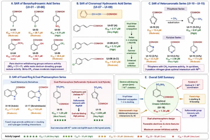
Figure 9: Overall Medicinal Chemistry SAR Summary
CONCLUSION
In the present study, a rational medicinal chemistry strategy was successfully employed to design, synthesize, characterize, and evaluate a series of novel Benzoic Acid derivatives as potential urease inhibitors. Structure-based virtual screening, molecular docking, and in silico ADMET analyses facilitated the identification of promising lead molecules prior to synthesis. The synthesized derivatives were obtained in satisfactory yields and their structures were unequivocally confirmed by FT-IR, ¹H NMR, ¹³C NMR, HRMS, TLC, and melting point analyses. Biological evaluation demonstrated that several synthesized compounds exhibited remarkable urease inhibitory activity, substantially outperforming the clinically used reference inhibitor acetohydroxamic acid. Among the evaluated derivatives, the sulfonamide–Benzoic Acidhybrid UI-18 displayed the highest inhibitory potency, supported by the strongest molecular docking score, excellent enzyme-binding affinity, favorable predicted pharmacokinetic properties, and competitive inhibition kinetics. The close agreement between computational predictions and experimental findings validates the adopted structure-based drug design strategy. Structure–activity relationship analysis revealed that electron-withdrawing substituents, extended conjugated systems, heteroaromatic rings, and dual-pharmacophore architectures significantly enhanced urease inhibition by improving catalytic nickel coordination, hydrogen-bonding interactions, and hydrophobic stabilization within the enzyme active site. These observations provide important medicinal chemistry insights for the future optimization of hydroxamic acid-based urease inhibitors. Overall, the present work identifies UI-18 as a promising lead compound for further development and highlights Benzoic Acid derivatives as attractive scaffolds for the discovery of next-generation anti-urease agents. Future investigations involving antimicrobial evaluation against Helicobacter pylori, cytotoxicity studies, pharmacokinetic profiling, and in vivo efficacy assessment will be essential to establish the therapeutic potential of these compounds and facilitate their progression toward preclinical drug development.
REFERENCES

Pooja Balkrushna Mairal1*, Mehraj Abukalam Kazi1, Mohammed Imran Siraj Ahmed2, Design, Synthesis, Spectral Characterization, Structure–Activity Relationship And Urease Inhibitory Evaluation Of Novel Benzoic Acid Derivatives, Int. J. Sci. R. Tech., 2026, 3 (8), 109-134. https://doi.org/10.5281/zenodo.21785832
 10.5281/zenodo.21785832