
We use cookies to ensure our website works properly and to personalise your experience. Cookies policy
RJS College of Pharmacy, Kokamthan, Kopargaon
Honey bee stings cause local pain, swelling, and inflammation due to venom containing melittin and phospholipase A2. The present study aims to formulate and evaluate an anti-inflammatory transdermal patch for effective management of honey bee stings. The patch was formulated using natural polymers HPMC, PVP K30, and PVA with Boswellia serrata extract as an anti-inflammatory agent. The formulated patches were evaluated for folding endurance, pH, adhesion test, and photosensitivity. The results indicated good physicochemical properties and potential for sustained drug delivery. The developed transdermal patch offers a patient-friendly, non-invasive approach for treatment of honey bee sting inflammation.
Introduction to Honey Bee
Honey Bee is a social insect belonging to the family Apidae. Honey bees are beneficial insects because they help in pollination and production of honey, beeswax, and royal jelly. However, they sting humans and animals as a defense mechanism.(1)
Types of Honey Bees
Only the female worker bee possesses a stinger and can sting.
Introduction to Honey Bee sting
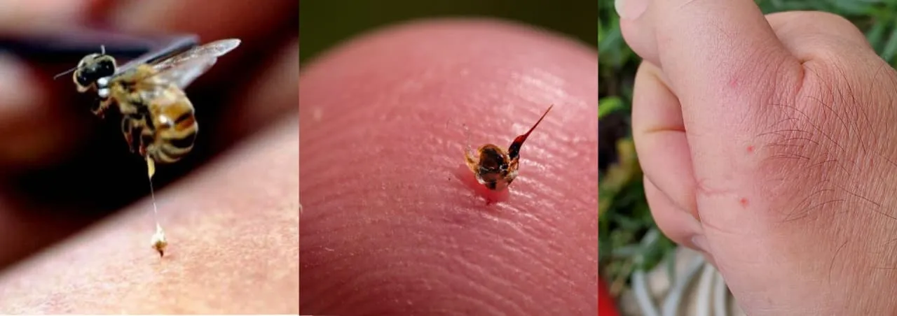
Fig no 1: Honey Bee sting
Honey bee sting is a common insect injury that injects venom into human skin. Bee venom contains melittin, histamine, phospholipase enzymes, and peptides that produce inflammatory reactions.(2)
A honey bee sting is a common defensive injury caused by the female worker honey bee when it feels threatened or disturbed. During the sting, the bee injects venom into the skin through a barbed stinger. This venom contains various biologically active substances that produce pain, redness, swelling, itching, and inflammation. In some individuals, bee stings may also cause severe allergic reactions known as anaphylaxis.(3)
Honey bee stings are common in rural and agricultural areas and may occur accidentally during outdoor activities, farming, gardening, or handling bee hives. Although a single sting usually causes mild local symptoms, multiple stings can lead to serious toxic effects and medical emergencies.(4)
Clinical Symptoms
Severe Reaction
Skin :
Skin is the largest and one of the most important organs of the human body. It forms the outer protective covering of the body and acts as a barrier between the internal organs and the external environment. The skin covers approximately 1.5–2 square meters of body surface area in an adult human and accounts for nearly 15% of total body weight.(5)
The skin performs several important physiological and protective functions such as protection against microorganisms, prevention of water loss, regulation of body temperature, sensation, excretion, immune defense, and absorption of drugs through the transdermal route.
In pharmaceutical and transdermal drug delivery systems, the skin plays a very important role because drugs can be administered through the skin to produce local or systemic therapeutic effects. (6)
Anatomy of Skin
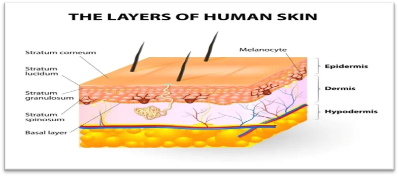
Fig no : 2 layers of human skin
1. Epidermis
2. Dermis
3. Hypodermis (Subcutaneous tissue)
Each layer has a distinct structure and specific functions.
1. Epidermis
The epidermis is the outermost layer of the skin. It acts as the first line of defense against physical injury, chemicals, microorganisms, and dehydration. It is made up of stratified squamous keratinized epithelium.(7)
The epidermis does not contain blood vessels and receives nutrients by diffusion from the dermis.
Layers of Epidermis
a) Stratum Corneum
b) Stratum Lucidum
c) Stratum Granulosum
d) Stratum Spinosum
e) Stratum Basale (Germinativum)
1. Protection against microorganisms and chemicals
2. Prevention of dehydration
3. Formation of keratin
4. Protection from UV radiation
5. Barrier for transdermal drug delivery (8)
2. Dermis
The dermis is the middle and thickest layer of the skin located beneath the epidermis. It is mainly composed of connective tissue containing collagen and elastin fibers.
The dermis provides strength, flexibility, nourishment, and sensory functions to the skin.
Layers of Dermis
a) Papillary Layer
b) Reticular Layer
1. Provides mechanical strength
2. Maintains elasticity of skin
3. Supports blood circulation
4. Contains sensory receptors for pain, pressure, touch, and temperature
5. Helps in wound healing and thermoregulation
3. Hypodermis (Subcutaneous Tissue)
The hypodermis is the deepest layer beneath the dermis. It mainly consists of adipose tissue and loose connective tissue.
1. Stores fat as energy reserve
2. Provides insulation against heat loss
3. Protects underlying organs from injury
4. Connects skin with muscles and deeper tissues
Factors Affecting Topical Absorption of Medications:
A-Physiological Factors of Skin
B-Pysiochemical Factors of Drug
Physiological Functions of Skin
1. Protective Function
2. Thermoregulation
3. Sensory Function
4. Excretory Function
Skin and Transdermal Drug Delivery
Transdermal Drug Delivery System (TDDS) is a method of delivering drugs through the skin in the form of a medicated patch. The drug penetrates through different layers of the skin and produces local or systemic therapeutic effects. TDDS provides controlled and sustained release of drugs over a prolonged period of time. It improves patient compliance, avoids first-pass metabolism, and reduces gastrointestinal side effects associated with oral drug delivery (9)
Principle of TDDS
Drug diffuses from the patch → Through Stratum Corneum → Via Epidermis & Dermis → Reaches dermal blood capillaries → Systemic circulation → Therapeutic effect. In transdermal drug delivery systems (TDDS), drugs are administered through adhesive patches applied on the skin surface.(10)
Fick's Law of Diffusion is the basic principle: (11)
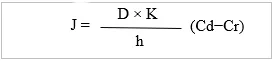
Where
J = Flux,
D = Diffusion coefficient,
K = Partition coefficient,
h = Skin thickness,
Cd = Donor concentration,
Cr = Receptor concentration
Applications of TDDS
The drug penetrates through:
1. Stratum corneum
2. Epidermis
3. Dermis
4. Blood circulation
This method provides:
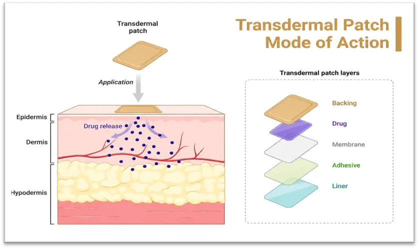
Fig no 3: Transdermal patch MOA
Introduction to Bos0wellia serrata
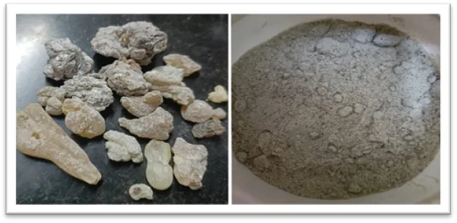
Fig no : 4 Boswellia serrata
Boswellia serrata is a medicinal herbal plant commonly known as Indian Frankincense or Salai Guggul.(14)
|
Biological Source. |
Gum resin obtained from Boswellia serrata tree |
|
Family |
Burseraceae |
|
Chemical Constituents |
|
|
Geographical Source |
It is mainly found in the dry hilly regions of India, North Africa, and the Middle East |
|
Uses |
|
Table No: 01 drug profile
MATERIALS AND METHODS
|
SR.NO |
INGREDIENT |
CATEGORY |
QUANTITY |
|
1. |
Boswellia serrata extract |
API |
0.5 gm |
|
2. |
Hydroxy polymethyl cellulose (HPMC) |
gel-forming agent. |
3 gm |
|
3. |
Polyvinyl Alcohol |
Provides strength to patch |
0.5 ml |
|
4. |
PVP K30 |
Enhances drug release |
1 gm |
|
5. |
Glycerin |
Plasticizer |
0.5 ml |
|
6. |
Propylene glycol |
Permeation enhancer and plasticizer |
0.5ml |
|
7 |
Peppermint Oil |
Cooling agent |
1 ml |
|
8 |
Polyethylene Glycol 400 (PEG 400) |
humectant and plasticizer. |
0.5 ml |
|
9 |
Distilled water |
Vehicle |
q.s to 100 ml |
Table no 02: Formulation Table
EXPERIMENTAL WORK
Method: Maceration Extraction method
Principle: Maceration is a cold extraction process where the powdered drug is kept in contact with the solvent for a prolonged period with occasional shaking. It allows softening and breaking of the cell walls so that the soluble phytoconstituents dissolve into the solvent without heat degradation. (18)
Materials Required:
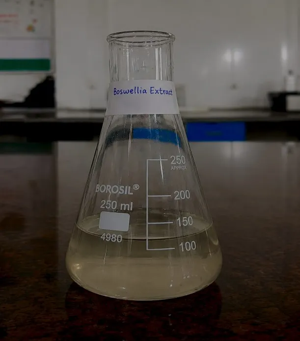
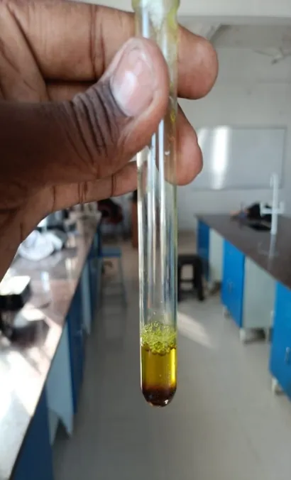
Fig no: 05 Extract
Procedure:
2. Ingredient Measurement:
Accurately weigh all ingredients using a precision balance. Common ingredients include:
3. Gel Formation:
4. Incorporation of Polymers and API:
5. Addition of Plasticizers and Permeation Enhancers
6. Incorporation of Cooling Agent
7. Final Adjustments:
Phase 2: Sheet Formation
This phase involves applying the prepared gel onto a backing substrate to form the actual patch.
1. Substrate Preparation:
Choose a suitable backing material such as non-woven fabric, polyethylene film, or polyurethane film. This material should be breathable, skin-safe, and flexible.(24)
2. Gel Application:
Spread the gel evenly onto the backing material using a coating machine, spreader, or doctor blade. The target thickness may range from 1 to 3 mm depending on the intended duration and intensity of cooling.(25)
3. Protective Layering:
Apply a release liner or protective film (such as PET or polyethylene sheet) over the gel surface to prevent drying, contamination, and sticking during storage and transport.
Phase 3: Drying and Curing
1. Controlled Drying:
Place the gel-coated sheets in a drying chamber or tunnel oven at controlled temperature (e.g., 40–50°C).
Maintain appropriate humidity to avoid over-drying which may affect gel tackiness and flexibility.(26)
Phase 4: Cutting and Packaging
Once the sheets are ready, they are cut and packed for use.
1. Cutting:
Use manual cutters to cut the dried gel sheets into child-friendly sizes (e.g., 2 cm x 2 cm).
Ensure uniformity in size and shape for consistent application.(27)
2. Packaging:
Pack each patch individually in sterile, airtight pouches to prevent moisture loss and microbial contamination.
Consider aluminum foil pouches with a resealable option for convenience.
3. Labeling and Storage:
Label each pack with product name, batch number, manufacturing and expiry dates, and usage instructions. Store in a cool, dry place away from direct sunlight.(28)
EVALUATION RESULT
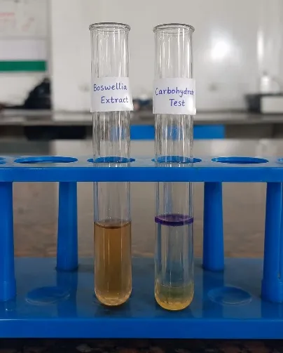
Preliminary Phytochemical Analysis
Prepared extracts of each drug were used for preliminary phytochemical analysis. In this, chemical tests for the detection of some primary metabolites (carbohydrate, amino acid, protein, lipid and starch) and secondary metabolites (alkaloids, flavonoids, tannins, saponins, glycosides) were done.(29)
|
Test for |
Test |
Observation |
Inference |
Pic of the test |
|
Primary Metabolites - |
||||
|
Carbohydrate |
Molisch's test (General test) To 2-3 ml aqueous extract, add few drops of alpha naphthol solution in alcohol, shake and add conc. H2SO4 from side of the test tube. |
Violet ring is formed at the junction of two liquids. |
Present |
|
|
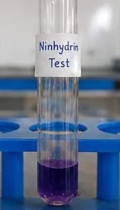
Amino Acid |
Ninhydrin test (General test) Heat 3 ml T.S. and 3 drops 5% Ninhydrin solution in boiling water bath for 10 min |
Purple / bluish colour appears. |
Present |
|
|
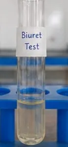
Protein |
Biuret test (General test) To 3 ml T.S. add 4% NaOH and few drops of 1% CuSO4 solution. |
Violet or pink colour not appeared. |
Absent |
|
|
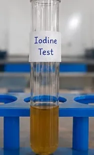
Starch |
Iodine test: Mix 3 ml test solution and few drops of dilute iodine solution. Blue colour appears, it disappears on boiling and reappears on cooling. |
Blue colour does not appear |
Absent |
|
|
Lipid |
Extract dropout on filter paper drought comes then lipids are present |
No permanent oily stain on filter paper |
Absent |
|
|
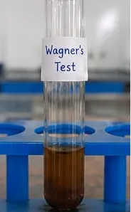
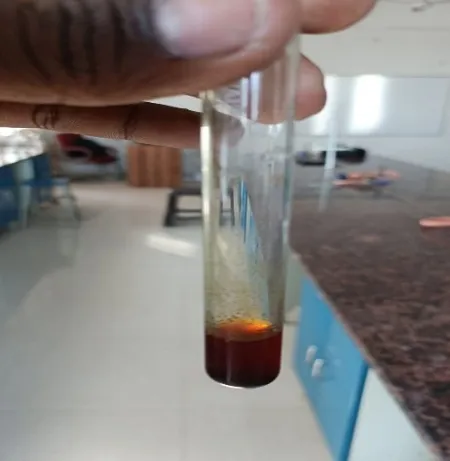
Secondary Metabolites Alkaloids: Evaporated the aqueous, alcoholic and chloroform extracts separately. To residue, dilute HCI added. After shaking well and filtration, using filtrate, following test was performed. |
||||
|
|
Wagner's test 2-3 ml filtrate with few drops Wagner's reagent gives reddish brown ppt Alpha naphthol solution in alcohol, shake and add conc. H2SO4 from side of the test tube. |
Reddish brown ppt |
Present |
|
|
Flavonoid |
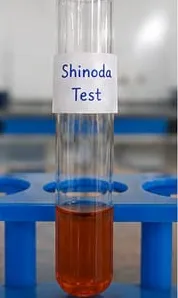
Shinoda Test To dry powder or extract, add 5 ml 95% ethanol few drops conc. HCI and 0.5 g magnesium turnings. Orange, pink, red to purple colour appears. |
Orange red colour observed |
Present |
|
|
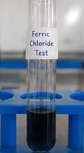
Tannins |
% FeCl3 solution |
Deep blue-black colour |
Present |
|
|
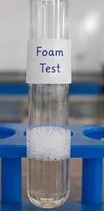
Saponins |
Add water into sample and shake for 15 sec. |
Foam is observed |
Present |
|
|
Glycosides |
||||
|
Legal's test (Test for cardenoloids) |
To aqueous or alcoholic extract, add 1 ml pyridine and 1 ml sodium nitroprusside. Alpha naphthol solution in alcohol, shake and add conc. H2SO4 from side of the test tube. |
Pink to red colour appeared |
Present |
|
|
Test for deoxy sugar (Keller-Killiani test) |
To 2 ml extract, add glacial acetic acid, one drop 5% FeCl3 and conc. H2SO4. Reddish brown colour appears at junction of the two liquid layers and upper layer appears bluish green. |
No reddish brown colour appears at junction of the two liquid layers. |
Absent |
|
Table no : 03 Preliminary Phytochemical Analysis
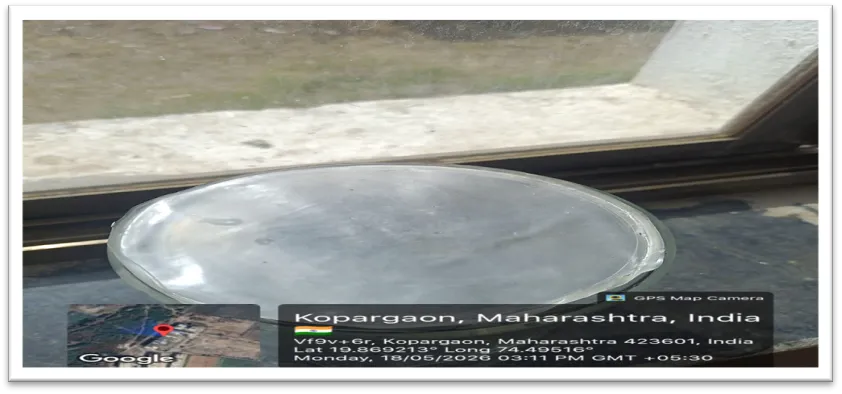
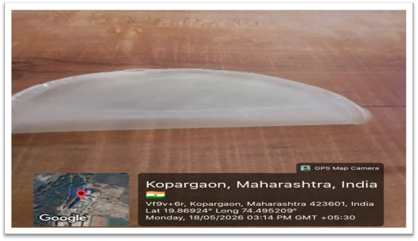
Fig no 6: Transdermal patch
Method:
Visual inspection of a representative number of patches under good light for continuity, brittleness, translucency, clarity, tackiness and any visible defects (cracks, bubbles, lumps). Record observations.
Colour: Transparent to slightly translucent, whitish appearance
Texture: Smooth and glossy surface, no visible cracks or wrinkles
Odor: Slight characteristic aromatic odor of Boswellia resin (30)
Method: Hold a strip between thumb and forefinger and fold repeatedly at the same place until it breaks; count number of folds. Perform in triplicate and report mean. Acceptance criteria: Folding endurance ≥200 folds (≥100 acceptable for brittle formulations). No crack or rupture during normal handling.
Soak patch in distilled water and measure pH using litmus or pH meter. pH should be close to skin pH (4.5–6.5)
Fig no 08: Ph test
Checks the sticking ability on skin without causing irritation. Apply patch on clean, dry skin for 8 hours. Observe if it stays adhered.(32)
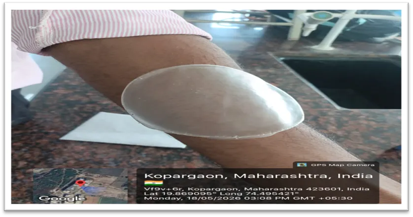
Fig no 09: Adhesion test
Determines if the patch causes skin reaction when exposed to sunlight. Apply patch on one arm and expose to UV/sunlight for 30 mins; compare with a protected/control area
Fig no 10: Photosensitivity test
|
Test |
Description |
Methodology |
Result |
|
1. Adhesion Test |
Checks the sticking ability on skin without causing irritation. |
Apply patch on clean, dry skin for 8 hours. Observe if it stays adhered. |
Patch should remain in place without peeling or causing discomfort. |
|
2. Durability Test |
Measures performance over a set duration. |
Keep patch on for recommended time (e.g., 8 hrs.) and observe physical condition and cooling effect. |
Cooling sensation should last as indicated (4–8 hrs.). |
|
3. Skin Irritation Test |
Assesses potential for skin irritation |
Apply patch on forearm or back for 24 hours. Check for redness, itching. |
No signs of irritation or sensitization. |
|
4. Photosensitivity Test |
Determines if the patch causes skin reaction when exposed to sunlight. |
Apply patch on one arm and expose to UV/sunlight for 30 mins; compare with a protected/control area. |
No erythema (redness) or pigmentation in exposed area. |
|
5. pH Stability Test |
Checks the pH balance to ensure compatibility with skin. |
Soak patch in distilled water and measure pH using litmus or pH meter. |
pH should be close to skin pH (4.5–6.5). |
|
6. Microbial Limit Test |
Ensures product is free from harmful microbes. |
Swab patch surface and culture on agar media. Check for bacterial/fungal growth. |
Should comply with pharmacopeial microbial limits. |
Table no 5.2 Evaluation test
Summary:
The present research work entitled Formulation and Evaluation of an Anti-Inflammatory Transdermal Patch for Honey Bee Stings was carried out to develop a natural and effective topical delivery system
The transdermal patch was formulated by solvent casting method using Boswellia serrata extract (0.5 gm) as API, HPMC (3 gm) as gel-forming agent, PVA (0.5 ml) to provide strength to patch, PVP K30 (1 gm) to enhance drug release, Glycerin (0.5 ml) as plasticizer, Propylene glycol (0.5 ml) as permeation enhancer, PEG 400 as humectant, Peppermint oil (1 ml) for cooling effect, and Purified water as solvent. The pH of the final formulation was maintained at 5.5-6.5 to match skin pH
The prepared patch was evaluated for various parameters and the following results were obtained:
The results confirmed that the formulated patch is stable, non-irritant, and effective for topical delivery

Shivam Eknath Limbore, Kadam Snehal*, Formulation And Evaluation Of An Anti-Inflammatory Transdermal Patch For Honey Bee Stings, Int. J. Sci. R. Tech., 2026, 3 (6), 642-659. https://doi.org/10.5281/zenodo.20610941
 10.5281/zenodo.20610941