
We use cookies to ensure our website works properly and to personalise your experience. Cookies policy
RJS College Of Pharmacy Kokamthan
Microsponges are highly porous, polymeric microspheres developed to enhance localized and controlled delivery of therapeutic agents to the skin. The present study was carried out to formulate and evaluate fluconazole-loaded microsponges incorporated into a topical gel for the treatment of fungal infections. Microsponges were prepared by the quasi-emulsion solvent diffusion method using suitable polymers such as ethyl cellulose and polyvinyl alcohol.to achieve controlled drug release and suatained release. The prepared microsponges were evaluated for particle size, production yield, entrapment efficiency, and zeta potential percentage yeild. The optimized microsponge formulation was further incorporated into a carbopol gel base. The formulated gel was evaluated for physical appearance, pH, spreadability, d, and antifungal activity. The results showed that the microsponge gel provided sustained release of fluconazole with good stability and satisfactory physicochemical properties. The formulation also exhibited effective antifungal activity against fungal pathogens. Thus, fluconazole-loaded microsponge gel can be considered a promising topical drug delivery system for improved therapeutic effectiveness and better patient compliance in fungal infections.
Fluconazole is a synthetic broad-spectrum antifungal agent belonging to the triazole class of azole drugs. It is one of the most widely used antifungal medicines in clinical practice due to its good efficacy, excellent bioavailability, predictable pharmacokinetics, and relatively low toxicity. Fluconazole works by inhibiting the fungal cytochrome P450 enzyme lanosterol 14-α-demethylase. This enzyme is essential in the ergosterol biosynthesis pathway. Ergosterol is a vital structural component of the fungal cell membrane. When fluconazole blocks its synthesis, ergosterol levels decrease, leading to defective cell membrane formation, increased membrane permeability, leakage of cellular contents, and ultimately inhibition of fungal growth or cell death.
Fluconazole is mainly active against Candida species (especially Candida albicans), Cryptococcus neoformans, and some other opportunistic fungi. Because of this spectrum, it is commonly used in the treatment of oropharyngeal candidiasis (oral thrush), vulvovaginal candidiasis, oesophageal candidiasis, urinary tract fungal infections, and cryptococcal meningitis. It is particularly important in immunocompromised patients such as those with HIV/AIDS, cancer chemotherapy, or post-transplant therapy
From a pharmacokinetic point of view, fluconazole has almost complete oral absorption and is not significantly affected by food or gastric pH. It distributes widely throughout body fluids and tissues, including cerebrospinal fluid, which makes it effective for central nervous system fungal infections. The drug is minimally metabolized and is excreted primarily unchanged through the kidneys, so dose adjustment is required in renal impairment.
In pharmaceutical formulation development, fluconazole is available in various dosage forms such as tablets, capsules, oral suspension, intravenous infusion, and topical preparations like gels and creams. In recent research, it is extensively incorporated into novel drug delivery systems such as microsponges, nanoparticles, liposomes, niosomes, and polymeric gels to improve its topical retention, sustain drug release, enhance therapeutic effectiveness, and reduce dosing frequency and side effects.
Because of its clinical importance and formulation flexibility, fluconazole is a key drug in antifungal therapy as well as in pharmaceutical research and development, especially in dermatological and controlled drug delivery systems.Drug delivery is the process of giving a medication to humans or animals in order to achieve a therapeutic result. For many years, pharmaceutical dosage forms such as tablets, pills, capsules, ointments, creams, liquids, injectables, suppositories, and gels—which are regarded as standard dosage forms—have been used to treat both acute and chronic illnesses.traditional dosing forms. Traditional transdermal applications of gels release the medication as soon as they are applied, which causes the medication to build up and irritate skin layers. The active ingredient can be spread out in a liquid or applied straight to the skin in a nearly pure form.
Recently new approaches in the novel drug delivery systems introduced are:
|
Carrier System |
TYPES |
|
|
|
Vesicular system |
Microparticulate system |
|
Colloidal carrier |
Liposome |
Nanocapsules |
|
Niosome |
Nanoparticles |
|
|
Immunoliposomes |
Microsponges |
|
Table No.1 Carrier System for Drug Delivery
MICROSPONGE: AN APPROACH FOR TOPICAL DELIVERY
Microsponges are a new, regulated medicinal product with a wide range of uses. They have the capacity to encapsulate a variety of medications, both liquid and solid. compliance. Overall, microsponges offer a promising approach in pharmaceutical formulations, enhancing drug delivery outcomes and patient satisfaction. By taking advantage of the special structure and physiology of human skin, microsponges offer a significant advantage in the administration of dermatological medicines. They provide increased effectiveness when applied to human skin. They minimize local side effects while providing increased efficacy. is essential to their topical Microsponges' remarkable entrapment effectiveness, which enables them to absorb more than six times their own weight, is one of their main advantages. Even at high temperatures and over a wide pH range of 1 to 11, these microsponges show remarkable stability. compatibility when added to liquid or semi-solid dosages and suspended in different carriers forms by giving the medication a regulated release. Microsponges add to the product's elegance by assuring stability and non-irritating medication release. qualities that enhance patient applicability.
DEFINITION:
The tiny sponge-like particles that make up microsponges are porous, polymeric, microscopic drug delivery systems that have the ability to trap active medicinal components within their network. They are usually between 5 and 300 µm in diameter and have many interconnected pores that enable targeted, regulated, and prolonged drug release. By releasing the medication gradually at the site of action, microsponges increase the stability of medications, lessen discomfort, and boost efficacy. These qualities make them popular in topical, oral, and cosmetic formulations to increase patient compliance and therapeutic efficacy.
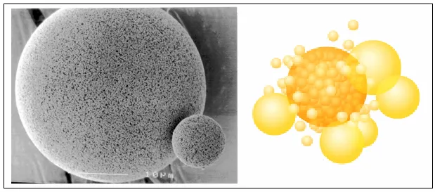
Fig no.1 Structure of Microsponges
Higher concentrations of active substances are needed to produce therapeutic effects because many conventional drug delivery methods are ineffective at delivering medications. Because of these restrictions, sophisticated drug delivery methods are being investigated more and more to accomplish site-specific and regulated drug release. A.Polymer-based microspheres with a substantially cross-linked structure are known as microsponge systems. They can encapsulate a wide range of active pharmacological substances since they are sufficiently flexible. These qualities make microsponges popular for applications involving controlled and extended drug administration.Microsponges are designed to effectively administer a pharmaceutically active drug at the lowest feasible dose, minimize adverse effects, and enhance stability, elegance, and flexibility in formulation. The drug release profile can also be changed with these. The microsponge system can stop an excess of a component from building up in the dermis and epidermis. These goods are often packaged in conventional ways and contain a very high concentration of active substances. Microsponges come in conventional forms like lotions, gels, or creams and have a fairly high concentration of active chemicals. A variety of active substances, including anti-infective, anti-fungal, and anti-inflammatory drugs, can be transported via microsponges, which are polymeric delivery systems made of porous microspheres.
|
Sr.no |
Active agent |
Application |
|
1 |
Sunscreens |
Long-lasting product efficacy, with improved protection against sunburns and sun-related injuries, even at elevated concentration and with reduced irritancy and sensitization |
|
2. |
Anti-acne, e.g., Benzoyl peroxide |
Maintained efficacy with decreased skin irritation and sensitization |
|
3. |
Anti-inflammatory e.g. hydrocortisone |
Long lasting activity with reduction of skin allergic response and dermatoses |
|
4. |
Anti-dandruffs, e.g., zinc pyrithione |
Reduced unpleasant odour with lowered irritation and extended safety and efficacy. |
Table No.2 List of marketed products
Objectives
MATERIAL AND METHODS:
Material: Fluconazole, Eudragit RS 100, Ethyl cellulose, PVA, Dichloromethane, Methanol, glycerol.
Methods:
Fluconazole microsponges were prepared by quasi emulsion solvent diffusion method by using different drug.
Quasi-Emulsion Solvent Diffusion Method:
The microsponges containing the anti-fungal drug Fluconazole as the core material was prepared by Quasi emulsion Method according to the formula given in table no. 3 the process involved formation of quasi-emulsion of two different phases i.e. internal phase and external phase similar to the emulsion.

Fig no.2.Process of stirring
|
Sr No. |
Ingredient |
F1 |
F2 |
|
1. |
Fluconazole |
1gm |
1gm |
|
2. |
Ethyl cellulose |
1gm |
2gm |
|
3. |
Polyvinyl Alcohol |
0.5gm |
1gm |
|
4. |
Dichloromethane |
10ml |
10ml |
|
5. |
Glycerol |
1ml |
1ml |
|
6. |
Water |
100ml |
100ml |
Table no.3.Formula for Microsponges formulation
|
Sr.no |
Ingredient |
Quantity |
|
1. |
Fluconazole Microsponges |
0.1gm drug |
|
2. |
Carbopol 940 |
0.7gm |
|
3. |
Triethanolamine |
0.04gm |
|
4. |
Methyl parabean |
0.06gm |
|
5. |
Propyl parabean |
0.02gm |
|
6. |
Distilled water |
q.s to 20ml |
Table No.4: Formula for Gel formulation
EVALUATION PARAMETER OF Fluconazole MICROSPONGES:
The formulation's physical characteristics, including color, consistency, and the existence of any obvious irregularities, were evaluated visually.
It is necessary to gather and weigh the microsponges produced from different formulations. In this case, the total weight of the medication and polymer utilized in the production procedure will be divided by the actual weight of the microsponges. This computation makes it possible to evaluate the proportion of medication contained in the microsponges, offering information on the formulation's effectiveness.
The yield percentage was computed using the formula that follows

Each batch of microsponges containing 100 mg of fluconazole should be accurately weighed and crushed. After that, the powder should be put in a 100 ml volumetric flask and adjusted with pH 7.4 phosphate buffer. To bring the solution up to par, phosphate buffer should be added after it has been filtered. Whatmann filter paper No. 44 was then used to filter the mixture. A precise amount of the solution (1 ml) should be taken after filtering and diluted up to 10 ml with pH 7.4. Following filtering, a precise amount of the solution (1 ml) should be obtained and diluted with pH 7.4 phosphate buffer up to 10 ml. A precise volume (1 ml) should be pipetted out of this diluted solution, and pH 7.4 phosphate buffer should be used to further dilute it up to 10 ml. A spectrophotometer should be used to measure the final solution's absorbance at 264 nm.
%Drug Content= Actual drug content of microsponges/Total amount of microsponges ×100
Microsponges' surface appearance and form are examined using microscopy. A tiny sample is put on a glass slide and examined under a microscope.
Actual Drug Content: 100 mg of precisely weighed microsponges were used. They were ground into a powder and extracted using a 100 ml technique. Additionally, it was serially diluted using phosphate buffer 7.8 at pH 7.4. Ibuprofen drug concentration was determined by measuring absorbance in a UV spectrophotometer at 260 nm using pH 7.4 phosphate buffer as a blank. The investigations were conducted in duplicate. The entrapment effectiveness and real drug content were purposefully chosen as:
where Mthe is the theoretical amount of ibuprofen in microsponges, Mms is the weighed quantity of microsponges, and Mact is the actual amount of ibuprofen in microsponges.
A digital pH meter was used to determine the formulation's pH. The formulation's pH was measured three times, and the average value was determined.
Optical microscopy was used to measure the prepared microsponges' particle size. An ocular micrometer and a stage micrometer were attached to the optical microsponges. The micrometer in the eyepiece was calibrated. Using an optical microscope, the diameters of over 200 microsponges were measured at random.
=(SM/EM) ×10
Were, µ- correction factor
SM- Reading of stage micrometer which coincides
with reading of eyepiece micrometer (EM).
To assess the surface charge and electrostatic stability of the prepared microsponges, zeta potential was calculated. The degree of repulsion between similarly charged particles scattered across the medium is revealed by the measurement. By preventing particle aggregation and sedimentation during storage, a sufficient zeta potential value denotes strong physical stability. A zeta potential analyzer based on electrophoretic mobility was used for the analysis. The formulation's stability and suitability for additional pharmaceutical uses are indicated by the acquired zeta potential values. Improved dispersion stability and homogeneity of the microsponge system are suggested by higher absolute zeta potential values.
EVALUATION OF MICROSPONGES LOADED GEL:
Evaluation of Gel Loaded with Microsponges
The, formulations were subjected to observed for color, consistency, quality, uniformity, homogeneity, and dispersion of gel loaded microsponges are examined by visual observation.
The gel loaded with Microsponges was subjected to determination of pH by utilizing digital pH meter. 5 g gel was suspended in 45 ml double distilled water, and solution pH was measured.
The gel loaded with Microsponges batch was subjected to spreadability studies by using and glass slide apparatus. It comprises of two slides upper movable slide and lower non-movable slide. The weight loading of about 20 gm were added to the pan and time was noted for upper slide to confine totally from the fixed slides. Spreadability was then determined by utilizing the formula.
S= M x L/T (g.cm/s)
Where,
S= Spreadability;
w= weight tide to upper slide;
L= length of glass slide;
T= time taken to separate the slide completely from each other.
The gel is applied on the skin surface and washed with water The ease of removal and residue remaining is observed A good gel should be easily washable without leaving significant residue.
5) In-vitro Antifungal Activity
The inoculum of the microorganism was prepared from the fungal cultures.
15ml of Saubroad agar (Hi media) medium was poured in clean sterilized Petri plates and allowed to cool and solidify.
100 µl of broth of fungal strain was pipette out and spread over the medium evenly with a spreading rod till it dried properly.
Wells of 6mm in diameter were bored using a sterile cork borer. Solutions of the compounds (100µl/ml) were prepared in DMSO and 100µl of prepared test solutions and standard was added to the wells. The petri plates incubated at 37 0C for 24 h. Miconazole (1mg/ml) was prepared as a positive control and DMSO was taken as negative control. Antifungal activity was evaluated by measuring the diameters of the zone of inhibitions (ZI) all the determination were performed in triplicates
RESULTS AND DISCUSSION:
The color and nature of the F1-F2 formed microsponges were determined to be white and powdered, as indicated in Table No. 4. This indicates that the resulting microsponges pass the visual test for the standards.
|
Formulation code |
Color |
Nature |
|
F1 |
White |
Powdered |
|
F2 |
White |
Powdered not rigid |
Table no: 4 Results of Visual Inspection of fluconazole microsponges
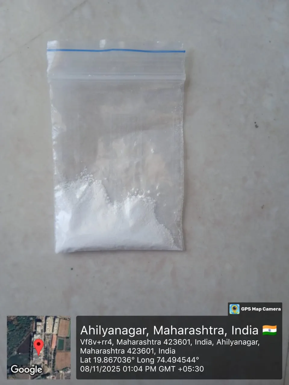
Fig no.3.Physical appearance of Microsponges
It was discovered that the Fluconazole drug's percentage yield in microsponges was 50%. For some applications, such topical or transdermal distribution, a PE of 50–70% is ideal.
The yield percentage was computed using the formula that follows:
% Yield= Practical yield of microsponges ×100
Total weight of drug and polymer
= 1.5/3.5×100
=50%
A UV-visible spectrophotometer was used to measure the drug content of the manufactured fluconazole-loaded microsponges. The drug content was represented as % w/w after the absorbance readings were transformed into concentration .The medication content of the formulation was 85%

Fig no 4: Determination of drug content
The microsponges solution's standard range was 1–11, and its optimal pH of 6.48 was discovered.
|
Sr no. |
Solution |
pH |
Mean |
|
1. |
Microsponges solution |
6.47 |
6.49 |
|
2. |
6.51 |
||
|
3. |
6.49 |

Fig No.5. Determination of pH
The prepared microsponges were consistently distributed and largely spherical in shape, according to optical microscopy. The particles had smooth surfaces and seemed distinct. There was no noticeable particle clumping or aggregation.

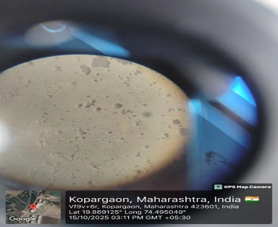
6)Results of particle size:
Zeta sizer analysis using the Microtrac Zeta sizer for microsponges. The Poly Dispersity Index (PDI) value was 0.566, which was appropriate for particle size determination, and the average microsponge particle size in the diluted sample with water was 5946(d) nm.
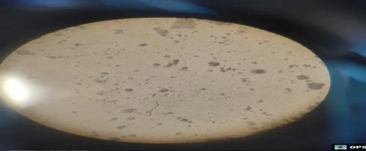
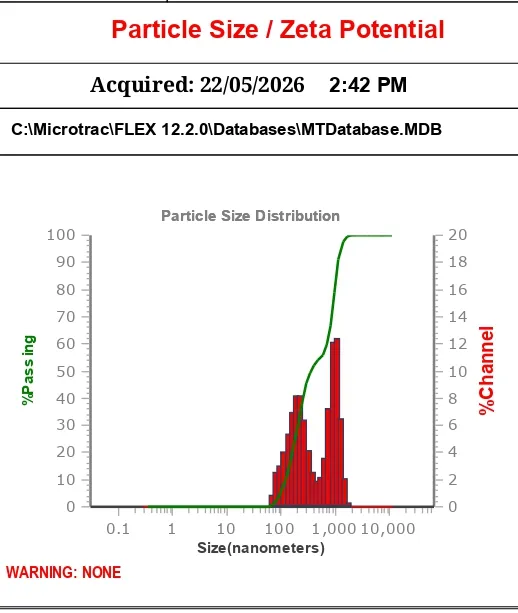
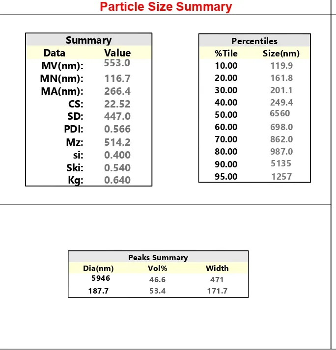
Fig No.6. Result of particle size
7) zeta potential:
Zeta potential was measured at a temperature of 20.07°C, a viscosity of 1 mPa.s, and an electrophoretic mobility of 0.48 µm2/Vs. Microsponges are stable in preparation because their zeta potential was discovered to be 28 mV.zeta potential used to determine the particle surface charge.

Fig No.7.zeta potential
8)Entrapment efficiency:
For all microsponges, the loading efficiency of Fluconazole microsponge formulations ranges from 33.17 to 82.76%, which is the maximum loading efficiency. This was discovered for the formulation F2, which contained more medication.Fluconzole and ethyl cellulose are suggested.
RESULTS OF GEL LOADED WITH FLUCONAZOLE MICROSPONGES
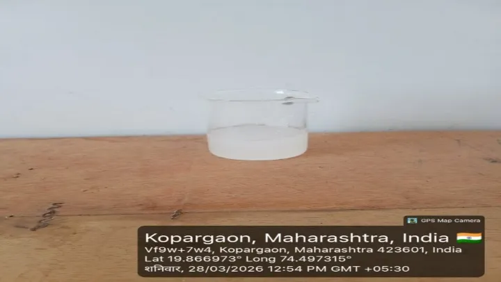
The formulated gel loaded with Fluconazole microsponges were inspected for their color and appearance. The gel loaded microsponges batches (F1) as shown in Table no: 7, were appearing transparent white with uniformly distributed microsponges,
|
F Code |
Color |
Consistency |
Homogeneity |
Appearance |
Uniformity |
|
F1 |
White |
Optimum viscous |
Good |
Transparent white |
Good |
Table No.8. Results of Visual inspection of gel
The gel formulations F1,loaded with microsponges were examined for color, consistency, homogeneity, appearance, uniformity Batch F1 showed white color, less viscous, good homogeneity, good uniformity and opaque appearance.
|
Sr. no. |
pH |
|
1 |
6.39 |
|
2 |
6.41 |
|
3 |
6.49 |
|
Average |
6.43 |
Table no.9.Result of microsponges loaded gel Ph
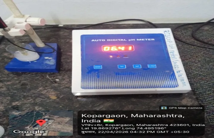
The pH of Gel was found to be 6.4. The pH of gel loaded microsponges was measured using pH meter. The formulation is considered as safe and non-irritating, since their pH was within the normal skin pH that is 6.7
|
Formulation |
Time |
Spreadability |
|
F1 |
51 Sec. |
7.5 g.cm/s |
|
Table no.10.Result of spreadability |
||

FIG NO.16.Determination of Spreadability
The spreadability values of microsponge loaded gel formulations were found in the range of 7.5 g·cm/sec. The optimized formulation showed good spreadability, indicating easy application on the skin and uniform distribution of the gel formulation.”
The prepared microsponge loaded gel formulations showed good washability. The gel was easily removed from the skin surface with normal water without leaving any sticky residue. This indicates good patient compliance and suitability for topical application.
|
Formulation |
Observation |
|
F1 |
Good washability and no residue observed |
Table no.10.Result of washability

Fig no.17.Result of antifungal activity
The antifungal profile of Microsponges loaded Fluconazole gel was evaluated by measuring the zone of inhibition against fungal strains C albicans (ATCC10231), Via well diffusion method. The compounds Microsponges loaded Fluconazole gel exhibited good activity as compared to the standard Miconazole
CONCLUSION
Spherical nanoparticles are used in microsponges, a polymeric delivery technique. Depending on the level of flattening or after-feel required in the final formulation, these systems are composed of porous microspheres that range in size from 5 to 300 micrometers. Emollients, fragrances, essential oils, sunscreens, anti-infectives, antifungals, anti-inflammatory medications, and some antibiotics are among the many active chemicals that can be found in polymeric delivery systems, which are composed of porous microspheres. Polymeric materials are used to create these adaptable microsponge devices. By selecting the right polymer for injection, the rate of medication release can be changed. Because of this, research is currently being done on the Microsponge Delivery System (MDS), a cutting-edge and developing technology for the delivery of routine medications. A. Originally developed for topical distribution, the microsponge delivery technology—which makes use of bioerodible polymers and tissue engineering—is being employed for controlled oral administration. As with other novel drug carriers, drug release from microsponge can be controlled by varying the temperature, pH, and polymer solubility of the medium. Additionally, they may improve drug release, lessen side effects, and improve pharmaceutical stability. The microsponge method is a reliable way to administer medication because of its numerous advantages. Furthermore, MDS has a bright future in a variety of therapeutic formulations due to its advantageous qualities, which include continuous release, decreased irritancy, small size, self-sterility, and interoperability with several vehicles and components.
REFERENCES

Vaishnavi Dhage*, Snehal Gondkar, Suvarna Sangale, Prerana Jamdhade, Tanvir Pathan, Kanchan Gursal, Formulation And Evaluation Of Fluconazole Loaded Microsponges Gel For Topical Drug Delivery System., Int. J. Sci. R. Tech., 2026, 3 (6), 794-808. https://doi.org/10.5281/zenodo.20676810
 10.5281/zenodo.20676810