

We use cookies to ensure our website works properly and to personalise your experience. Cookies policy
Rashtrasant Janardhan Swami College Of Pharmacy, Kokamthan, Kopargaon.
The aim of this study was to develop and assess fluconazole-loaded niosomes to boost drug stability and enhance the delivery of the antifungal medication. Niosomes were prepared using non-ionic surfactants and cholesterol, and the developed formulation was evaluated for key physicochemical parameters. Particle size analysis revealed the formation of vesicles in the nanometer range with a bimodal size distribution and a mean volume diameter of 1878 nm, indicating effective vesicle formation. Zeta potential analysis showed a positive surface charge of approximately +28 mV, confirming good electrostatic stability and reduced aggregation tendency of the formulation. The presence of sufficient surface charge contributes to improved colloidal stability and prolonged shelf life of the niosomal suspension. Overall, the results demonstrate that fluconazole-loaded niosomes possess suitable physicochemical characteristics and can serve as a promising vesicular carrier for controlled and effective antifungal drug delivery.
Fungal infections of the skin and mucosal surfaces remain a major therapeutic challenge due to the limited penetration and poor retention of conventional topical antifungal formulations. Fluconazole, a synthetic triazole derivative, is widely used for treating dermatophytes and Candida infections; however, its therapeutic effectiveness is often restricted by short retention time on the skin, variability in drug absorption, and inability to achieve adequate drug levels at deeper layers of the stratum corneum. These limitations indicate a need for an improved drug-delivery system that can enhance local drug availability while minimizing systemic exposure.
Niosomes, which are closed bilayer vesicles made from non-ionic surfactants and cholesterol, have emerged as a promising alternative for topical drug delivery. They enhance the solubility, stability, and permeation characteristics of encapsulated drugs. Due to their flexible structure and ability to entrap both hydrophilic and lipophilic molecules, niosomes improve drug retention at the site of application and provide controlled release, leading to improved therapeutic response.
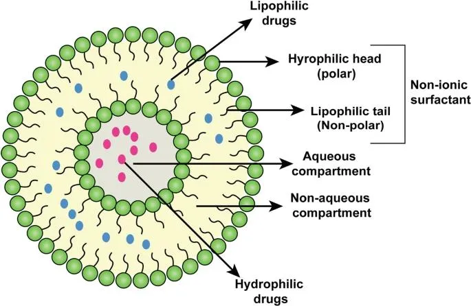
Niosomes are vesicles composed of non-ionic surface-active agent bilayers, which serve as novel drug delivery systems. Niosomes are non-ionic surfactant-based microscopic lamellar structures. Niosomes are vesicles composed of non-ionic surfactants, amphipathic compounds with an overall neutral charge. These non ionic surfactant are cheap and safe for use in biomedicine. Niosomes are obtained on admixture of non-ionic surfactant of the alkyl or dialkyl polyglycerol ether class and PEG with subsequent hydration in aqueous media. Niosomes have more penetrating property, due to the incorporation of non-ionic surfactants. Niosomes can entrap both hydrophilic and lipophilic drugs, either in aqueous layer or in vesicular membrane made of lipid materials. It is reported to attain better stability than liposomes. It can prolong the circulation of the entrapped drugs. Because of the presence of non-ionic surfactant with the lipid, there is better targeting of drugs to tumour, liver and brain. It may prove very useful for targeting the drug for treating cancers, parasitic, viral and Fungal infections more effectively. Niosomes are promising vehicle for drug delivery and being non-ionic, it is less toxic and improves the therapeutic index of drug by restricting its action to target cells. They are vesicular systems similar to liposomes that can be used as carriers of amphiphilic and lipophilic drugs. Compared to liposomes, niosomes have the advantage that the components are extremely cheap compared to phospholipids, and both the lipids and non-ionic surfactants are similarly stable.( 9,10)
For fluconazole, niosomal encapsulation offers the benefits of increased drug loading, better permeation across the skin barrier, and reduced dosing frequency. The physicochemical properties of niosomes, including vesicle size, entrapment capacity, and drug content, directly influence their performance and stability. Hence, evaluating these parameters is essential to ensure formulation quality.
The present study focuses on the formulation and evaluation of fluconazole-loaded niosomes, with emphasis on key parameters such as drug content, vesicle size, and entrapment efficiency. These evaluation tests provide a scientific basis for determining the suitability, stability, and effectiveness of the prepared niosomal formulation for enhanced topical delivery of fluconazole.(2,3)
|
|
Fig no.01.structure of noisome (11)
ADVANTAGES OF NIOSOMES:
Can Encapsulate both hydrophilic and lipophilic drugs due to their amphiphilic bilayer structure.
GENERAL CHARACTERISTICS OF NIOSOME:
APPLICATIONS OF NIOSOME:
METHODS OF PREPARATION:
1.Ether Injection method
2.film method/Hand Shaking method
3.Sonication
4.Heating method
5. Multiple membrane Extrution method
6.Reverse phase evaporation
7.Bubble method
1. ETHER INJECTION METHOD:
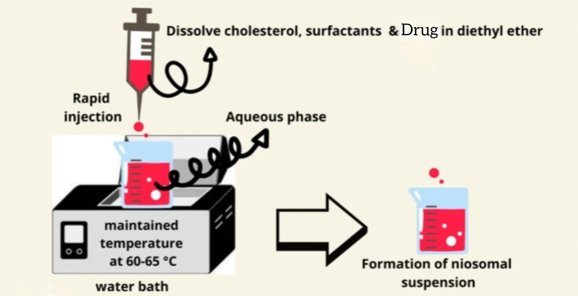
Fig.no.02. Ether injection method (1)
2. FILM METHOD/HAND SHAKING METHOD:
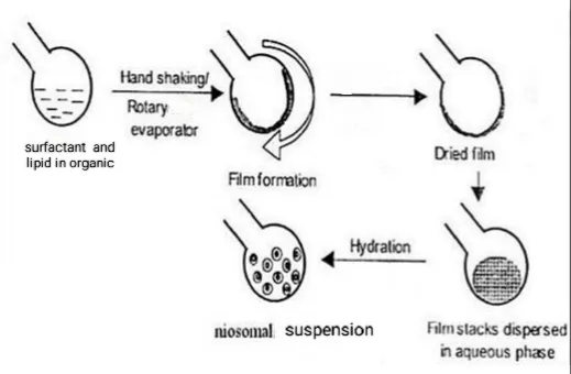
Fig.No.03. Hand Shaking Method (12)
3. SONICATION:
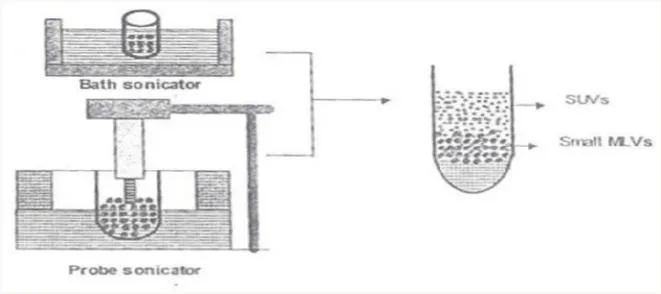
Fig..no.04. Sonication Method (10)
4. HEATING METHOD:
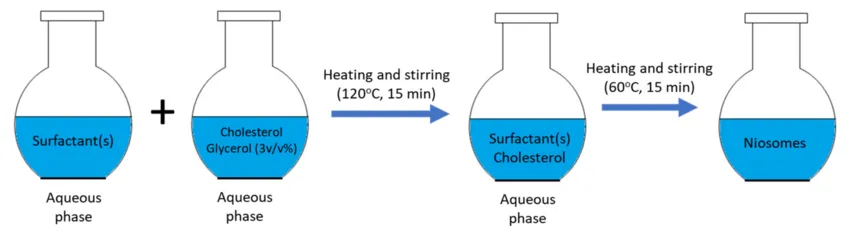
Fig.No.05. Heating Method (12)
5. MULTIPLE MEMBRANE EXTRUSION METHOD:
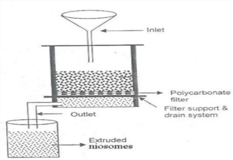
Fig.No.06. Multiple Extrution Method (11)
6. REVERSE PHASE EVAPORATION:
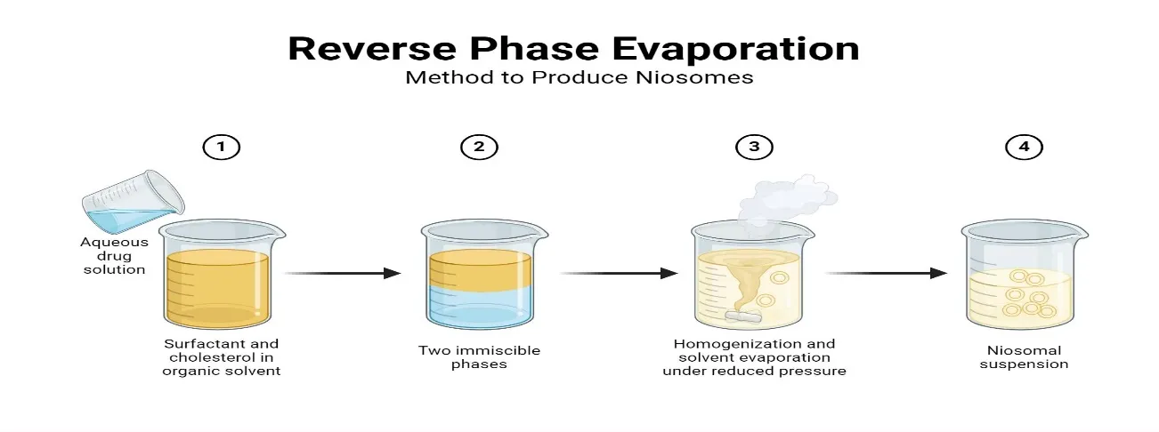
Fig.No.07. Reverse Phase Evaporation (9)
7.BUBBLE METHOD:
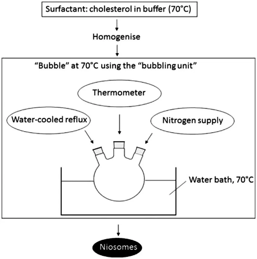
Fig.No.08. Bubble Method (13)
2. MATERIAL AND METHOD:
Material:
METHOD:
The ether injection method is a technique used to prepare niosomes, which are vesicular drug delivery systems composed of non-ionic surfactants and PEG. The method involves injecting a solution mixture of ether, surfactant, PEG, and drug into a hot aqueous solution, resulting in the formation of niosomes. (1,9)
Advantages:
Disadvantages:
EXPRIMENTAL WORK:
|
Sr.no |
Ingredients |
F1 |
F2 |
F3 |
|
1. |
Polyethylene Glycol (PEG) |
200mg |
200mg |
200mg |
|
2. |
Span 80 |
400mg |
200mg |
150mg |
|
3. |
Fluconazole |
200mg |
200mg |
200mg |
|
4. |
Ethanol |
2ml |
2ml |
2ml |
|
5. |
Diethyl ether |
8ml |
8ml |
8ml |
|
6. |
Phosphate buffer |
15ml |
15ml |
15ml |
Table no.01.formula for noisome preparation
PROCEDURE:
|
Polyethylene Glycol (PEG)+Surfactant |
|
Dissolved in 6ml diethyl ether |
|
2ml of ethanol containing weighed quantity of fluconazole |
|
Resulting solution was slowly injected using micro syringe at a rate of 1ml/min into 15ml of hydrating solution phosphate buffer (pH 7.4) |
|
Solution was stirred continuously on magnetic stirrer and temp. was maintained at 60-65°C |
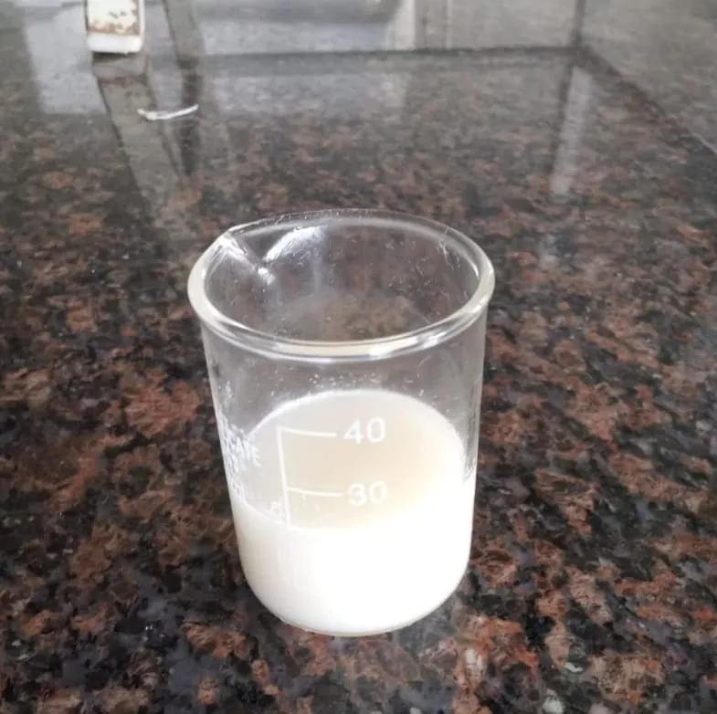
Fig.No.09. Niosomal Dispersion
Optimization: To optimize the ether injection method, several parameters can be adjusted, including:
1. Surfactant concentration.
2. Injection rate.
3. Temperature of the aqueous solution.
4. Stirring speed.
By adjusting these parameters, it is possible to optimize the ether injection method for specific applications and improve the quality and characteristics of the resulting noisome.
EVALUTION TEST –
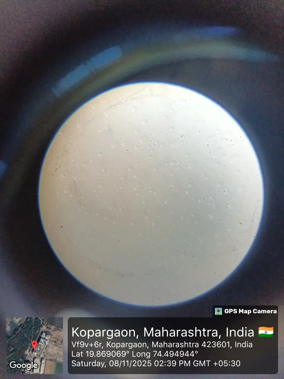
Fig.No.10. particle shape
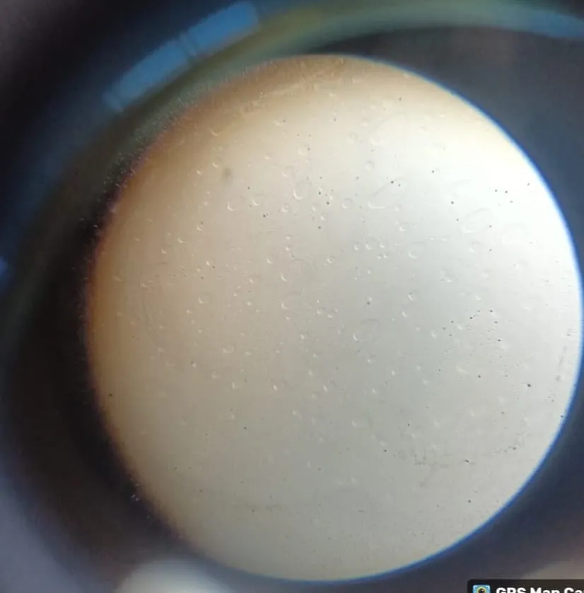
Fig.No.11. Particles Shape
niosomal suspension equivalent to 10mg taken in a volumetric flask of 100ml and volume was make up by phosphate buffer pH 7.4, after that 1ml of this mixture was diluted to 10ml by phosphate buffer 7.4 and the % drug content was calculated or observed at using UV spectrophotometer.
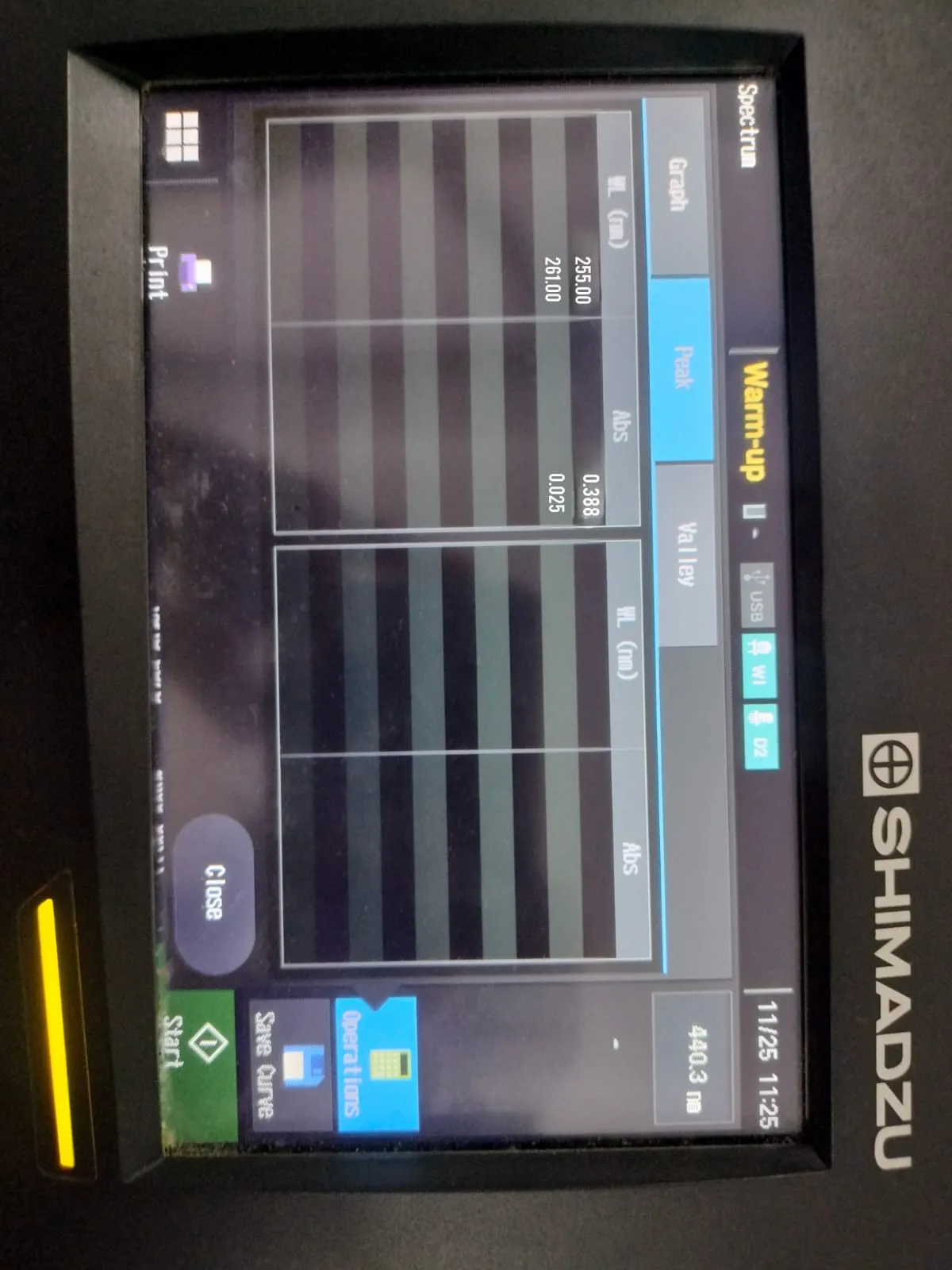
Fig.No.12. Drug Content by UV-Spectroscopy
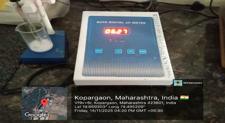
Fig.No.13.PH Meter
EE (Amount of trapped drug/Total amount of initially added-drug) × 100% after preparing niosomal dispersion unentrapped drug is separated by dialysis, gel filtration and centrifugation.(6)
Centrifugation-
3. Separate the supernatant: Carefully collect the supernatant, which contains the free drug.
4. Determine the amount of free drug: Measure the absorbance of the supernatant using a UV-Vis spectrophotometer.
5. Calculate the entrapment efficiency: Using the formula:
Entrapment Efficiency (%) = (Total amount of drug added-Amount of free drug)/Total amount of drug added×100
Particle size shows the average diameter of the niosomal vesicle. Zeta potential represents the surface charge of the vesicles, which helps to predict their stability. These parameters were measured using a Zetasizer instrument
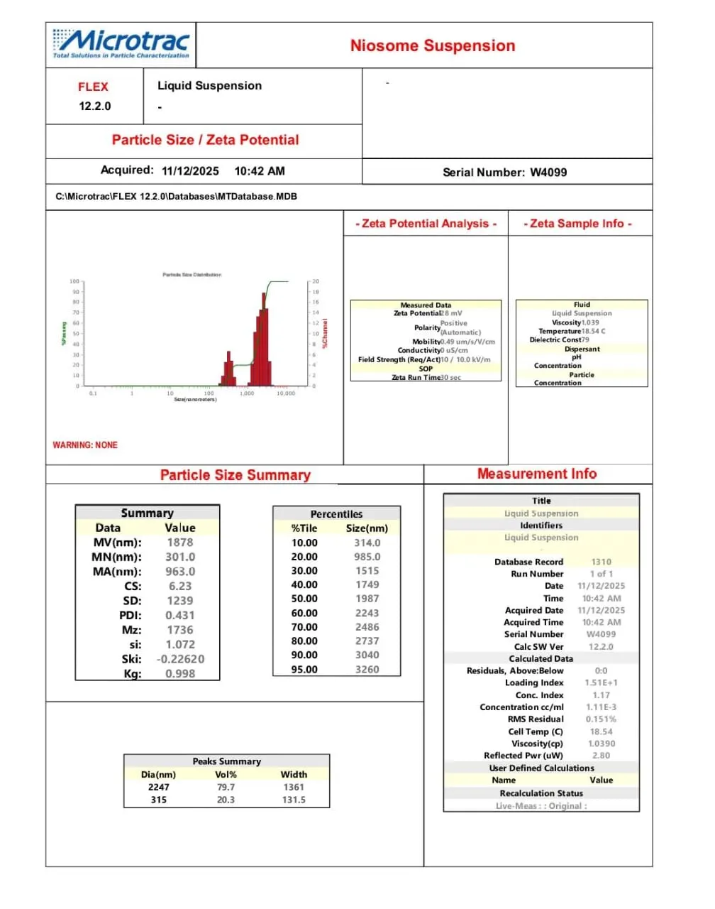
Fig.no.14. Particle size and Zeta potential
RESULT AND DISCUSSION
1. 1. Particle shape and size analysis-
The niosomes prepared using fluconazole drug was studied under microscope to observe the formation of niosomal vesicles. The niosomal vesicles were found to be uniform in size and shape. The shape of the niosomes was observed spherical.
2. Drug content:
The drug content was studied for the formulation prepared by ether injection method. The drug content was found to be (72.3%), which may have optimum surfactant Polyethylene Glycol (PEG) ratio to provide highest drug content of Fluconazole in niosomal vesicles.
The standard range of the pH of the niosomal solution was 5.5-7.5 and the pH of the niosomal solution was found to be 6.39 which is ideal pH.
|
Sr.No. |
SOLUTION |
PH |
Mean |
|
1. |
Niosomal suspension |
6.27 |
6.44 |
|
2. |
6.78 |
||
|
3. |
6.27 |
Table No.02.PH of niosomal suspension
|
Formulation |
%Entrapment Efficiency |
|
F1 |
75.71 |
|
F2 |
81.10 |
|
F3 |
90.6 |
Table.No.03. Entrapment efficiency of formulation
The prepared fluconazole-loaded niosomes showed an average particle size of 2 µm (2000 nm) . The zeta potential was found to be 28 mV, confirming good stability of the niosomal suspension.
SUMMARY & CONCLUSION
Summary:
Niosomes are advanced vesicular drug delivery systems composed of non-ionic surfactants and PEG, designed to enhance the bioavailability and therapeutic effectiveness of various drugs. In recent years, they have gained considerable attention due to their stability, biocompatibility, and ability to encapsulate both hydrophilic and lipophilic drugs. Among the several techniques used for niosome preparation, the ether injection method is one of the most simple, reproducible, and efficient methods for forming uniform vesicles.
In the ether injection method, the selected surfactant and PEG are dissolved in diethyl ether, and the drug is incorporated either in powder form or as a solution. If the drug is insoluble in ether, it may first be dissolved in a small amount of ethanol or another suitable solvent before adding it to the ether phase. This organic mixture is then slowly injected into a warm aqueous phase, typically phosphate buffer pH 7.4, maintained at 60–70°C with continuous stirring. As the ether rapidly evaporates, the surfactant and PEG self-assemble to form niosomal vesicles, successfully entrapping the drug within their bilayers.
The prepared niosomal suspension is allowed to cool to room temperature, and any remaining organic solvent is removed by evaporation. The final formulations are evaluated for vesicle diameter, particle shape, entrapment efficiency, drug content, and pH. Vesicle size and morphology are examined microscopically, while entrapment efficiency is commonly assessed using centrifugation followed by UV-Vis spectrophotometric analysis. Drug content is also determined using UV-Visible spectroscopy to ensure accurate drug loading.
Overall, the study highlights the successful formulation of fluconazole-loaded niosomes using the ether injection method and demonstrates the potential of niosomal delivery systems to improve drug performance, stability, and therapeutic outcomes.
Conclusion:
Fluconazole-loaded niosomes were successfully prepared using the ether injection method, showing good stability and suitability for topical delivery. The optimized formulation exhibited high entrapment efficiency, acceptable pH, spherical vesicles, and good drug content, confirming effective niosomal formation. Overall, the ether injection method proved to be a simple and efficient technique for preparing niosomes, and the choice of surfactant and drug ratio played an important role in achieving a stable and effective formulate
REFERENCES

Sakshi Ghorpade*, Aparna Sawant, Shruti Gosavi, Pooja Lohakane, Sachin Chavan, Formulation And Evalution Of Fluconazole Loaded Niosomes By Ether Injection Method, Int. J. Sci. R. Tech., 2026, 3 (5), 955-965. https://doi.org/10.5281/zenodo.20410081
 10.5281/zenodo.20410081