

We use cookies to ensure our website works properly and to personalise your experience. Cookies policy
1Department of Biotechnology, St. Peter’s College of Engineering and Technology Avadi, Chennai-6000054, Tamil Nadu, India.
2Department of Biotechnology ,Rajalakshmi Institute of Technology, Kuththambakkam ,Chennai -600124 Tamil Nadu, India
3Encapscifi Life Sciences Pvt Limited, Jigani industrial area, Bengaluru 560105, Karnataka.
Black rot of cauliflower, caused by Xanthomonas campestris pv. campestris (Xcc), is increasingly difficult to manage with conventional copper sprays due to environmental concerns and emerging bacterial resistance. This study explores a sustainable alternative via the green synthesis of copper nanoparticles (CuNPs) using agricultural waste—onion (Allium cepa) peel extract—as a reducing and capping agent. CuNPs were synthesized at room temperature using 0.3 M and 0.5 M CuSO? solutions. Particle formation was marked by a distinct color change and a UV–Vis absorption band at 400–460 nm; however, the exact phase (metallic versus oxide) remains unconfirmed without X-ray diffraction data. FTIR confirmed successful phytochemical capping, while SEM and EDX indicated a copper-rich core surrounded by an organic shell containing minor oxygen and sulfur. In vitro agar well-diffusion assays demonstrated concentration-dependent antibacterial activity against Xcc. The 0.5 M CuNPs produced larger zones of inhibition than the 0.3 M variant, though the total copper dose was not normalized between preparations. While these preliminary findings highlight a low-toxicity, ambient-temperature route for valorizing agri-waste, extensive future work is required. Phase confirmation, primary particle sizing, minimum inhibitory/bactericidal concentration (MIC/MBC) values, and in-planta phytotoxicity testing are essential before these CuNPs can be advanced for practical black rot management.
Cauliflower (Brassica oleracea var. botrytis) is a widely cultivated cruciferous vegetable valued for its nutritional richness in dietary fibre, vitamins, minerals, and health-promoting phytochemicals such as glucosinolates and sulforaphane (Shinali et al., 2024). In addition to its dietary importance, cauliflower holds significant economic value in many regions, including India, where growing market demand contributes to farmer livelihoods (Sharma et al., 2023). However, cauliflower production faces major losses from disease, among which black rot caused by Xanthomonas campestris pv. campestris is the most devastating. This seed-borne bacterial pathogen invades through hydathodes or wounds, leading to V-shaped chlorotic lesions, vascular blackening, and yield losses that can reach 50–60% under conducive conditions (Geat et al., 2023; Vicente and Holub, 2012). The pathogen’s ability to survive in crop debris, seeds, and soil complicates control, and resistance is scarce in commercial cultivars (Cruz et al., 2017).
Historically, black-rot management has relied on chemical bactericides such as copper compounds, but these pose environmental and health risks, increase production costs, and pressure pathogens to develop resistance. Indeed, copper-resistant strains of Xanthomonas have emerged, undermining conventional sprays (Carvalho et al., 2019). Sustainable approaches including biological control, integrated pest management, and breeding for resistance have been explored (Baker et al., 2019), yet none alone provides fully effective or long-term control. This scenario underscores the need for innovative, eco-friendly solutions for black rot.
Nanotechnology has recently gained traction in plant pathology as a means to manage microbial disease. Metal nanoparticles, by virtue of their nanoscale size and high surface area, possess antimicrobial properties and can interact with and disrupt microbial cell membranes (Carvalho et al., 2019). Among these, copper nanoparticles (CuNPs) are promising bactericides owing to their broad-spectrum activity, lower cost, and comparatively lower toxicity to non-target organisms than noble metals such as silver (Shandila et al., 2025). Conventional chemical synthesis of CuNPs, however, often requires hazardous reagents and harsh conditions unsuitable for agricultural use (Saleem et al., 2024). Green synthesis has therefore emerged as an attractive alternative, using plant-derived biomolecules to reduce metal ions under mild, aqueous conditions. Plant extracts are rich in flavonoids, phenolic acids, alkaloids, and other phytochemicals that can act as natural reducing agents and stabilisers for nanoparticle formation (Dikshit et al., 2021).
Onion (Allium cepa) peels, a common agricultural waste, are particularly abundant in quercetin, phenolics, and sulfur compounds, conferring antioxidant and antimicrobial activity (Kumar et al., 2021). Using onion-peel extract for CuNP synthesis provides an eco-friendly route to antimicrobial nanomaterials while valorising agri-food waste, aligning with circular-bioeconomy principles (Singh et al., 2025; Rashid et al., 2023). Numerous studies report that green-synthesised nanoparticles from plant extracts exhibit antimicrobial activity against diverse plant and human pathogens (Vanlalveni et al., 2021). In particular, CuNPs derived from onion-peel extract have shown growth-inhibitory effects on bacteria, including phytopathogens, indicating potential as biocontrol agents in sustainable agriculture (Islam et al., 2024). Recent work continues to highlight the promise of biogenic copper nanomaterials in crop protection; copper-based nanoproducts have been studied as alternatives to traditional copper bactericides and have achieved disease suppression in planta (Varympopi et al., 2020; Varympopi et al., 2022).
By integrating nanotechnology with green chemistry, a new strategy emerges for managing black rot of cauliflower in an eco-conscious manner. The present study aims to synthesise CuNPs using onion-peel extract and to evaluate, as a first step, their in vitro activity against Xanthomonas campestris pv. campestris. We hypothesise that green-synthesised CuNPs can serve as an environmentally friendly antibacterial agent, simultaneously addressing plant-disease management and agricultural-waste utilisation.
MATERIALS AND METHODS
2.1 Sample collection
Onion peels were collected from discarded onion waste at local vegetable markets, and visibly black-rot-infected cauliflower leaves were gathered from nearby fields/market sources. The onion peels were washed thoroughly with distilled water to remove dirt and air-dried at room temperature. Infected cauliflower-leaf samples were transported to the laboratory for pathogen isolation.
2.2 Preparation of onion-peel extract
Dried onion peels were cut into small pieces. Approximately 10 g of peel were boiled in 1000 mL of distilled water for 30 min to extract bioactive compounds. The mixture was cooled to room temperature and filtered through Whatman No. 1 filter paper. The resulting clear reddish-brown extract was collected and stored at 4 °C for use as a natural reducing and capping agent. The extract had an approximately neutral pH (~7) and remained stable (no precipitation or spoilage) under refrigeration.
2.3 Copper sulfate solution
Copper (II) sulfate pentahydrate (CuSOâ‚„·5Hâ‚‚O) was used as the precursor at two concentrations, 0.3 M and 0.5 M. For each concentration, the calculated mass of CuSOâ‚„·5Hâ‚‚O was dissolved in distilled water with stirring and brought quantitatively to a final volume of 200 mL. Fresh solutions were prepared to avoid hydrolysis or degradation of the salt.
2.4 Green synthesis of copper nanoparticles
CuNPs were synthesised by green reduction using the onion-peel extract. For each reaction, 100 mL of CuSOâ‚„ solution (0.3 M and 0.5 M respectively) was mixed with 100 mL of onion-peel extract, giving a 1:1 (v/v) metal:extract ratio, and stirred continuously on a magnetic stirrer at room temperature (~20 °C) for 3 h. A colour change from pale blue (Cu²âº) through reddish-brown to dark brown/black accompanied particle formation. After incubation, the mixtures were centrifuged (10,000 rpm, 15 min); the pellets were washed two to three times with distilled water to remove unreacted ions and loosely bound metabolites and were oven-dried at 60–80 °C for ~4 h to obtain a dry CuNP powder. The dried product was weighed and stored in sealed vials for characterisation and testing (Peiris et al., 2022).
2.5 Characterisation of nanoparticles
UV–Vis spectroscopy. Absorbance of the colloids was recorded over 250–700 nm. Samples from the 0.3 M and 0.5 M syntheses were monitored at 30, 60, 90, 120, and 180 min to follow formation. The onion-peel extract alone and the CuSOâ‚„ solution alone should be recorded as baselines so that the colloid spectrum can be distinguished from the intrinsic absorption of the extract (see Section 3.3.1).
FTIR spectroscopy. Spectra of the dried CuNP powders were acquired over ~500–4000 cm⻹ to identify functional groups from the onion-peel extract capping the particles, with attention to O–H/N–H, C–H, carbonyl, and C–O/C–N bands.
Scanning electron microscopy (SEM) and EDX. Dried CuNP samples were mounted on stubs, gold-sputter-coated, and imaged by SEM for morphology, dispersion, and aggregation. Elemental composition was assessed by EDX coupled to the SEM. Because gold coating was applied, EDX in the ~2.1–2.3 keV region overlaps the Au M lines; the sulfur assignment is therefore treated as tentative.
2.6 Bacterial isolation and identification
The pathogen was isolated from diseased cauliflower leaves by standard methods. Symptomatic tissue was surface-sterilised in 70% ethanol (30 s) and 1% sodium hypochlorite (1 min), rinsed in sterile distilled water, macerated, and streaked onto nutrient agar. After incubation at 28 °C for 48–72 h, yellow, mucoid, round colonies typical of X. campestris pv. campestris were selected and sub-cultured to purity. Gram staining produced pink, Gram-negative rods, and a 3% KOH string test was positive (formation of a viscous thread), consistent with a Gram-negative cell wall. As per literature (Massomo et al., 2003; Popović et al., 2014), these features are consistent with X. campestris pv. campestris.
2.7 Antibacterial assay
Antibacterial activity against the isolate was evaluated by agar well diffusion. An overnight nutrient-broth culture was adjusted to OD₆₀₀ ≈ 0.1, and ~100 µL was spread on nutrient agar to form a lawn. Wells (6 mm) were punched with a sterile cork borer, and 50 µL of each treatment was dispensed: (1) CuNPs from 0.3 M CuSOâ‚„, (2) CuNPs from 0.5 M CuSOâ‚„, (3) a positive control (streptomycin), (4) a negative control (sterile distilled water), (5) Vehicle controls 0.3M and 0.5M CuSOâ‚„ and (6) Allium cepa extract. Plates were incubated at 28 °C for 48 h and the zone of inhibition was recorded using digital vernier calipers. All treatments were tested in triplicate.
2.8 Statistical analysis
Zone-of-inhibition data are expressed as mean ± standard deviation of three independent replicates.
RESULTS AND DISCUSSION
3.1 Onion-peel extract as a bioreductant and stabiliser
The aqueous onion-peel decoction (10 g peel in 1000 mL distilled water, 30 min) presented a uniform brownish-red hue and neutral pH (~7) and remained stable without precipitation at 4 °C over short-term storage. The colour intensity is consistent with a high load of water-extractable phenolics and flavonoids — molecules known to donate electrons and to chelate or cap nascent metal nuclei (Vallejo et al., 2025). This profile is advantageous because (i) polyphenolic –OH and carbonyl groups can furnish reducing equivalents for Cu²âº → Cuâ° conversion, and (ii) the same groups can adsorb onto copper surfaces, imparting steric/electrostatic stabilisation. The homogeneous colour and absence of visible particulates indicated batch uniformity, supporting reproducible synthesis (Mülhopt et al., 2018; Akinniyi, 2025).
3.2 Synthesis of copper nanoparticles
Mixing onion-peel extract with CuSOâ‚„ at a 2:1 (v/v) metal:extract ratio (200 mL CuSOâ‚„ + 100 mL extract; ~20 °C; 3 h, continuous stirring) produced a progressive colour transition from reddish-brown to dark brown/black, consistent with copper nanoparticle formation. Mechanistically, polyphenolic/hydroxylated constituents reduce Cu²âº to Cuâ°, enabling nucleation, while concurrent adsorption of biomolecules on nascent surfaces limits uncontrolled growth and agglomeration, yielding a capped colloid:
Cu2+ —(onion-peel polyphenols; reduction)→ Cu0 (nucleation) —(phytochemical capping)→ stabilised CuNPs
Post-reaction recovery by centrifugation (10,000 rpm, 15 min), iterative washing (two to three times with distilled water), and oven drying (60–80 °C, ~4 h) afforded a dry powder. The use of a neutral, food-grade aqueous medium and ambient-temperature synthesis reduces energy input and avoids toxic reagents, which is favourable for scale-up (Peiris et al., 2022). The dry-mass yield and conversion efficiency are reported per concentration in Section 2.4.
3.3 Characterisation
3.3.1 UV–Vis spectroscopy
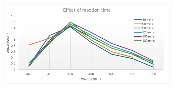
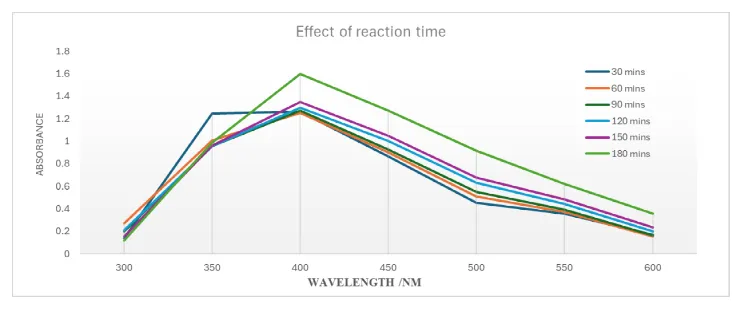
For the 0.3 M synthesis, the colour changed from pale blue to brown over 3 h, and the UV–Vis spectra (Figure 1a) show a broad absorption band centred near ~400–450 nm that intensified with reaction time, plateauing by ~150–180 min. At 0.5 M (Figure 1b), a comparable but slightly red-shifted (~450–460 nm), broader, and more intense band developed, consistent with a higher particle concentration and a broader size distribution. The growth of the band with time and the dark coloration indicate progressive reduction of Cu²âº and accumulation of copper-based particles, and the broader envelope at 0.5 M is consistent with reduced capping efficiency and greater aggregation at the higher precursor loading (Siddiqi and Husen, 2020; Patcharawit et al., 2025).
An important caveat applies to the phase interpretation. The surface-plasmon resonance most frequently reported for metallic copper nanoparticles lies at appreciably longer wavelengths (commonly ~560–590 nm, and generally above ~520 nm depending on size and medium). A band at ~400–460 nm is therefore not, on its own, diagnostic of metallic Cuâ°: absorption in this region is also exhibited by cuprous oxide (Cuâ‚‚O) and by onion-peel polyphenols such as quercetin, which absorb strongly between ~370 and ~450 nm. Consequently, the present UV–Vis data should be read as evidence of copper-based particle formation rather than as proof of a metallic phase. Distinguishing metallic Cuâ° from Cuâ‚‚O/CuO, and from residual extract chromophores, requires (i) an extract-only baseline spectrum and (ii) X-ray diffraction; the latter was not performed in this study and is identified as a priority for revision (see Limitations).
(a) 0.3 M CuSOâ‚„
(b) 0.5 M CuSOâ‚„
Figure 1: UV–Vis Absorption Spectra of Onion-Peel CuNPs Synthesised at (a) 0.3 M and (B) 0.5 M CuSOâ‚„ over 30–180 min at ~20 °C. the Band Near ~400–460 nm Grows with Reaction Time; This Position Is Below the Plasmon Region Typically Reported for Metallic Copper and May Include Contributions from Cuâ‚‚O and/or Onion-Peel Polyphenols
3.3.2 Scanning electron microscopy
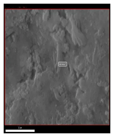
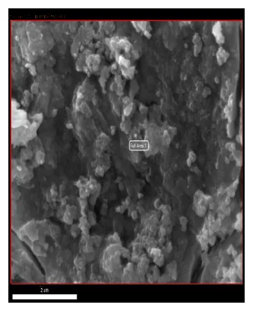
SEM images of the 0.3 M sample (Figure 2a) show comparatively dispersed material within an organic matrix with moderate clustering, whereas the 0.5 M sample (Figure 2b) appears densely clustered into larger, irregular aggregates. These observations are consistent with the UV–Vis trend and with reports that higher metal-salt concentrations favour larger, more aggregated structures and reduced colloidal stability (Akinniyi, 2025; Patcharawit et al., 2025).
Two limitations of the imaging should be stated plainly. First, at the magnification used (scale bar 2 µm) individual nanoparticles in the tens-of-nanometres range are not resolved; the micrographs therefore characterise aggregate morphology and the surrounding organic matrix rather than primary particle dimensions. Accordingly, statements about “smaller” versus “larger” particles describe dispersion/aggregation state and are inferred, not measured. Second, no quantitative size distribution was extracted. Primary particle size and shape should be established by transmission electron microscopy (TEM), and hydrodynamic size and polydispersity by dynamic light scattering (DLS); these were not performed here.
(a) 0.3 M CuSOâ‚„ (b) 0.5 M CuSOâ‚„
Figure 2: SEM Micrographs of Onion-Peel CuNPs from (a) 0.3 M and (B) 0.5 M CuSOâ‚„ (Scale bar = 2 Μm). at This Magnification the Images Show Aggregate Morphology within an Organic Matrix; Individual Nanoparticles Are Not Resolved and Primary Size Was Not Measured
3.3.3 Energy-dispersive X-ray analysis (EDX)
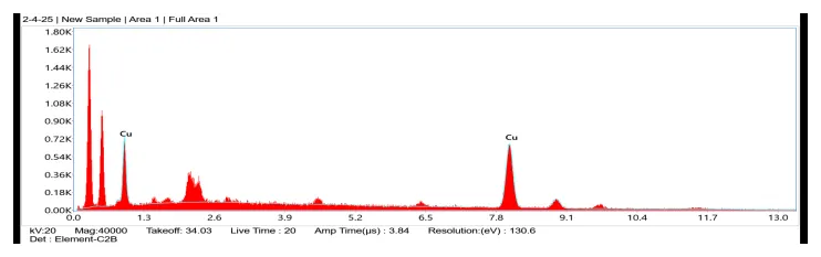
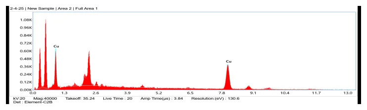
EDX (Figure 3) identified copper as the dominant element in both samples, with Cu L and K lines (the Kα/Kβ features near ~8.0–8.5 keV) clearly present, together with a strong low-energy signal attributable to carbon/oxygen from the organic capping layer and a minor feature near ~2.2–2.3 keV. The prominence of Cu and the absence of other inorganic elements indicate that the washing protocol removed most soluble residues. The oxygen signal is consistent with a thin surface oxide and/or adsorbed oxygenated phytochemicals rather than complete bulk oxidation, since the latter would be expected to depress the Cu signal markedly relative to O.
Three interpretive cautions are warranted. (i) EDX is at best semi-quantitative, especially for light elements (C, O); comparisons of Cu/O ratio between the 0.3 M and 0.5 M samples are therefore qualitative only and should not be used to claim a defined difference in capping density. (ii) Because the specimens were gold-sputter-coated, the ~2.1–2.3 keV region overlaps the Au Mα line; the assignment of this feature to sulfur from onion-peel residues is consequently tentative and should be confirmed by a coating-free analysis (e.g., carbon coating) or by X-ray photoelectron spectroscopy (XPS). (iii) EDX cannot establish crystalline phase; the metallic-versus-oxide question raised in Section 3.3.1 cannot be resolved from these data. With these caveats, the EDX results support the presence of copper-based particles bearing an O- (and probably N-/S-) containing organic shell, in agreement with the FTIR data below (Siddiqi and Husen, 2020; Akinniyi, 2025).
(a) 0.3 M CuSOâ‚„
(b) 0.5 M CuSOâ‚„
Figure 3: EDX Spectra of Onion-Peel CuNPs from (a) 0.3 M and (B) 0.5 M CuSOâ‚„. Strong Cu Signals Confirm the Copper Nature of the Particles; the Low-Energy C/O Signal Reflects the Organic Capping Layer. the ~2.1–2.3 keV Region Overlaps Au M Lines from the Sputter Coating, So the Sulfur Assignment Is Tentative (See Section 3.3.3).
3.3.4 Fourier-transform infrared spectroscopy (FTIR)
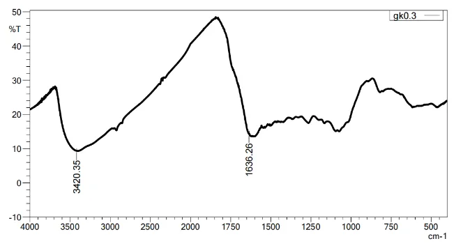
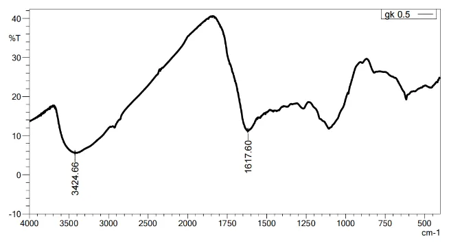
Both batches display the same phytochemical “fingerprint,” supporting a role for onion-peel constituents as reducing and capping agents (Figure 4). A broad band at ~3420–3425 cm⻹ (0.3 M: 3420.35 cm⻹; 0.5 M: 3424.66 cm⻹) is assigned to O–H/N–H stretching from phenolics and amino groups; weaker features near ~2925 and ~2850 cm⻹ correspond to aliphatic C–H stretches; and bands within ~1260–1000 cm⻹ arise from C–O/C–N stretching of alcohols, ethers, glycosidic linkages, and amino functionalities. The clearest difference between concentrations is in the ~1620–1640 cm⻹ region: 1636.26 cm⻹ at 0.3 M versus 1617.60 cm⻹ at 0.5 M (Δ ≈ 18–20 cm⻹).
This shift is consistent with stronger coordination of carbonyl/conjugated groups to surface copper at the higher precursor loading; however, the interpretation should remain cautious. The 1620–1640 cm⻹ region also contains the O–H bending of adsorbed water and aromatic/amide C=C/C=O contributions, so a shift of ~18–20 cm⻹ may partly reflect differences in hydration or baseline rather than metal–ligand bonding alone. Neither spectrum shows a sharp Cu–O lattice band in the ~530–580 cm⻹ region characteristic of bulk CuO, which argues against extensive bulk oxide but does not by itself confirm a metallic phase. Overall the spectra are consistent with a two-step pathway — reduction of Cu²âº by phenolics, followed by anchoring of oxidised products and amino/sugar moieties to the particle surface via O/N donors — and with a somewhat more abundant, less oxidised capping layer at 0.3 M (Siddiqi and Husen, 2020; Akinniyi, 2025).
(a) 0.3 M CuSOâ‚„
(b) 0.5 M CuSOâ‚„
Figure 4: FTIR Spectra of Onion-Peel CuNPs from (a) 0.3 M and (B) 0.5 M CuSOâ‚„. Major Bands: O–H/N–H (~3420–3425 cm⻹), Aliphatic C–H (~2925/2850 cm⻹), the ~1620–1640 cm⻹ Carbonyl/Aromatic Region (0.3 M: 1636 cm⻹; 0.5 M: 1618 cm⻹), and C–O/C–N (~1260–1000 cm⻹).
3.3.5 Antibacterial activity
In agar well-diffusion assays against Xanthomonas campestris pv. campestris, the onion-peel CuNPs inhibited growth in a concentration-dependent manner, with the 0.5 M-derived particles producing larger inhibition zones than the 0.3 M-derived particles (Figure 5; Table 1). The streptomycin positive control produced the largest zone, and the negative control (sterile distilled water) produced none, confirming that the suppression in the test wells was attributable to the applied CuNPs while remaining below the activity of the antibiotic under identical conditions. This dose dependence is consistent with the general behaviour of antibacterial nanoparticles, in which higher effective concentration and greater ion availability increase growth suppression (PeÄenka et al., 2016; Fatima et al., 2023).
Table 1: Inhibition of Xanthomonas Campestris pv. Campestris by Onion-Peel Copper Nanoparticles in the Agar Well-Diffusion Assay (28 °C, 48 H).
| Treatment | Description | n | Zone of inhibition (mm, mean ± SD) |
|---|---|---|---|
| A – 0.5 M CuNPs | Green synthesized CuNPs | 3 | 10.8 ± 0.9 |
| B – 0.3 M CuNPs | Green synthesized CuNPs | 3 | 8.6 ± 0.8 |
| C – 0.5 M CuSOâ‚„ | Copper sulphate solution | 3 | 8.3 ± 0.7 |
| D – 0.3 M CuSOâ‚„ | Copper sulphate solution | 3 | 6.7 ± 0.5 |
| E – Onion peel extract | Aqueous extract | 3 | 8.5 ± 0.6 |
| PC – Positive control | Streptomycin (10 µg) | 3 | 25.9 ± 0.8 |
| NC – Negative control | Sterile distilled water | 3 | 0.0 ± 0.0 |
Values are mean ± SD of three replicates; .
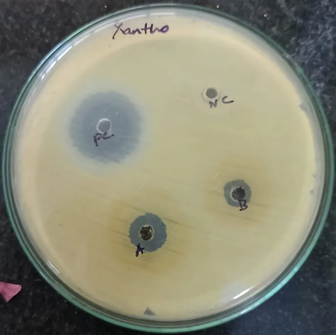
Figure 5: Agar Well-Diffusion Assay Against Xanthomonas campestris pv. campestris. Wells: a = 0.5 M CuNPs, B = 0.3 M CuNPs, PC = Positive Control (Streptomycin), NC = Negative Control (Sterile Distilled Water). Clear Zones Indicate Growth Suppression; Measured Diameters Are Given in Table 1.
Benchmarked against other nano-systems targeting Xanthomonas, the zones observed here are modest. Silver nanoparticles frequently outperform copper in plate assays: Ulva-derived AgNPs inhibited X. campestris pv. malvacearum with larger zones and low MICs (Rajesh et al., 2012), and Moringa-synthesised AgNPs produced larger zones against X. vesicatoria than copper- or zinc-based particles (Fatima et al., 2023). Copper can nonetheless be strongly inhibitory when appropriately formulated, with substantial in vitro suppression of Xanthomonas reported for optimised CuNPs (Peiris et al., 2022). Differences among studies reflect synthesis route, particle size and dispersion, capping chemistry, and ion release, which collectively govern efficacy (PeÄenka et al., 2016; Varympopi et al., 2022). The present onion-peel CuNPs may have physicochemical attributes (size, surface charge, capping density, aggregation) that limited diffusion and bioavailability in agar and thereby constrained zone sizes.
Several mechanisms could contribute to, or modulate, the observed activity but were not tested here and are noted only as context for future work. Onion-peel phenolics and flavonoids (notably quercetin derivatives) have intrinsic antimicrobial and redox properties and could, as surface residues, influence interaction with bacterial envelopes (Joković et al., 2024). For other plant-mediated systems, capping phytochemicals have acted as elicitors and improved disease outcomes in planta — for example, noni-peel AgNPs inhibited Xcc in vitro and primed host defences in cabbage (Santos et al., 2025) — and polyphenol-rich extracts have reduced Xanthomonas motility and biofilm formation and attenuated xanthan/exoenzyme production (Muawiya et al., 2025; Qian et al., 2006; Adonizio et al., 2006). Copper ions and reactive oxygen species released from CuNPs can likewise perturb membranes and respiratory enzymes. Whether any of these anti-virulence or host-priming effects operate for onion-peel CuNPs against Xcc remains to be determined.
These considerations make formulation optimisation central. Comparative work on CuNP products against X. vesicatoria showed that synthesis protocol and resulting bioavailability — not size alone — governed bacterial suppression and could even exceed a conventional copper-hydroxide product, with improved copper delivery verified in planta (Varympopi et al., 2022). In the present system, tuning extract:precursor ratio, pH, temperature, and reaction time to reduce size, narrow polydispersity, increase colloidal stability, and control ion release could enhance activity; composite strategies (e.g., minor Ag or ZnO fractions) have increased zones in related models, although not all bimetallics outperform Ag alone (Fatima et al., 2023; PeÄenka et al., 2016).
3.4 Limitations
The conclusions of this study are constrained by its scope, and the following gaps should be addressed before the material is advanced. (i) Crystalline phase was not determined; X-ray diffraction is needed to establish whether the product is metallic Cuâ°, Cuâ‚‚O/CuO, or a mixture, particularly given that the UV–Vis band lies below the metallic-Cu plasmon region. (ii) Primary particle size, shape, hydrodynamic size, and surface charge were not measured; TEM, DLS, and zeta-potential analyses are required to support the size, polydispersity, and colloidal-stability statements. (iii) Antibacterial assessment relied on a single diffusion method; MIC and MBC, and ideally time-kill kinetics, would quantify potency, and a broth-based method would reduce the diffusion artefacts that disadvantage nanoparticles. (iv) The applied copper dose was not normalised between preparations, and no ionic-copper (CuSOâ‚„) control was included; a dose-matched comparison and an ionic control are needed to attribute activity to the nanoparticle form rather than to total copper delivered. (v) A single batch per concentration was characterised; independent replicate syntheses would demonstrate reproducibility. (vi) Pathogen identification was presumptive; molecular confirmation is recommended. (vii) Efficacy and safety were not evaluated on the plant; in-planta trials and phytotoxicity assessment on cauliflower are essential before any claim of biocontrol.
CONCLUSION
Onion-peel extract enabled a simple, room-temperature synthesis of phytochemical-capped copper nanoparticles that inhibited Xanthomonas campestris pv. campestris in vitro. Spectroscopic and microscopic analyses are consistent with copper-based particles bearing an organic corona whose character varied with precursor concentration, and the antibacterial response was concentration dependent, with the 0.5 M preparation producing larger inhibition zones than the 0.3 M preparation but remaining below the streptomycin control. Because phase was not confirmed and the applied dose was not normalised, these findings are best regarded as preliminary evidence of antibacterial potential rather than proof of a metallic-nanoparticle-specific effect. Given the sustainability advantages — valorisation of agri-waste, aqueous processing, and avoidance of hazardous reagents — onion-peel CuNPs nonetheless represent a promising starting point. Priority next steps are phase confirmation (XRD), primary-size and surface-charge characterisation (TEM, DLS, zeta potential), dose-matched and ionic-copper comparisons, MIC/MBC and time-kill kinetics, evaluation of anti-virulence and biofilm effects, and in-planta efficacy and phytotoxicity trials on cauliflower. With such optimisation, green CuNPs could complement integrated disease management while lowering bulk copper inputs.
Declaration of generative AI in scientific writing
The authors used Grammarly PRO version for proof reading and correcting syntax errors in the present article. After the proof read stage, the authors read the entire paper and take responsibility.
Funding
No Funding was received for this project
CRediT authorship contribution statement
Conceptualization and Methodology - Roseline Mary
Investigation – Student name
Data Curation – Student name and Roseline Mary
Writing – Original Draft: Student Name
Writing – Review & Editing: Roseline Mary
All authors have read and approved the final manuscript
Declaration of competing interest
The Authors declare no competing interests.
Acknowledgments
Data availability
All the data pertaining to this study is available in this paper. No data is withheld.
REFERENCES



Gokula Krishnan M., Rosseline Mary D., Gaanappriya Mohan, Green Synthesis of Copper Nanoparticles Using Allium Cepa (Onion) Peel Extract and Their in Vitro Antibacterial Activity Against Xanthomonas Campestris pv. Campestris, the Black Rot Pathogen of Cauliflower, Int. J. Sci. R. Tech., 2026, 3 (10), 578-590. https://doi.org/10.5281/zenodo.23259616
 10.5281/zenodo.23259616