

We use cookies to ensure our website works properly and to personalise your experience. Cookies policy
Department of Biotechnology, St. Peter’s College of Engineering and Technology
Venomous snakes harbor significantly more diverse microbial communities than their non-venomous counterparts, and these oral microbiomes remain largely unexplored in terms of their biotechnological potential. This study investigated the oral microbiota of the Indian cobra (Naja naja), focusing on isolating pigment-producing microorganisms of environmental and applied significance. Microbial isolation was performed using aseptic techniques, including streak plate and agar slant methods. Morphologically distinct colonies exhibiting yellow, white, and orange pigmentation were obtained and characterized based on their cultural and morphological properties. Molecular identification via 16S rRNA gene sequencing revealed that the isolates were Staphylococcus arlettae, Enterobacter hormaechei, and Exiguobacterium sp., each demonstrating ? 97% sequence similarity to their respective reference strains in the NCBI database. Phylogenetic relationships were established through BLAST analysis, which confirmed the taxonomic placement. These findings highlight the oral cavity of Naja naja as a reservoir of pigment-producing bacteria with potential applications in environmental biotechnology, including bioremediation and the production of natural pigments.
Microorganisms associated with the oral flora of snakes have emerged as promising candidates for pharmaceutical applications. Among these, chromogenic bacteria have gained wide attention due to their production of metabolites.(Sengupta, 2021). These secondary metabolites exhibit antibacterial and antioxidant properties that are useful in drug discovery. (SinghP, 2024) Characterisation presents an underexplored reservoir that harbors diverse bacterial communities of pharmaceutical significance. These types of oral bacteria are often influenced by venom composition, feeding environmental behavior ,and factors (Mc Laughlin, 2019). The oral cavity of snakes contains a complex microbial community, including both pathogenic and beneficial bacterial species, some of which may possess unique pharmaceutical and therapeutic properties. (Tasoulis,2019) Despite increasing interest in microbial bioprospecting, the oral microflora of snakes, especially Indian species such as Naja naja, remains underexplored. Isolation and Characterization of such bacteria are important preliminary steps in identifying novel bacteria. Conventional methods, including culture-based isolation and biochemical characterization, provide preliminary identification. However molecular approaches such has 16s rRNA Gene sequencing offers reliable taxonomic and phylogenetic confirmation of bacterial isolates.(Janda,2007) Therefore, the present study aimed to isolate chromogenic bacterial strains from the oral flora of Naja naja using cultural and morphological methods, followed by molecular identification through 16S rRNA gene sequencing. This study aims to contribute to the understanding of microbial diversity and provide a basis for future exploration of oral bacteria in the pharmaceutical and cosmetic industries.
2. MATERIALS AND METHODS
2.1 Sample Collection
The Indian Cobra (Naja naja), a highly venomous snake belonging to the family Elapidae, was selected as the target species for oral cavity swab collection. To ensure adherence to ethical guidelines, the snake was handled carefully by a trained professional with proper safety measures. The oral cavity of the snake was gently opened, and a sterile cotton swab was carefully rubbed against the oral mucosa and inner mouth region to collect samples. The collected swabs were immediately transferred to sterile tubes under aseptic laboratory conditions to avoid external contamination (Blaylock, 2001; Lam et al., 2011).
2.2 Isolation and Purification of the Bacterial Isolates
The oral swab obtained from the Indian cobra was inoculated onto nutrient agar containing NaCl, yeast extract, beef extract, and peptone, with the medium adjusted to pH 7.2. The purpose of inoculating the swab culture was to identify different microorganisms under sterile laboratory conditions. The medium was sterilized using an autoclave for 15 min at 121°C and 15 PSI pressure for the proper sterilization. After sterilization, the media were poured into sterile Petri plates under aseptic conditions. The poured plates were allowed to solidify under sterile conditions for 15 min. Subsequently, the swab sample was inoculated onto nutrient medium using the streak plate technique. The pure cultures of bacterial isolates were repeatedly subcultured to identify the specific strains of microbes present in the swabs (Cappuccino & Sherman, 2014).
2.3 Colony Morphology and Cultural Characterization
Colony morphology is used to determine the physical characteristics of bacteria. Gram staining was performed to determine the morphological characteristics of the bacteria. Using an inoculation loop, a drop of the suspended culture was transferred to a microscopic slide. The culture was spread in an even circle with the inoculation loop, and the slide was heated from a gentle flame to prepare the proper smear on the slide. After proper smear fixation, crystal violet stain was added to the fixed culture for 10-60 sec and then poured off. Excess stain was gently rinsed with water. Subsequently, Gram’s iodine was added for 1 minute, and the samples were gently rinsed with water. A few drops of a decolorizer with 95% ethanol were added to the slide and rinsed within 5 s. Finally, the slides were counterstained with safranin for 30 seconds and rinsed with water. The slides were air-dried after removing excess water. To observe the morphological characteristics, the slide was examined under a microscope using oil immersion. The slide should be viewed with a 40x objective to assess the smear distribution and with a 100x objective to observe the cell with high magnification to determine the morphological characteristic feature of bacteria accurately (Beveridge, 2001; Cappuccino & Sherman, 2014).
2.4 Biochemical Characterization Biochemical characterization of bacteria is a method used to identify and classify bacteria based on their metabolic and enzymatic activities. Some of the biochemical tests performed in our study were growth of microbes in differential media, catalase test, and KOH string assay. All three tests helped identify the unique features of bacteria in the snake swab.
2.4.1 Growth of Microbes in Differential Media
Differential media are used to differentiate closely related organisms or groups of organisms based on their biochemical characteristics. In the present study, MacConkey and Mannitol Salt agars were selected for the isolation of different microbial isolates. The prepared media were sterilized by autoclaving at 121°C for 15 minutes under aseptic conditions. After sterilization, the media were poured into sterile Petri plates, allowed to solidify for 15 min, and maintained under sterile conditions before inoculating the microbes. The microbial colony was inoculated into the prepared differential media using the streak plate technique and incubated for 24 h at 37°C. After incubation, the growth and morphological characteristics on the respective media plates were observed. This reveals the unique feature of bacterial culture present in snake swabs (Forbes et al., 2007).
2.4.2 Catalase Test
To survive, microbes must rely on defense mechanisms that allow them to survive the oxidation of hydrogen peroxide. The catalase test is essential for differentiating catalase-positive bacteria from catalase-negative bacteria. In this study, the catalase test was performed using the slide drop method. A clean and dry slide was used. A few drops of the suspended culture were added to the slide using an inoculum loop and allowed to settle properly. A few drops of hydrogen peroxide were added, and the slide was covered with a proper glass shield to prevent contamination. to avoid any external The bubble formation happens, when the bacteria produce catalase enzyme. If the bacteria do not form bubbles, they do not produce the catalase enzyme (MacFaddin, 2000).
2.4.3 KOH String test
The KOH String test is a rapid biochemical test used to differentiate gram-negative bacteria from gram-positive bacteria. The main principle behind this test is that the bacterial culture is mixed with Potassium Hydroxide (KOH) to observe the lysis of the bacterial cell wall, which occurs based on the density of the peptidoglycan layer in bacteria. A clean and dry glass slide was used for this test. Add few 3 drops of Potassium Hydroxide (KOH) solution were added to the center of the slide. Using an inoculum loop, a visible amount of bacterial culture was picked from a fresh culture plate and transferred to the KOH drop. The mixture was stirred in a circular motion for 5-10 seconds. The loop is then gently lifted upward. If the bacteria are gram-negative with a thin layer peptidoglycan cell wall, KOH lyses the cell wall and forms a mucoid viscous string. If the bacteria are gram-positive, KOH does not lyse the cell wall, and no string formation occurs (Holt et al., 1994), (Benson, 2002).
2.5 16SrRNA Sequencing
16SrRNA sequencing is a molecular technique used for the identification and classification of bacteria based on the sequencing of the 16s ribosomal RNA gene. The 16SrRNA sequence consists of two regions: the highly conserved region and the hypervariable region. These regions help identify unknown bacteria from various sources. In this present work, the unknown bacterial culture obtained from the snake swab was given to Lupex Biotechnologies Pvt.Ltd for performing the 16SrRNA sequencing in the obtained bacterial culture. The purpose of this process was to identify unknown bacteria in snake swabs. Sequencing was performed using the Sanger sequencing method (Clarridge, 2004; Janda & Abbott, 2007).
2.6 BLAST Analysis
BLAST (Basic Local Alignment Search Tool) was used to compare the two nucleotide sequences. In this study, the unknown 16SrRNA sequence obtained from bacterial culture of Snake swab was compared with the known sequence in the NCBI database. To perform this, we obtained the fasta format of the 16SrRNA sequence of unknown bacterial cultures from snake swabs. As it is based on nucleotide sequencing, Blastn was used to compare the two sequences. Open Blastn from the NCBI database and copy the fast format obtained from the unknown 16SrRNA sequence in the query box. The program was set to select the sequence with the highest similarity for bacterial identification. Run the Blastn. After the sequence blast, it gives the closest organism match, % similarity, E Value, alignment score, and query coverage. This helps to identify bacteria with 100% accuracy (Altschul et al., 1990), (Johnson et al., 2008).
3. RESULTS
3.1 Sample Collection and Isolation of Bacteria
The oral swab collected from the Indian cobra (Naja naja) identified three morphological distinct bacterial colonies which were identified by their pigmentation: Yellow, White and Orange colour. The isolated colonies were inoculated in Nutrient agar, Casitose Soya Blood agar, MacConkey Agar and Mannitol Salt Agar using streak plate method.
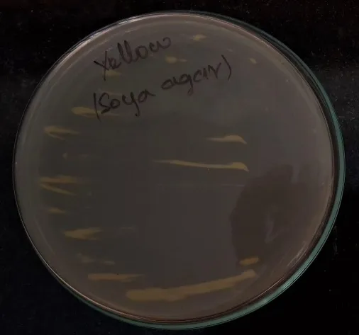
Figure 3.1 Isolated Yellow colony streaked plate in casitose soya blood agar
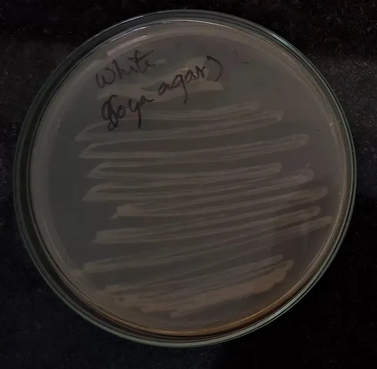
Figure 3.2 Isolated White colony streaked plate in casitose soya blood agar
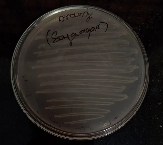
Figure 3.3 Isolated Orange colony streaked plate in casitose soya blood agar
3.2 Colony Morphology
Gram’s Staining was used to identify the colony morphology of the isolated bacteria. The yellow and orange colony showed a purple coloured cytoplasm, which indicates as gram positive bacteria; whereas the white colony showed red coloured cytoplasm, which can be identified as gram negative bacteria.
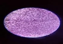
Figure 3.4 Isolated Yellow colony gram staining which shows purple coloured cytoplasm
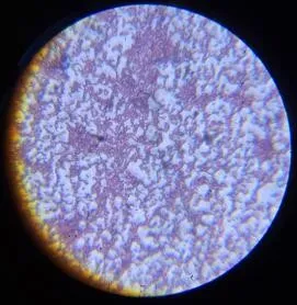
Figure 3.5 Isolated White colony gram staining which shows pink coloured cytoplasm
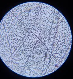
Figure 3.6 Isolated Orange colony gram staining which shows purple coloured cytoplasm
3.3 Biochemical Characterization
3.3.1 Catalase Test
The catalase test was performed to determine the ability of the isolated bacterial colonies to produce catalase enzyme, which decomposes hydrogen peroxide into water and oxygen which causes rapid bubbling reaction. All three isolates showed positive reaction towards catalase test. These results indicate that the colonies are either aerobic or facultative anaerobic nature of the organisms.
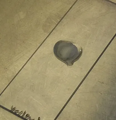
Figure 3.7 Catalase reaction of Isolated yellow colony
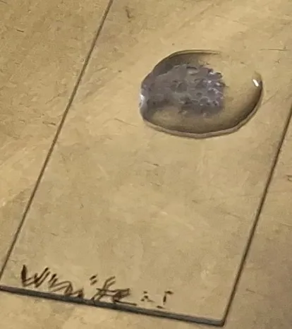
Figure 3.8 Catalase reaction of Isolated white colony
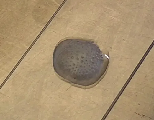
Figure 3.9 Catalase reaction of Isolated orange colony
3.3.2 KOH String Test
The KOH string assay was performed to differentiate Gram-Positive and Gram-Negative bacterial isolates based on the lysis of bacterial cell walls in potassium hydroxide solution. Formation of a viscous mucoid string indicated a Gram-Negative bacteria, whereas absence of string formation indicated a Gram-Positive reaction. Yellow and Orange colony did not produce any viscous string formation when mixed with KOH solution whereas White colony showed clear mucoid string formation during the assay, confirming its Gram-negative characteristic.
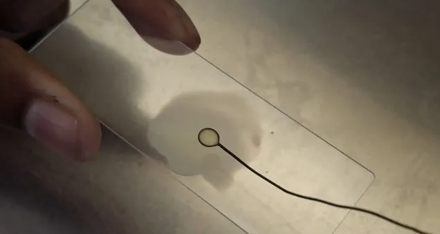
Figure 3.10 KOH string test of Isolated yellow colony
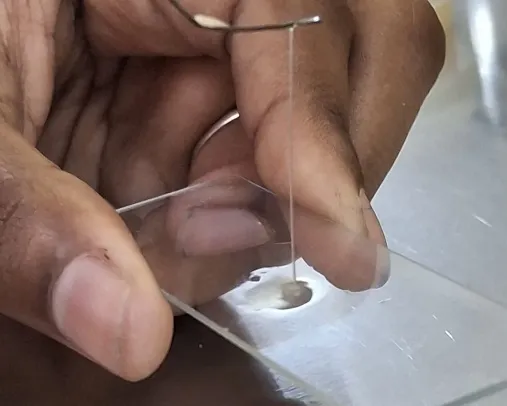
Figure 3.11 KOH string test of Isolated white colony with clear mucoid string
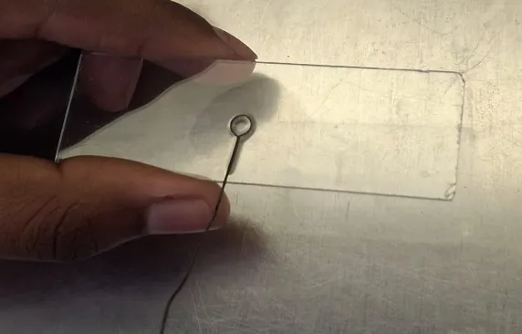
Figure 3.12 KOH string test of Isolated orange colony
3.4 16SrRNA Sequencing and Blast Analysis
The molecular identification of the bacterial isolates obtained from the oral cavity of Indian Cobra was carried out using 16S rRNA gene sequencing followed by BLAST analysis against sequences available in the NCBI database. The sequencing results confirmed the identity of the bacterial isolates with high sequence similarity and query coverage, providing accurate taxonomic classification of the isolates.
The yellow pigmented isolate was identified as Staphylococcus arlettae based on the close sequence homology obtained through 16S rRNA and BLAST analysis. The sequence alignment showed high similarity with previously reported S. arlettae strains available in the NCBI database, confirming the isolate as a member of the genus Staphylococcus. The molecular identification correlated well with the observed colony morphology and biochemical characteristics.
The white colony isolate was identified as Enterobacter sp. through 16S rRNA sequence analysis. The obtained nucleotide sequence exhibited significant similarity with reference sequences of E. hormaechei in the database, confirming its taxonomic identity. The isolate also showed biochemical characteristics typical of Gram-negative enteric bacteria, supporting the sequencing results.
Similarly, the orange pigmented isolate was identified as Exiguobacterium sp. based on sequence similarity obtained through BLAST analysis. The isolate demonstrated close phylogenetic relationship with previously reported Exiguobacterium species. The distinct orange pigmentation and Gram-positive nature of the isolate further supported its identification.
The sequences from all three isolates were uploaded in NCBI database in the ID code of: Staphylococcus arlettae PX457860 and PZ007714, Enterobacter hormaechei PX980609, Exigunobacterium sp. PX930901.
Sequence of PX457860:
GGGGCGAAAGGGAACCAGTAGAGAGCAATTGATTCCTGGAGCGAACAGATGAGGAGACGTTGCTCCTTTGACGTTAGCGGCGGACGGGTGAGTAACACGTGGGTAACCTACCTATAAGACTGGAATAACTCCGGGAAACCGGGGCTAATGCCGGATAACATTTAGAACCGCATGGTTCTAAAGTGAAAGATGGTTTTGCTATCACTTATAGATGGACCCGCGCCGTATTAGCTAGTTGGTAAGGTAATGGCTTACCAAGGCAACGATACGTAGCCGACCTGAGAGGGTGATCGGCCACACTGGAACTGAGACACGGTCCAGACTCCTACGGGAGGCAGCAGTAGGGAATCTTCCGCAATGGGCGAAAGCCTGACGGAGCAACGCCGCGTGAGTGATGAAGGGTTTCGGCTCGTAAAACTCTGTTATTAGGGAAGAACAAACGTGTAAGTAACTGTGCACGCTTGACGGTACCTAATCAGAAAGCCACGGCTAACTACGTGCCAGCAGCCGCGGTAATACGTAGGTGGCAAGCGTTATCCGGAATTATTGGGCGTAAAGCGCGCGTAGGCGGTTTCTTAAGTCTGATGTGAAAGCCCACGGCTCAACCGTGGAGGGTCATTGGAAACTGGGAAACTTGAGTGCAGAAGAGGAAAGTGGAATTCCATGTGTAGCGGGAAATGCGGGAAGATATGGGGGAACACAATCGGTGAAGGCGACTTTCTGGTCTGTACCTGACACTGATGTGCGAACGCGGGGGGCATCAAACGGA
Sequence of PZ007714:
AGCCGCACTAAGCGGTGGGACCATGTGGTGTAACTCGAAGCAACGCGAAG AACCTTACGACATCTTGACATCCTTTGTCCACTCTAGAGATAGAGCATTCGCCTTCGGGGGACAAAGTGACAGGTGGTGC ATGGTTGTCG TCAGCTCGTG TCGTGAGATGTTGGGTTAAGTCCCGCAACGAGCGCAACCC TTAAACTTAG TTGCCAGCATTTAGTTGGGCACTCTAGGTTGACTGCCGGTGACAAACCGGAGGAAGGTGGGGATGACGTCAAATCATCATGCCCCTTATGATTTGGGCTACACACGTGCTACAATGGACAATACAAAGGGCAGCTAAACCGCGAGGTCATGCAAATCCCATAAAGTTGTTCTCAGTTCGGATTGTAGTCTGCAACTCGACTACATGAAGCTGGAATCGCTAGTAATCGTAGATCAGCATGCTACGGTGAATACGTTCCCGGGTCTTGTACACACCGCCCGTCACACCACGAGAGTTTGTAACCCTAGAATTTCGTCGGAGTTCACAAAGTCGAACCAAGGTACCTCGGATAGGAGGTTGTC
Sequence of PX980609:
GGGGGGAAAGGGTGACCAATATGGAAGTAATTGCTCCGTCGATCGGTAACAGGAAGCAGCTTGCTGCTTGGCTGACGAGTGGCGGACGGGTGAGTAATGTCTGGGAAACTGCCTGATGGAGGGGGATAACTACTGGAAACGGTAGCTAATACCGCATAACGTCTCAAGACCAAAGAGGGGGACCTTCGGGCCTCTTGCCATCGGATGTGCCCAGATGGGATTAGCTAGTAGGTGGGGTAACGGCTCACCTAGGCGACGATCCCTAGCTGGTCTGAGAGGATGACCAGCCACACTGGAACTGAGACACGGTCCAGACTCCTACGGGAGGCAGCAGTGGGGAATATTGCACAATGGGCGCAAGCCTGATGCAGCCATGCCGCGTGTATGAAGAAGGCCTTCGGGTTGTAAAGTACTTTCAGCGGGGAGGAAGGCGATAAGGTTAATAACCTTGTCGATTGACGTTACCCGCAGAATAAGCACCGGCTAACTCCGTGCCAGCAGCCGCGGTAATACGGAGGGTGCAAGCGTTAATCGGAATTACTGGGCGTAAAGCGCACGCAGGCGGTCTGTCAAGTCGGATGTGAAATCCCCGGGCTCAACCTGGGAACTGCATTCGAAACTGGCAGGCTAGAGTCTTGTAGAGGGGAGTA GAATTCCAGGTGTAGCGGTGAAATGCGTAGAGATCTGGAGGAATACCGGT GGCGAAGGCGGCCCCCTGGACAAAGACTGACGCTCAGGTGCGAAAGCGTGAGGAGCAAACAGGATTAGATACCCTGGTAGTCCACGCCGTAAACGATGTCGACTTGGAGGTTGTGCCCTTGAGGCGTGGCTTCCGGAGCTAACGCGTTAAGTCGACCGCCTGGGGAGTACGGGCGCAAGGTTTGAACTCAAATGAATTGACGGAGGCCCGCCCAACCGGTGAAACATGTTGGTTTAATTCGATGCAACCGAAAAAACCTTACCTACCCTTGACATCCGGAGAAATTTATCCGAAAAGCTTTGGGGCCTTCTGGAACTTTGAAACAGGGCTGGATGGGTTT
Sequence of PX930901:
ATTTTTACACAATGGAGGAAAGTTTGACGGAGGAACGGCGGAGAAACGCTGAAGCCTTTCGGGTCGTAAAGTTCGGTTGTAAGGGAAGACCCAGCCCCGCAGGCAATGGGGGCACTTTCATCGTTCCTTGCGAGAAAGGCACGGTTAAGTACGTGCCAGCAGCCGCGGTAATAGGTAGGTGGCAAGCGTTGTCCGGAATTATTGGGCGTAAAGCGCGCGCAGCGGCTTTTTTAAGTCTGATGTGAAAGCC CCCGGCTCAGGCGGGGAGGTCCATTGGAAACTGCGAGGCTTGAGTATAGGAGAGAAGAGTGGAATTCCTCGTGTAGCGGTGAAATGCGTAGAGATGTGGA GGAACATCAGTGGCGAAGGCGACTCTCTGGCCTATAACTGACGATGAGGCGCGAAAGCGTGGGGAGCAAACAGGATTAGATACCCTGGTAGTCCACGCCGTAAACGATGAGTGCTAGGTGTTGGAGGGTCTCCGCCCTTCAGTGCTGAAGCTAACGCATTAAGCACTCCGCCTGGGGAGTACGGTCGCAAGGCTGAAACT CAAAGGAATTGACGGGGACCCGCACAAGCGGTGGAGCATGTGGTTTAATTCGAAGCAACGCGAAGAACCTTACCAACTCTTGACATCCCCCTGACCGGTA CAGAGATGTACCTTCCCCTTCGGGGGCAGGGGTGACAGGTGGTGCATGGTTGTCGTCAGCTCGTGTCGTGAGATGTTGGGTTAAGTCCCG CAACGAGCGC AACCCTTGTCCTTAGTTGCCACCATTCAGTTGGGCACTCTAAGGAGACTGCCGGTGACAAACCGGAGGAAGGTGGGGATGACGTCAAACATCATGCCCCT TATGAGTTGGGCTACACACGTGCTACAATGGACAAAGGGCAGCGAAGCCGCGAGGTGGAGCCAATCCCAGAAAGCCGTTCTCAGTTCGGATTGCAGGCTGCAA
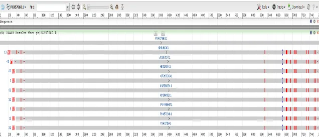
Figure 3.16 Blast analysis result of Isolated yellow colony
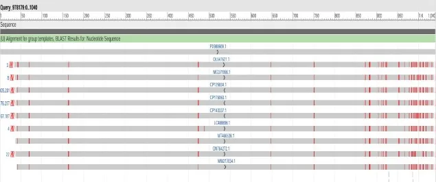
Figure 3.17 Blast analysis result of Isolated white colony
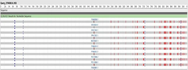
Figure 3.18 Blast analysis result of Isolated orange colony
4. DISCUSSION
The present study reports the isolation and molecular characterization of three pigment-producing bacterial species from the oral cavity of the Indian cobra (Naja naja), namely Staphylococcus arlettae, Enterobacter hormaechei, and Exiguobacterium sp. These findings are consistent with prior observations that the oral microbiota of venomous snakes is markedly more diverse than that of non-venomous species, likely due to the selective pressures imposed by venom constituents, dietary habits, and environmental interactions (Blaylock, 2001; McLaughlin et al., 2019; Tasoulis & Isbister, 2019).
The isolation of Staphylococcus arlettae as a yellow-pigmented, Gram-positive coccus from the cobra oral cavity is noteworthy. Although S. arlettae is primarily described as an environmental and animal-associated coagulase-negative staphylococcus, its detection in the reptilian oral environment aligns with reports of staphylococcal species being consistently recovered from both venomous and non-venomous snake oral swabs (Nemeghaire et al., 2021). The yellow pigmentation observed in this isolate is associated with carotenoid biosynthesis, which confirms protection against oxidative stress and UV radiation, traits advantageous for microorganisms inhabiting ecologically variable niches (Kumar et al., 2023). The catalase-positive nature of the isolate further supports its aerobic or facultatively anaerobic lifestyle, consistent with the physiological conditions of the snake oral cavity.
The white colony identified as Enterobacter hormaechei is a Gram-negative, facultatively anaerobic bacterium of the family Enterobacteriaceae. Its presence in the oral microbiota of Naja naja is consistent with earlier studies reporting Enterobacteriaceae as prevalent constituents of reptilian oral flora, likely introduced through the ingestion of prey (Lam et al., 2011). E. hormaechei is a metabolically versatile species known for its capacity to colonize diverse ecological environments, including hospital settings, soil, and animal hosts (Kamathewatta et al., 2020). Of particular environmental relevance, recent studies have demonstrated that certain strains of E. hormaechei exhibit phosphate-solubilizing activity and can mitigate heavy metal toxicity in contaminated soils, suggesting significant potential in bioremediation applications (Chakraborty et al., 2026). The KOH string test and MacConkey agar growth results obtained in the present study were concordant with the Gram-negative identity confirmed by 16S rRNA sequencing, reinforcing the reliability of the integrative phenotypic-molecular approach employed.
The orange-pigmented isolate identified as Exiguobacterium sp. is among the most ecologically significant findings of this study. Members of the genus Exiguobacterium are recognized for their remarkable physiological versatility, tolerating extreme temperatures, pH ranges, salinity levels, and heavy metal concentrations (Kasana & Pandey, 2021). The characteristic orange pigmentation in Exiguobacterium species is attributed to the production of carotenoid pigments, which function as photoprotective and antioxidant compounds, facilitating survival under oxidative stress conditions encountered in the reptilian oral microenvironment (Kumar et al., 2023). The recovery of an Exiguobacterium isolate from the oral flora of Naja naja expands the known ecological repertoire of this genus and underscores the snake oral cavity as an unexplored niche for extremotolerant, biotechnologically relevant bacteria.
The application of 16S rRNA gene sequencing provided robust taxonomic resolution for all three isolates, with sequence similarities exceeding 97% relative to reference strains in the NCBI database. This threshold is widely accepted as sufficient for reliable genus-level and species-level identification in diagnostic and environmental microbiology (Janda & Abbott, 2007; Clarridge, 2004). The use of BLAST analysis and phylogenetic tree construction further corroborated the taxonomic assignments and demonstrated the evolutionary relatedness of the isolates to previously characterized strains. This integrative molecular approach overcomes the limitations of phenotypic identification alone, which can be ambiguous for morphologically similar species, and is increasingly recommended as a gold standard in microbial characterization (Janda & Abbott, 2007).
From an environmental biotechnology perspective, the pigment-producing capacity of all three isolates is of considerable interest. Bacterial pigments, particularly carotenoids, are increasingly sought for applications in bioremediation, natural colorant production, and antioxidant formulation (Kumar et al., 2023; Sengupta, 2021). The catalase-positive profiles of all isolates indicate their ability to neutralize reactive oxygen species, a trait that enhances survival under environmental stress and may have relevance in industrial bioprocesses requiring oxidative stress tolerance (Panda et al., 2022). Furthermore, the secondary metabolites produced by chromogenic bacteria from animal microbiomes have been reported to exhibit antimicrobial and antioxidant properties, broadening the scope of their pharmaceutical and cosmetic applications (Singh, 2024).
CONCLUSION
The present study successfully isolated and characterized diverse bacterial species from the oral cavity of Indian Cobra through conventional microbiological and molecular approaches. Three morphologically distinct bacterial colonies were identified as Staphylococcus arlettae, Enterobacter hormaechei, and Exiguobacterium sp. using colony morphology, biochemical characterization, 16S rRNA sequencing, and BLAST analysis.
The Gram staining and KOH string assay effectively differentiated Gram-positive and Gram-negative isolates, while the catalase test confirmed the oxidative stress tolerance ability of all isolates.
Molecular identification through 16S rRNA sequencing provided accurate taxonomic confirmation of the bacterial isolates and demonstrated the effectiveness of integrating phenotypic and genotypic methods for microbial characterization. The study highlights that the oral cavity of Indian Cobra harbors phylogenetically diverse microorganisms with distinct biochemical and physiological properties.
Furthermore, the presence of pigmented and environmentally adaptable bacteria such as Exiguobacterium sp. and Enterobacter hormaechei suggests their possible significance in biotechnology, environmental remediation, and bioactive metabolite production. Overall, the findings contribute to the understanding of reptilian oral microbiota and provide a foundation for future investigations on the biomedical, ecological, and industrial applications of snake-associated bacterial isolates.
ACKNOWLEDGEMENT
The authors sincerely acknowledge Dr. B. A. Gowri Shankar, Head of the Department, for his valuable guidance, continuous encouragement, and academic support throughout the course of this research work. His insightful suggestions and motivation greatly contributed to the successful completion of the study.
The authors also express their heartfelt gratitude to Mr. Mohandoss, Laboratory Assistant, for his technical assistance, laboratory support, and cooperation during the experimental work and microbial analysis.
The authors further thank the Department for providing the necessary laboratory facilities and support required to carry out this research successfully.
We also extend our gratidute to Lupex Laboratory, Ambattur for their help in 16SrRNA sequencing which helped us to find the species of the organism.
REFERENCES





Thilagavathy Udhayakumar*, Sakthi Rathai Nellai Manigandan, Ushadevi Balakrishnan, Vismaya Sajan, Gowri Shankar Bagvanantham Anadavan, Isolation And Characterization Of The Oral Flora Of Indian Cobra (Naja naja), Int. J. Sci. R. Tech., 2026, 3 (8), 845-855. https://doi.org/10.5281/zenodo.22054580
 10.5281/zenodo.22054580