

We use cookies to ensure our website works properly and to personalise your experience. Cookies policy
Dept of MLT, Haldia Institute of Health Sciences, Haldia.
Background: A major concern in public health, iron deficiency anemia affects the world's neonates, babies, and young children more than any other nutritional condition. Early-life populations are more vulnerable to iron deficiency due to their fast growth and elevated iron needs and can have significant impacts on physical growth, neurodevelopment and immune function. Clinical Significance: Untreated IDA is linked with poor cognitive functioning, delayed psychomotor development, behavioral disorders, increased susceptibility to infection, and diminished quality of life. It is therefore crucial to recognise and respond to a child's needs at an early stage to avoid permanent damage to their development. Epidemiology: IDA is a global problem affecting children to a greater extent than in middle- and low-income nations like India, with maternal anaemia, malnutrition, prematurity, low birth weight and poor complementary feeding as significant drivers of IDA. Public health efforts have had a long-term impact on IDA, but the disease continues to have a significant negative effect on health and social outcomes. Advances in Diagnosis: The diagnosis has shifted from classical hematological to biochemical and molecular diagnostics. New indicators of iron metabolism (such as soluble transferrin receptor, reticulocyte hemoglobin concentration, and hepcidin) complement traditional blood tests, peripheral blood smears, serum ferritin, and iron profiles, artificial intelligence (AI) based analysis, digital microscopy, and new point-of-care (POC) technologies have improved diagnosis accuracy and early detection. Hematological Characteristics: IDA is defined by reduced hemoglobin concentration, microcytosis, hypochromia, increased red cell distribution width, decreased serum ferritin and impaired erythropoiesis, representing progressive loss of body iron stores. Current Management and Future Directions: Management involves oral or intravenous iron supplementation, maternal nutrition optimisation, delayed cord clamping, diet modification, food fortification, and following international clinical guidelines. Future studies should seek to confirm new biomarkers, use AI in laboratory diagnosis, expand national screening programmes and promote the development of inexpensive precision diagnostic technologies to enhance early diagnosis and the outcome of children.
Worldwide, individuals of all ages are susceptible to iron deficiency anemia , the most frequent kind of anemia and nutritional deficit. It disproportionately affects pregnant women, newborns, and young children. Anaemia affects over 30% of reproductive-age women and 40% of children aged 6-59 months, as reported by the World Health Organization (WHO). Iron shortage is the underlying cause of about half of all cases of anemia. Problems with insufficient iron consumption in the diet are more prevalent in low- and middle-income nations, frequent illnesses, parasite infestations, and limited access to health care are persistent [1]. There is strong evidence that maternal malnutrition and socioeconomic variables contribute to the development of neonatal and childhood anemia, particularly in sub-Saharan Africa and South Asia. Because it happens during the baby and toddler years, when there is a high need for iron due to rapid growth, neurodevelopment, and other factors, neonatal and pediatric IDA is clinically very important. Deficiencies in iron are more common in the first few days after birth among kids who were born prematurely, had a low birth weight, or were born to moms who did not get enough iron throughout pregnancy [2]. After the body's iron stores are depleted in the first few months of life, anemia can develop due to insufficient iron intake or postponed iron replacement. The cognitive, psychomotor, immune, growth and learning capacity and behavioural effects of chronic iron deficiency during these critical periods may not be completely overcome by treatment later in life, and many of these effects are associated with impaired cognitive development and/or delayed psychomotor performance. Public health-wise, IDA has a significant socio-economic cost as it results in higher healthcare usage, lower educational levels, lower productivity at work, and inter-generational transmission of malnutrition. Neonatal and childhood anemia is further compounded in developing countries by maternal anemia, nutritional deficiencies, infectious diseases and the lack of adequate ante-natal care. Thus, early detection and preventive measures have become important elements of national maternal and child health programmes. Depletion of iron reserves, reduced hemoglobin synthesis, and defective erythropoiesis are the hallmarks of iron-deficiency anemia's pathophysiology, which in turn causes generation of microcytic, hypochromic RBCs (red blood cells) with less ability to transport oxygen [3]. Changes in the blood, such as an increase in hemoglobin, MCH, red blood cell distribution width, serum ferritin levels, and mean corpuscular volume, occur as iron deficiency disease progresses become more distinct. Soluble transferrin receptor, reticulocyte hemoglobin concentration, hepcidin, and iron metabolism indicators are just a few examples of how recent advances in laboratory medicine have increased diagnosis accuracy. Early diagnosis is especially crucial to avoid irreversible neurodevelopmental impairments and to reduce the complications of the disease. Comprehensive haematological tests, with suitable biochemical parameters and risk-based screening, aid in timely diagnosis and tailored treatment interventions [4]. While there has been significant advances in diagnostic technologies and clinical management, there are still a lot of challenges in resource limited areas including lack of awareness, limited laboratory infrastructure, and delayed diagnosis which now make it difficult to control the disease. The present review attempts to critically summarize the existing evidence on epidemiology, pathophysiology, hematological parameters, diagnostic approach, differential diagnosis and clinical implications, and therapeutic options for IDA in neonates and young children. In addition, it discusses recent developments in laboratory diagnostics, new biomarkers, and the future directions of potential improvements in the early detection, monitoring, and treatment of children.
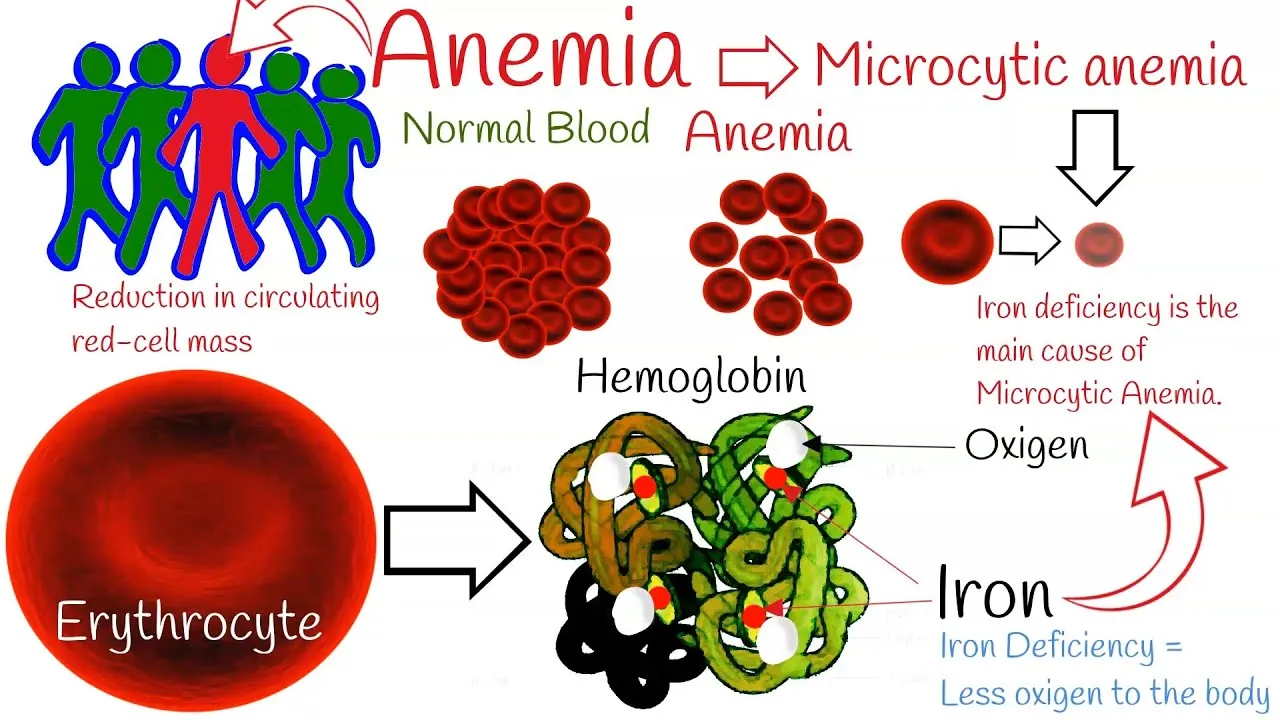
Figure 1: Global overview of iron metabolism and development of microcytic anemia [5]
2. EPIDEMIOLOGY AND RISK FACTORS
Neonates, babies, One of the most common nutritional disorders worldwide, iron deficiency anemia (IDA) is especially dangerous for young children. Nearly half of the anaemia cases in children aged 6–59 months are caused by an iron deficiency, and the percentage is around 40%. Poverty, food instability, recurrent infections, and inadequate healthcare facilities are recognized risk factors for chronic iron insufficiency; as a result, the burden is disproportionately high in low and middle income nations. Growth, neurodevelopment, immunity, and future educational results are all negatively impacted by IDA, raising serious public health concerns, even though there have been global initiatives to curb the use of baby vitamins and maternal nutrition. In terms of iron deficiency anemia, India is among the world's most afflicted nations. Infants and preschoolers, as well as pregnant women and nursing moms, have a high prevalence of anemia, according to national reports [6]. Despite the national nutrition programmes, iron and programs that provide folic acid supplements and enhanced prenatal care, children anaemia persists, attributed to maternal undernutrition, lack of dietary diversity, frequent infections and low socioeconomic status, and limited knowledge about infant feeding practices. In the absence of an intervention, maternal anemia has a significant impact on the iron status of the baby, extending the cycle of malnutrition and anemia from generation to generation. Iron status of newborns is greatly influenced by maternal factors. Anemia in pregnant women leads to decreased transfer of iron to the fetus and, consequently, lower iron stores at birth. Low maternal dietary iron intake, micronutrient deficiencies and poor nutritional care during pregnancy further reduces fetal iron accretion. Premature delivery disrupts the time when the fetus is accumulating the most iron in the third trimester, making it more likely that the infant will be iron deficient at birth [7]. The same applies to multiple pregnancies because there is a greater requirement for iron in the mother and there may be a clinging of maternal iron to the twins and thus reduced iron stores in multiple births. A number of factors are also linked to infant and childhood iron deficiency anemia. Low-birth-weight infants have lower iron reserves and also grow quickly after birth, which causes the iron reserves to be depleted early in life. Preterm infants are especially susceptible due to their lack of the last trimester of fetal iron accumulation. Breast milk is a relatively low iron containing food and infants with low iron stores at birth might need iron supplementation according to clinical guidelines, even though exclusive breastfeeding is strongly recommended in the first 6 months. Moreover, infants who are fed complementary food beyond six months of age that is low in iron face a much greater risk for iron deficiency in infancy. Other factors are recurrent infection, chronic inflammation, parasitic infestation of the gut, low dietary diversity and lack of adequate access to preventive health care services.
|
Risk Factor |
Mechanism |
Clinical Significance |
|
Maternal anemia |
Reduced placental iron transfer |
Low neonatal iron stores and early-onset anemia |
|
Poor maternal nutrition |
Inadequate fetal iron accumulation |
Increased risk of neonatal iron deficiency [8] |
|
Prematurity |
Incomplete third-trimester iron deposition |
Reduced iron reserves and rapid postnatal depletion |
|
Multiple pregnancy |
Increased maternal iron demand and fetal competition |
Lower birth iron stores in twins or multiples [9] |
|
Low birth weight |
Limited iron storage at birth |
Higher susceptibility to early iron deficiency |
|
Preterm birth |
Reduced fetal iron acquisition |
Increased requirement for early iron supplementation [10] |
|
Exclusive breastfeeding without iron supplementation (high-risk infants) |
Insufficient iron intake after depletion of neonatal stores |
Development of iron deficiency during infancy |
|
Delayed complementary feeding |
Delayed introduction of dietary iron |
Persistent iron deficiency and impaired growth [11] |
|
Recurrent infections |
Increased iron utilization and inflammation |
Worsening anemia and delayed recovery |
|
Low socioeconomic status |
Poor nutrition and limited healthcare access |
Increased prevalence and delayed diagnosis of IDA [12] |
Table 1. Major Risk Factors for Iron Deficiency Anemia in Neonates and Young Children
3. IRON METABOLISM AND PATHOPHYSIOLOGY
Iron is a vital trace mineral needed for oxygen transport, cellular respiration, DNA synthesis and many enzyme reactions. Iron homeostasis is important because iron deficiency and overload can both have a detrimental effect on physiological functions. Iron demands are especially high in neonates and young children due to the accelerated growth, blood volume expansion and erythropoietic activity. The processes of uptake, transport, storage, use, in erythropoiesis, and recycling by macrophages are tightly coupled to control iron metabolism. Any of these processes may cause iron deficiency and eventually microcytic hypochromic anemia. The site of highest iron absorption is in the proximal jejunum and duodenum. To be transported by divalent metal transporter-1 in enterocytes, non-heme iron from plants must first be reduced from Fe³âº to Fe³âº, although enterocytes easily absorb heme iron from animals via specialized transporters [13]. Intestinal epithelial cells can temporarily store iron as ferritin or use the transmembrane protein ferroportin to export iron into the bloodstream. Attachment to transferrin, the principal plasma iron transport protein, hephaestin or ceruloplasmin transforms the exported iron to its ferric form in the plasma. Transferrin keeps iron soluble and also non-toxic, and transports it to metabolically active tissues by endocytosis mediated by the transferrin receptor. The majority of iron in the bloodstream reaches the bone marrow for incorporation into and the growing erythroblasts that produce hemoglobin. Skeletal muscles, the liver, and spleen receive less, meaning they have less to store or use for metabolism. In normal physiological settings, the amount of circulating iron available for erythropoiesis is correlated with transferrin saturation, making it a significant indicator of iron status. The majority of cellular iron is stored by ferritin, the most abundant iron storage protein [14]. The liver is primarily responsible for storing iron, and the reticuloendothelial system's macrophages retrieve iron from spent erythrocytes and reintroduce it to the bloodstream. One of the first laboratory indicators for iron shortage, serum ferritin concentrations are typically associated with total body iron reserves. However, because ferritin is also an acute-phase reactant, it should be interpreted with caution in individuals with infections and/or chronic inflammation. Iron homeostasis is regulated in many parts of the body by hepcidin, a peptide hormone that is mostly produced by the liver. Hepcidin controls the amount of iron in the plasma by triggering the internalization and degradation of ferroportin. Low blood iron levels are caused by hepcidin, which is high and hinders iron absorption from the intestines. Instead, it causes iron to be reproduced from macrophages or hepatic reserves. In contrast, hepcidin levels are low in cases of iron deficiency or high erythropoiesis demands because the body absorbs more iron from food and mobilizes more iron from reserves to support hemoglobin production [15]. Because erythroid precursor cells require an ongoing supply of iron to produce hemoglobin, erythropoiesis primarily occurs in the bone marrow. Low hemoglobin levels can be caused by a shortage of iron, which in turn slows the creation of hemoglobin. This slows down the maturation of the cells that make up red blood cells. Microcytic and hypochromic red blood cells, which are progressively smaller in size, are produced as a result of this. Morphological alterations in iron deficiency anemia include anomalies in the distribution width of red blood cells, low serum ferritin, reduced transferrin saturation, and decreased mean corpuscular hemoglobin.
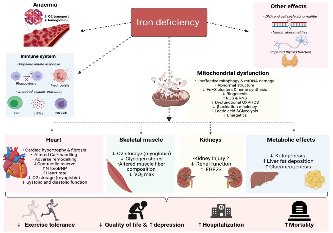
Figure 2: Pathophysiology of iron deficiency anemia [16]
4. HEMATOLOGICAL CHARACTERISTICS
The hematological profile is one of the basic tools for the diagnosis, classification and monitoring of neonates and young children with iron deficiency anemia. The hallmark of iron deficiency is changes in the morphology and haematological parameters of erythrocytes, indicating impairment of hemoglobin synthesis and inefficient erythropoiesis. Laboratory evaluation, which consists primarily of a complete blood count and peripheral blood smear examination and reticulocyte analysis, is the basis of ongoing assessment. Anemia is diagnosed using the level of haemoglobin (Hb) [17]. In iron deficiency, less hemoglobin is synthesized and the level of hemoglobin gradually decreases, causing less oxygen to be carried in the blood. While hemoglobin is very sensitive to detecting anemia, it does not have specificity for iron deficiency; other disorders such as thalassemia, chronic inflammatory diseases, vitamin deficiencies, and hemolytic disorders all also cause decreased hemoglobin. So more haematological parameters are needed to make an accurate diagnosis. The red blood cell count is useful in assessing erythropoietic activity. In iron deficiency anemia the number of mature erythrocytes is generally decreased or is in the lower end of the normal range because iron deficiency hinders the production of mature erythrocytes. Thalassemia trait, on the other hand, may have a fairly normal or elevated RBC count, even though hemoglobin levels are low, and this factor may help to make a differential diagnosis. As anemia becomes more severe, the percent of blood volume taken up by red blood cells, called hematocrit (Hct), tends to fall. Anemia (low hematocrit) is a sign of decreased mass of erythrocytes, which can lead to decreased delivery of oxygen to peripheral tissues [18]. But the measurements of hematocrit must always be considered with that of hemoglobin concentration and erythrocyte indices. Erythrocyte indices give information about the detailed Red blood cell shape. A decrease in mean corpuscular volume is one of the initial signs of developing iron deficiency anemia when microcytosis is present. The average concentration of hemoglobin in one red blood cell, known as the mean corpuscular hemoglobin, decreases when hemoglobin synthesis declines. Iron deficiency is characterized by hypochromic erythrocytes, which occur when the total concentration of hemoglobin in red blood cells drops. Microcytic hypochromic anemia is characterized by low MCV, MCH, and MCHC levels in the laboratory. When the red cell distribution width (RDW) varies in size, anisocytosis results. One of the earliest abnormal blood parameters noted in iron deficiency and usually rises before a significant decrease in MCV occurs. An elevated RDW suggests that other erythrocytes are also present in the bloodstream: some are normal size and others are smaller and smaller, as the iron deficiency progresses; this explains why RDW is a useful parameter to differentiate IDA from thalassemia trait, where the RDW is usually within the normal range or is only slightly elevated [19]. The bone marrow is actively producing red blood cells (RBCs) if the Reticulocyte count is high. Because there isn't enough iron for erythrocyte maturation in untreated iron deficiency anemia, reticulocyte output usually drops. A satisfactory response and good bone marrow response are indicated by reticulocytosis, which is a rise in the reticulocyte count, which typically comes 5-10 days after starting iron therapy. An integral aspect of the hematological evaluation is the study of peripheral blood smears. The hallmarks of these conditions include microcytic and hypochromic erythrocytes, elevated central pallor, anisocytosis, and poikilocytosis, elliptocytes and pencil-shaped cells. Target cells may also be seen in severe cases, but are more commonly seen in hemoglobinopathies. These morphological characteristics, along with hematological indices and iron studies, are good indicators of iron deficiency anemia. Age, nutritional status, gestational age, inflammatory conditions and associated clinical findings should always be taken into consideration in the clinical interpretation of hematological parameters. None of the hematological parameters are specific enough to make the diagnosis alone, and a combined assessment of CBC parameters, peripheral smear morphology, biochemical iron studies, and clinical history are most accurate for diagnosis and allow for timely therapeutic intervention.
|
Parameter |
Normal |
Iron Deficiency |
Clinical Significance |
|
Hemoglobin (Hb) |
Age-dependent normal range |
↓ Decreased |
Primary indicator of anemia severity and reduced oxygen-carrying capacity [20] |
|
RBC Count |
Normal |
↓ Reduced or low-normal |
Reflects impaired erythropoiesis; useful in differentiating IDA from thalassemia |
|
Hematocrit (Hct) |
Normal |
↓ Decreased |
Indicates reduced red cell mass and severity of anemia [21] |
|
Mean Corpuscular Volume (MCV) |
80–100 fL* |
↓ Low (Microcytic) |
Identifies small-sized erythrocytes; characteristic of IDA |
|
Mean Corpuscular Hemoglobin (MCH) |
27–33 pg* |
↓ Low |
Indicates reduced hemoglobin content per red blood cell [22] |
|
Mean Corpuscular Hemoglobin Concentration (MCHC) |
32–36 g/dL* |
↓ Low |
Demonstrates hypochromia due to decreased hemoglobin concentration |
|
Red Cell Distribution Width (RDW) |
11.5–14.5%* |
↑ Increased |
Early marker of anisocytosis; helps distinguish IDA from thalassemia trait [23] |
|
Reticulocyte Count |
0.5–2.5%* |
↓ Low before treatment; ↑ after iron therapy |
Evaluates bone marrow response and treatment effectiveness |
|
Peripheral Blood Smear |
Normocytic, normochromic RBCs |
Microcytic, hypochromic RBCs with anisocytosis, poikilocytosis, pencil cells, elliptocytes |
Confirms characteristic morphological changes and supports diagnosis [24] |
Table 2. Hematological Characteristics of Iron Deficiency Anemia
5. DIAGNOSTIC APPROACHES
Neonates and young children deserve the best care possible including early diagnosis of iron deficiency anemia to prevent progression of disease, avoid irreversible neurodevelopmental impairment and long-term health consequences. No single laboratory test is sufficient to diagnosis iron deficiency under all clinical conditions, so a mode of diagnosis needs to be integrated, based on hematological parameters, biochemical markers of iron metabolism and clinical evaluation. New laboratory diagnostic technologies have added to the precision of diagnosis recently as new biomarkers have been developed to diagnose iron deficiency before overt anemia has started. The first laboratory test ordered in patients who are suspected to be anemic is the complete blood count (CBC) [25]. Information on hemoglobin concentration, hematocrit, red blood cell count, and erythrocyte indices (MCHC, red cell distribution width, mean corpuscular hemoglobin, and mean corpuscular volume) are included in this. Common lab abnormalities in IDA are decreased hemoglobin, low MCV and MCH, increased RDW, and low hematocrit. While cheap, fast and readily available, these abnormalities are not iron-deficiency specific and can also be seen in thalassaemia, anaemia of chronic disease and sideroblastic anaemia. Peripheral blood smear examination will supplement the CBC results, and it will give direct visualization of the erythrocyte morphology. Red blood cells that are microcytic and hypochromic, with an increased central pallor, anisocytosis, poikilocytosis, elliptocytes, and pencil-shaped cells are the identifiable features. Peripheral smear also helps to rule out other hematological diseases like hemolytic anemia, hereditary spherocytosis and leukemia. However, interpretation is subjective and should always be accompanied by quantitative values from the laboratory. Biochemical investigations, Being the most reliable way to gauge the body's iron storage capacity, serum ferritin is one of the first laboratory measures to drop when iron levels drop [26]. Before hemoglobin levels start to decline, an iron deficit is strongly indicated by a ferritin level below 12 ng/mL. Nevertheless, ferritin levels can rise due to infections, inflammation, liver illness, or cancer since it is an acute-phase reactant, resulting in a masked iron-deficiency. Therefore, ferritin should be considered with inflammatory tests like C-reactive protein in case of any suspicion of inflammation. Serum iron is the iron that is bound to transferrin in the blood and tends to be low with iron deficiency. There is however, significant diurnal variation in serum iron levels, and the recent food intake also affects it, which makes it an inadequate test on its own. Therefore it is typically used in conjunction with total iron binding capacity and transferrin saturation. In IDA, TIBC is elevated, because synthesis of transferrin is stimulated by low levels of iron and circulating levels of transferrin are decreased by low levels of iron [27].
Soluble transferrin receptor (sTfR) has proved as an important a biomarker for the evaluation of functional iron toxicity. The expression of TfR on erythroid precursor cells rises, leading to larger circulating levels of sTfR, in response to increased erythropoietic activity and cellular iron deprivation. When distinguishing iron deficiency anemia from chronic illness anemia, sTfR is invaluable because, in contrast to ferritin, it is not significantly impacted by inflammation. The sTfR-to-log ferritin index has proven to have high diagnostic accuracy, particularly when used in conjunction with patients with inflammatory diseases. ZPP is a substance that is stored in the erythrocytes when iron is not available for heme production. High levels of ZPP are associated with poor incorporation of iron into protoporphyrin IX and could be used to detect iron deficient erythropoiesis before anemia is clinically evident. ZPP measurement is relatively cheap and advantageous for population screening, but non-specific because high levels are also found in lead poisoning, chronic inflammatory diseases, and some hemoglobinopathies [28]. Reticulocyte hemoglobin content (CHr or Ret-He) has been a topic of great interest as an early marker of Fe-restricted EPO. The hemoglobin content of reticulocytes reflects recent availability of iron for red blood cells, since they can only circulate for 1-2 days. When the iron circulation parameters are reduced, it is sometimes observed before the changes in conventional hematological parameters and is sensitive and fast to respond after iron therapy, hence it can be utilized for treatment monitoring and early diagnosis. One promising area of research in the field of iron metabolism is hepcidin, the primary regulator of systemic iron homeostasis. Enhanced iron intake and intestinal iron store mobilization are hallmarks of absolute iron shortage, which is characterized by a low hepcidin level. On the other hand, high hepcidin levels are seen in inflammatory anemia, where an increase in hepcidin causes iron to become sequestered even when it is there in the body [29]. Measurement of hepcidin has a great potential for the diagnosis of anemia. However, owing to factors such as restricted laboratory availability, unstandardized reference ranges, and test variability, it is not commonly utilized in ordinary clinical practice. The gold standard for determining iron reserves is the staining of bone marrow with Prussian blue. Absolute iron shortage can be confirmed by the presence of non-staining hemosiderin. But the bone marrow aspiration is invasive, painful, expensive and impractical for routine assessment especially in paediatrics. It is therefore generally used in cases that are difficult to diagnose using non-invasive methods. In recent years, the emergence of molecular diagnosis has led to the identification of a number of new biomarkers such as erythroferrone, ferroportin expression, transferrin receptor gene polymorphisms and hepcidin signaling pathway iron-regulatory proteins. The use of high throughput proteomic and metabolomic techniques and an artificial intelligence based diagnostic algorithm that combines multiple laboratory parameters are also being explored for diagnostic clarification. These technologies are mostly still limited to research environments, but have great potential for personalized diagnosis and early detection of iron deficiency [30].
|
Diagnostic Test |
Advantages |
Limitations |
Clinical Utility |
|
Complete Blood Count (CBC) |
Rapid, inexpensive, widely available |
Low specificity |
Initial screening and anemia classification |
|
Peripheral Blood Smear |
Evaluates RBC morphology |
Observer-dependent |
Confirms microcytic hypochromic anemia and excludes other disorders [31] |
|
Serum Ferritin |
Best indicator of iron stores |
Elevated in inflammation and liver disease |
Early diagnosis of iron deficiency |
|
Serum Iron |
Measures circulating iron |
Diurnal variation; influenced by diet |
Used with TIBC and TSAT |
|
Total Iron-Binding Capacity (TIBC) |
Reflects transferrin availability |
Altered in liver disease and malnutrition |
Supports diagnosis of iron deficiency [32] |
|
Transferrin Saturation (TSAT) |
Indicates iron available for erythropoiesis |
Requires combined interpretation |
Functional assessment of iron status |
|
Soluble Transferrin Receptor (sTfR) |
Not significantly affected by inflammation |
Higher cost; limited availability |
Differentiates IDA from anemia of chronic disease [33] |
|
Zinc Protoporphyrin (ZPP) |
Early marker of iron-deficient erythropoiesis |
Reduced specificity |
Population screening and early detection |
|
Reticulocyte Hemoglobin Content (Ret-He/CHr) |
Detects early iron-restricted erythropoiesis; monitors therapy |
Requires modern hematology analyzers |
Early diagnosis and treatment monitoring [34] |
|
Hepcidin |
Reflects systemic iron regulation |
Lack of assay standardization |
Differentiates absolute and functional iron deficiency |
|
Bone Marrow Iron Staining |
Gold standard for iron stores |
Invasive and impractical |
Reserved for complex or inconclusive cases |
|
Molecular Biomarkers |
High diagnostic potential; supports precision medicine |
Mostly research-based; expensive |
Future applications in personalized diagnosis [35] |
Table 3. Diagnostic Tests for Iron Deficiency Anemia
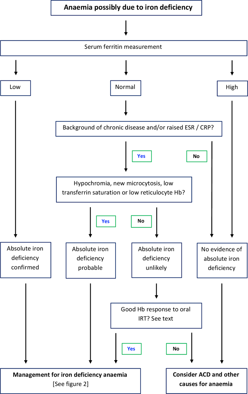
Figure 3: Diagnostic workflow of iron deficiency anemia [36]
6. DIFFERENTIAL DIAGNOSIS OF MICROCYTIC ANEMIA
Microcytic anemia is characterized by extremely small red blood cells with a typical corpuscular volume (MCV<80 fL in adults and age adjusted in children). While iron deficiency anemia accounts for the vast majority of cases, several additional diseases share similar hematological features. Since the causes, treatments, and outcomes of each of these diseases are unique, correct categorization is of the utmost importance. Thus, it is essential to do a comprehensive examination that includes a patient's medical history, a peripheral blood smear, an iron study, inflammatory markers, a full blood count, and any other specialist tests that may be necessary. Since hemoglobin cannot be produced when iron is not present, the most common kind of anemia, microcytic anemia, results from iron deficiency anemia. Results from laboratory tests for hemoglobin, MCV, MCH, serum iron, total IBC, and transferrin saturation tend to be on the low side. Anisocytosis, poikilocytosis, pencil cells, and microcytic, hypochromic erythrocytes are all visible in the peripheral blood smear. Clinical evidence suggests that iron supplementation can help confirm a diagnosis [37]. Thalassemia, a hereditary disorder of globin chain production, is another major cause of microcytic anemia. It is common for patients with IDA to have a markedly reduced MCV and a normal or high RBC count. In cases where β-thalassemia trait is present, aberrant hemoglobin fractions, specifically higher HbAâ‚‚, are revealed by hemoglobin electrophoresis or high-performance liquid chromatography, while iron investigations typically return normal results. To avoid needless iron therapy and to facilitate genetic counseling, it is critical to be knowledgeable about thalassemia. Anemia due to chronic illness (ACD) occurs when an inflammatory disease, cancer, chronic infection, or autoimmune disorder persists over an extended period of time. Even though iron reserves are normal or even raised, functional iron insufficiency can occur because this increased hepcidin production leads to lower intestinal iron absorption and iron sequestration in macrophages. Serum ferritin levels should be within normal ranges, total iron binding capacity should be within normal ranges, serum iron and transferrin saturation should be lowered, and so on. It is especially important to distinguish ACD from IDA as treatment is directed toward the underlying inflammatory condition. A variety of diseases known as sideroblastic anemia cause erythroid precursors to accumulate iron in their mitochondria and fail to produce heme. The diagnosis is made by staining bone marrow for Prussian blue and by measuring serum ferritin and transferrin saturation levels, which indicate inadequate iron consumption rather than availability. It can be caused by a lack of vitamin B₆, drugs, alcohol, myelodysplastic syndromes or environmental toxins, or may occur at birth. Microcytic anemia, especially in children exposed to contaminated paint, water or industrial pollutants, is an uncommon but clinically significant cause of microcytic anemia [38]. Lead interferes with enzymes in the heme synthesis pathway, causing microcytic anemia and neurological, gastrointestinal and developmental effects. An high blood lead level and the presence of basophilic stippling on the peripheral blood smear both lend credence to the diagnosis. The signs and symptoms of these disorders often overlap, so laboratory testing is essential in making a diagnosis. This systematic approach combining hematological indices, iron profile, inflammatory markers, hemoglobin analysis and disease-specific tests is good enough to differentiate with accuracy and provide an appropriate therapeutic management.
|
Parameter |
Iron Deficiency Anemia |
Thalassemia |
Anemia of Chronic Disease |
Sideroblastic Anemia |
Lead Poisoning |
|
Primary cause |
Iron deficiency |
Inherited globin gene defect |
Chronic inflammation/infection |
Defective heme synthesis |
Lead toxicity |
|
Hemoglobin |
↓ |
↓ |
↓ |
↓ |
↓ |
|
MCV |
↓↓↓ |
↓↓↓ |
↓/Normal |
↓ |
↓ |
|
RBC Count |
↓ or Normal |
Normal/↑ |
↓ |
Variable |
Variable |
|
Serum Ferritin |
↓ |
Normal/↑ |
Normal/↑ |
↑ |
Normal |
|
Serum Iron |
↓ |
Normal |
↓ |
↑ |
Normal/↓ |
|
TIBC |
↑ |
Normal |
↓/Normal |
Normal |
Normal |
|
Transferrin Saturation |
↓ |
Normal/↑ |
↓ |
↑ |
Variable |
|
Peripheral Smear |
Microcytic, hypochromic RBCs, anisocytosis, pencil cells |
Target cells, marked microcytosis |
Mild microcytosis |
Dimorphic RBCs, Pappenheimer bodies |
Basophilic stippling |
|
Confirmatory Test |
Low ferritin and response to iron therapy |
Hb electrophoresis/HPLC or genetic testing |
CRP/ESR with iron profile |
Bone marrow ring sideroblasts |
Blood lead level |
|
Treatment |
Iron supplementation and dietary correction |
Supportive care, transfusion (selected cases), genetic counseling |
Treat underlying disease ± iron when indicated |
Vitamin B₆ (selected cases), treat underlying cause |
Remove lead exposure and chelation therapy when indicated [40] |
Table 3. Comparison of Major Causes of Microcytic Anemia [39]
7. CLINICAL MANIFESTATIONS AND COMPLICATIONS
The age of the affected child, the extent and duration of iron loss, and other factors determine the clinical manifestations of iron deficiency anemia. Iron-deficiency anemia can be manifested as asymptomatic in neonates and infants, but as iron levels become increasingly depleted, hematological findings and multisystem complications appear. Iron is critical for oxygen transport, cellular metabolism, myelination, synthesizing neurotransmitters, and immunity, making prolonged deficiency have significant – and sometimes irreversible – consequences. Iron deficiency is most common in the neonatal period in term infants whose mothers have severe anemia or are under- or over-nutrient, low-birth-weight infants, and preterm infants. Clinical signs are pallor, decreased feeding, lethargy, increased heart rate and failure to gain weight [41]. If severe, decreased oxygen delivery can lead to apnea, inadequate physiologic adaptation after birth and susceptibility to neonatal infection. Haemoglobin levels in the newborns are closely linked to the iron status of their mothers, and placental transfer during the third trimester is the most important source of iron to the neonate, meaning that poor iron acquisition is likely to lead to early onset iron deficiency. Clinical symptoms become more prominent during infancy and early childhood because there is rapid growth and therefore a high iron requirement. Symptoms are common such as fatigue, irritability, loss of appetite, delayed motor development, concentration difficulties, recurrent infections and decreased physical activity. Chronic iron deficiency can also affect linear growth and weight gain leading to growth retardation [42]. Children who have an anemic condition for an extended period often exhibit late developmental stages due to low exercise tolerance and reduced oxygen carrying capacity. Children with iron deficiency often experience cognitive impairment, which is a serious adverse effect. Myelination, neurotransmitter synthesis, hippocampus function, and brain development are all activities that rely on iron. Deficiency at critical times of neurodevelopment has been linked to learning disability, inability to pay attention, memory loss, decreased IQ and poor school performance. Early detection and treatment is important as several longitudinal studies have shown that some neurodevelopmental deficits can remain even after correction of anemic status. Iron deficiency also has a negative effect on immunity by reducing the activity of neutrophils, impairing function of the macrophages and reducing the proliferation of lymphocytes, which leads to an increased susceptibility to infections of the respiratory tract, gastrointestinal tract, etc. Affected children are also often seen to be behaving differently, such as being irritable, socially withdrawn, less responsive, having problems with their sleep, being anxious and less interacting with their carers which may have a negative impact on their psychosocial development. Rare in mild anemia, cardiovascular complications may develop in moderate-to-severe anemia. Compensatory tachycardia, increased cardiac output, systolic flow murmurs, and left ventricular hypertrophy/high-output cardiac failure occurs in severe untreated cases in which oxygen-carrying capacity has been chronically reduced. Infants with severe, chronic anemia, or any other cardiopulmonary disease are at increased risk of these complications. The chronic effects of iron-deficiency anemia are not limited to the hematological disorders [43]. Long-term deficiency during the early years of life is associated with cognitive dysfunction, psychomotor delay, low achievement in education, decreased immunity, and decreased productivity in the adult population. The importance of maternal nutrition as a preventative measure, regular screening of high-risk infants, early laboratory diagnosis and iron supplementation can be seen as a result of these lifelong consequences. Early intervention not only helps restore hematological parameters, but also helps to avoid irreversible neurological damage and better overall child health and developmental consequences.
8. CURRENT TREATMENT STRATEGIES
The main goals of IDA treatment in neonates and young children are to replenish iron stores, normalize hemoglobin levels, ensure adequate growth and neurodevelopment, and to avoid long term complications. Treatment should be directed towards both the cause of iron deficiency and correction of hematologic abnormalities. It is recommended to diagnose early, to provide the necessary iron supplementation, to optimize the nutritional status and to implement preventive action from the ante-conception period.
Iron Supplementation
Iron supplementation continues to be the mainstay of the treatment of IDA. Therapy depends upon child's age, severity of the anemia, underlying etiology, gastrointestinal tolerance, and urgency of treatment. In cases of simple IDA, oral iron therapy is preferred over intravenous iron therapy, which is reserved for specific clinical circumstances [44].
Oral Iron Therapy
The effective and highly bioavailable ferrous salt formulations, including ferrous sulfate, fumarate, and gluconate, are the ones to choose. In children with confirmed IDA, the recommended therapeutic amount daily, in one or two doses, the recommended amount of elemental iron is 3-6 mg/kg. Treatment should be maintained for an additional three months following normalization of hemoglobin levels to make sure the body has enough iron stores. After a week or two of treatment, patients should start to feel better, and in the weeks that follow, they may see reticulocytosis and a gradual increase in hemoglobin levels. Regrettably, certain gastrointestinal side effects, including sickness, abdominal discomfort, irregular bowel movements, dark colored feces might make it harder to stick to treatment plans. While consuming vitamin C-containing foods or beverages may enhance iron bioavailability, it is not advisable to do so at the same time as consuming milk, calcium supplements, tea, or foods rich in phytates, as these substances have the reverse effect [45].
Intravenous Iron Therapy
When iron sucrose or ferric carboxymaltose cannot be taken or is not tolerated or is not suitable for oral iron therapy, or when it is clinically imperative to address the iron shortage quickly, intravenous iron preparations like these should be administered. Cases of severe persistent anemia, chronic renal disease, severe malabsorption syndromes, and chronic inflammatory bowel illness in children, or a poor oral iron response to therapy will particularly benefit from IV iron. The safety profile of the modern forms of IV iron has been enhanced, but they must be administered under medical supervision due to the possible occurrence of hypersensitivity reactions, although quite rare [46].
Maternal Iron Supplementation
During pregnancy, it is possible to avoid neonatal iron insufficiency. Increases in maternal hemoglobin, placental iron transfer, and newborn iron storage are all outcomes of iron supplementation during pregnancy. Pregnant women should get tested for anemia regularly and take iron and folic acid supplements are essential components of maternal health care. Premature birth, low birth weight, and newborn anemia are all greatly reduced when nutritional deficits in the mother are treated properly.
Dietary Interventions
A simple way to avoid iron shortage again is to alter one's eating habits. During the first six months of a baby's life, they should only eat breast milk. When the time is appropriate, you can start introducing iron-rich complementary foods. Consuming lean meat, liver, fish, egg yolk, legumes, green leafy vegetables, fortified cereals, pulses, and vitamin C-rich fruits might improve your body's absorption of non-heme iron. It is recommended that cows only drink milk up to the age of one year since it can prevent iron absorption and "silent" gastrointestinal hemorrhage [47].
Delayed Cord Clamping
The delayed clamping of the umbilical cord, where the cord is not clamped for about 30-60 seconds after birth has become a simple, effective intervention to increase neonatal iron stores. Delayed cord clamping results in increased neonatal hemoglobin concentration and a decreased prevalence of iron deficiency in infancy, especially that of preterm and LBW infants, due to placental transfusion. Current recommendations are for its use in vigorous term and preterm newborns, except in those that are contraindicated.
Fortified Foods
Food fortification is a good population-based approach to mitigate the problem of IDAs. Iron fortified infant cereals, complementary foods and fortified staple foods have been shown to significantly improve the hemoglobin concentration and iron status in children from high prevalence areas. National fortification programs are especially helpful in countries with low dietary diversity and high prevalence of malnutrition [48].
International and National Recommendations
The World Health Organization suggests routine iron supplementatfor children who do not have known contraindications, as well as for those living in areas where anemia is common. A nutritious diet and folic acid supplementation are essential for pregnant women. Starting at 4 months of age and continuing until they start eating iron-containing complementary meals, which usually takes around 6 months, the American Academy of Pediatrics (AAP) recommends supplementing the diet of full-term, exclusively breastfed newborns with 1 mg/kg/day of iron. Because their bodies have fewer iron stores, infants born prematurely need higher iron doses for prophylaxis. Screening for anemia regularly, providing nutritional counseling, deworming as needed, and communicating the need of behavior change are all part of the National Iron Plus Initiative (NIPI) and the Anemia Mukt Bharat programs' efforts to lower the incidence of anemia in India, especially among pregnant women, infants, and children. Pediatric IDA requires a multi-disciplinary approach including nutritional, timely, adequate iron therapy, maternal health optimization and public health strategies [49]. The restoration of normal hematological parameters and the prevention of irreversible complications of development requires early diagnosis, regular follow-up and compliance with evidence-based guidelines.
|
Treatment Strategy |
Recommendation |
Clinical Indication |
Key Benefits |
|
Oral iron supplementation |
Ferrous sulfate/fumarate/gluconate; 3–6 mg/kg/day elemental iron |
First-line treatment for uncomplicated IDA |
Restores hemoglobin and replenishes iron stores |
|
Intravenous iron |
Iron sucrose, ferric carboxymaltose |
Oral iron intolerance, malabsorption, severe deficiency, chronic disease |
Rapid correction of iron deficiency |
|
Maternal iron supplementation |
Daily iron–folic acid during pregnancy |
Prevention of maternal and neonatal anemia |
Improves fetal iron stores and birth outcomes |
|
Exclusive breastfeeding |
Recommended for first 6 months |
Healthy term infants |
Optimal nutrition and immune protection |
|
Iron-rich complementary feeding |
Introduce at approximately 6 months |
All infants |
Prevents depletion of iron stores |
|
Delayed cord clamping |
Delay clamping by 30–60 seconds after birth |
Vigorous term and preterm neonates |
Increases neonatal iron reserve and reduces infant anemia |
|
Iron-fortified foods |
Fortified cereals and complementary foods |
High-risk populations |
Improves population iron status |
|
WHO recommendations |
Iron supplementation in high-prevalence settings; maternal supplementation |
Public health programs |
Reduces anemia prevalence globally |
|
AAP recommendations |
Iron supplementation for exclusively breastfed infants from 4 months; higher doses for preterm infants |
Infant health care |
Prevents early iron deficiency |
|
Indian guidelines (NIPI/Anemia Mukt Bharat) |
Universal iron–folic acid supplementation, screening, dietary counseling |
National anemia control programmes |
Reduces childhood and maternal anemia burden |
Table 4. Current Treatment Recommendations for Iron Deficiency Anemia [50]
9. EMERGING DIAGNOSTIC TECHNOLOGIES
The diagnosis of iron deficiency anemia is rapidly changing, as advanced laboratory technologies and digital health solutions play an integral role. Conventional haematological investigations remain essential, but newer technologies aim to enhance diagnostics, decrease turnaround time and aid in early detection, especially in resource-limited environments. In laboratory hematology, artificial intelligence (AI) is a promising tool. Complete blood count (CBC) parameters, clinical information and imaging data can be analyzed using AI-based algorithms to uncover subtle patterns that are characteristic of iron deficiency and differentiate IDA from other microcytic anemias. For the first time in machine learning, models learned from large clinical databases have shown to have high diagnostic accuracy with reduced variability in observers and assisted clinical decisions [51]. The combination of automated peripheral blood smear capturing with computer assisted interpretation has given a great boost to the power of digital microscopy. These systems are able to precisely detect erythrocyte morphology (microcytosis, hypochromia, anisocytosis, poikilocytosis, and target cells) which decreases manual labor and increases the repeatability. In a similar manner, machine learning based automated morphology analyzers combine image recognition and pattern analysis to classify red blood cells based on their size, shape and hemoglobin content. These technologies help to maintain a consistent diagnosis and can help laboratories that do not have extensive hematology knowledge. Point-of-care testing (POCT) devices have facilitated the use of anemia screening in remote, underserved areas. Portable hemoglobin analyzers, finger-prick ferritin assays, and rapid diagnostic platforms allow for “on-the-spot” testing without the need for high-tech laboratory facilities. Portable hematology analyzers also aid in the care of neonates and pediatrics by giving rapid CBC results with minimal blood volume—an advantage for neonates and critically ill patients. In recent years, there has also been a focus on novel biomarkers to make it easier to detect iron deficiency before it results in anemia. Biomarkers like hepcidin, reticulocyte hemoglobin content (Ret-He), In the context of coexisting inflammatory conditions, soluble transferrin receptors, erythroferrone, growth differentiation factor-15, and iron-regulatory proteins exhibit increased sensitivity and specificity [52]. In the coming years, the rapidly evolving field of genomics, proteomics, metabolomics, and multi-omics technologies promise to uncover more biomarkers related to iron metabolism and to uncover individual disease risk. The future of laboratory medicine promises to involve AI-driven interpretation, digital pathology, molecular diagnostics, cloud-based data processing, and wearable health technologies in everyday diagnostics. These innovations may have the potential to allow earlier diagnosis, better treatment monitoring, and help facilitate precision medicine strategies in order to cure children's iron deficiency anemia [53].
10. CHALLENGES AND FUTURE PERSPECTIVES
There has been a lot of success in learning about and treating iron deficiency anemia, but there are still a lot of obstacles to successful anemia control, particularly in low- and middle-income countries. Delayed diagnosis continues to be one of the most important obstacles, with early iron deficiency often being undiagnosed and not routinely performed in all cases. For this reason, many children only receive a diagnosis when their anemia is clinically apparent, which puts them at risk to suffer permanent neurodevelopmental damage. Another significant problem is poor compliance with oral iron therapy. Many of these side effects, including gastrointestinal, undesirable taste, treatment length and limited awareness by caregivers, often limit compliance resulting in incomplete correction of iron stores and reoccurring anemia. Furthermore, inappropriate empirical iron therapy, without laboratory confirmation, could lead to overdiagnosis and/or unnecessary treatment, especially in patients with thalassemia trait or anemia of chronic disease. Lack of laboratory facilities also limits access to more sophisticated laboratory investigations, such as serum ferritin, soluble transferrin receptor, hepcidin, and molecular tests. Most primary health care offices still use only hemoglobin estimation to diagnose anemia without specificity as to the cause of anemia. In developing countries, insufficient training, limited access to health services and financial limitations are still major challenges. AI-driven diagnostic tools that combine hematological data, clinical history, and blood smear analysis for enhanced diagnostic accuracy should be a focus in future strategies. Future validation of novel markers, such as hepcidin and Ret-He, can help in earlier detection of iron deficiency when significant hematological changes have not occurred. Establishing uniform reference intervals and increasing the number of assays will be key for broader clinical use. Increasing coverage of national screening programmes for pregnant women, neonates, infants, and other high-risk groups, can significantly decrease the disease burden by detecting and intervening early. Personalized medicine that takes into account genetic susceptibility, nutritional assessment and biomarker profiling could help develop individual prevention and treatment plans. Moreover, telehematology, cloud-based laboratory reporting, and digital health platforms can enhance the specialist consultation, remote diagnosis, and healthcare services in remote areas. Multicenter prospective studies, cost effectiveness analysis of new diagnostic technologies and the creation of a low-cost point-of-care diagnostic platform for easy use in clinical practice should be considered for future research. These efforts will help to promote access to health and health outcomes for IDA-affected children, which should be equitable.
CONCLUSION
The prevalence of iron deficiency anemia, the most frequent kind of nutritional anemia worldwide, is a major issue in children's and toddlers' public health. It is more common in countries with low or medium incomes due to factors such as maternal anaemia, reduced iron consumption during pregnancy, early delivery, low birth weight, and socioeconomic status. Because of its negative effects on hemostatic state, immunological function, growth, neurodevelopment, and long-term cognitive performance, iron deficiency must be diagnosed and treated appropriately in early childhood. Currently, a series of tests including a full blood count, peripheral blood smear, biochemical iron studies, and newly discovered biomarkers such as serum ferritin, soluble transferrin receptor, reticulocyte hemoglobin content, and hepcidin are the gold standard for diagnosing iron deficiency anemia. Technological progress in the fields of artificial intelligence, digital microscopy, and automation of haematology and point-of-care diagnostics will lead to a better diagnostic accuracy, earlier diagnosis and widening of the access to laboratory services. A multi-disciplinary strategy for optimal maternal nutrition, evidence-based iron supplementation, dietary counseling, delayed cord clamping, food fortification, and compliance to the international and national clinical guidelines are necessary to implement effective prevention and treatment. However, there are still many challenges to be addressed: late diagnosis, small laboratory facilities, non-compliance with treatment and inequalities in the access to health care. Further studies are needed to confirm novel biomarkers; take the use of artificial intelligence to the routine laboratory; create cost-effective diagnostic technologies and improve population-based screening programmes. Ongoing investments in precision diagnostics, translation research and public health interventions are still critical to decrease the burden of IDA and to improve health outcomes for vulnerable paediatric sub-populations around the world.
REFERENCES

Shrabani Jana Maity*, Shweta Choudhary, Biswajit Chakraborty, Microcytic Iron Deficiency Anemia In Neonates and Young Children: Current Diagnostic Approaches, Hematological Characteristics, Clinical Challenges, And Future Perspectives, Int. J. Sci. R. Tech., 2026, 3 (7), 956-974. https://doi.org/10.5281/zenodo.21641634
 10.5281/zenodo.21641634