

We use cookies to ensure our website works properly and to personalise your experience. Cookies policy
KMCH college of Pharmacy, Coimbatore – 641048, India
Previously known as polycystic ovarian syndrome (PCOS), polyendocrine metabolic ovarian syndrome (PMOS) is a common endocrine and metabolic condition linked to insulin resistance, abnormal lipid metabolism, irregular or missing ovulation, and increased androgen production. PMOS is increasingly recognized as a more comprehensive systemic syndrome that may have long-term impacts on metabolic health and cancer risk, despite the fact that it was once primarily thought of as a reproductive disorder. The association with ovarian and breast cancers is still unclear, but the strongest and most reliable evidence has been documented for endometrial cancer. The potential link between PMOS and cancer may be explained by a number of biological processes. Without enough progesterone, persistent anovulation can expose the endometrium to estrogen for extended periods of time, which could promote ongoing endometrial development. Through the insulin/IGF system and signaling pathways like PI3K/Akt/mTOR and MAPK/ERK, insulin resistance and increased insulin levels can also stimulate cell growth. Abnormal cellular activity may also be caused by other factors, including as excess androgens, oxidative stress, chronic inflammation, genetic and epigenetic alterations, and disruptions in the gut flora. However, as obesity, diabetes, infertility, and reproductive variables can all influence cancer risk, these processes by themselves do not prove that PMOS causes cancer. Therefore, more study is required to elucidate these relationships, find trustworthy biomarkers, and develop practical methods for early risk assessment and cancer prevention in women with PMOS.
Previously known as polycystic ovarian syndrome (PCOS), polyendocrine metabolic ovarian syndrome (PMOS) is a heterogeneous endocrine-metabolic disorder that primarily affects women in their reproductive years. Hyperandrogenism, ovulatory dysfunction, polycystic ovarian morphology, and metabolic abnormalities like insulin resistance, obesity, and dyslipidemia are among its main clinical characteristics [3,21–24]. Because reproductive abnormalities often coincide with metabolic, inflammatory, endocrine, and genetic issues, the illness is becoming more widely acknowledged as a systemic condition [4,12,21,23].
Abnormalities in hypothalamic-pituitary-ovarian signaling, ovarian and adrenal steroidogenesis, insulin action, adipose tissue function, and genetic vulnerability are all part of the complex pathophysiology of PMOS [3,4,21–24]. Because they can interact in both directions, hyperandrogenism and insulin resistance are especially significant. While hyperinsulinemia may increase ovarian androgen production and decrease sex hormone-binding globulin (SHBG), increasing circulating free androgens, androgen excess may further impair metabolic and reproductive function [21–23,27].
PMOS's systemic effects have raised interest in its potential connection to chronic conditions other than infertility and irregular menstruation. Epidemiological research has specifically examined whether women with PMOS are at a higher risk of hormone-responsive cancers. While results regarding ovarian and breast cancer are still inconsistent, the most consistent link has been shown for endometrial cancer [7,13,14].
There is a compelling biological basis for the link with endometrial cancer. Endometrial hyperplasia and eventual malignant transformation may be encouraged by chronic anovulation, which exposes the endometrium to estrogen for extended periods of time without the cyclical differentiating effects of progesterone [11,15,16]. Through altered estrogen metabolism, hyperinsulinemia, and growth-factor signaling activation, obesity and insulin resistance may exacerbate this situation [8,15,47].
The PI3K/Akt/mTOR and MAPK/ERK pathways, which control cellular proliferation, metabolism, and survival, may be activated at the molecular level by insulin and insulin-like growth factor signaling [15,47,48]. Moreover, NF-κB and MAPK-related pathways may be activated by chronic inflammation and oxidative stress, which can lead to cellular stress and genomic instability [33–44,52]. Steroidogenesis, insulin signaling, inflammatory control, and tissue-specific responses may all be further impacted by genetic and epigenetic changes [1,44–53].
Recent studies have also looked at the gut microbiota as a possible cause of endocrine and metabolic dysfunction linked to PMOS. Through the estrobolome, changes in microbial composition may affect intestinal permeability, inflammation, and estrogen metabolism [5,54–56]. It is unclear, therefore, how much these microbial changes directly affect cancer susceptibility.
Thus, the objective of this review is to critically integrate the molecular and epidemiological evidence that connects PMOS to breast, ovarian, and endometrial malignancies. Distinguishing proven epidemiological connections from mechanistic ideas and highlighting areas that require more research are given special attention.
2. EPIDEMIOLOGICAL ASSOCIATION BETWEEN PMOS AND CANCER
2.1 Endometrial Cancer
The most frequently reported cancer link is that between PMOS and endometrial cancer. Women with PCOS/PMOS have a higher relative risk of endometrial cancer than women without the syndrome, according to systematic reviews and meta-analyses [7,13,14]. Previous estimates have indicated a roughly two- to four-fold increase in relative risk, however the exact amount varies between studies due to variations in population characteristics, diagnostic criteria, and confounding variable correction [7,8].
Because chronic ovulatory failure can lead to extended estrogenic stimulation without sufficient cyclical progesterone exposure, the elevated risk is biologically reasonable [11,15]. In vulnerable people, persistent endometrial growth may raise the risk of hyperplasia and malignant transformation. Since estrogenic and metabolic variables have a significant impact on endometrioid endometrial cancer, this pathway is especially pertinent [11,15].
This link could be further altered by metabolic disorders. Obesity is a known risk factor for endometrial cancer and can raise peripheral estrogen production. Additionally, estrogen metabolism and insulin/IGF-mediated proliferative signaling may be influenced by insulin resistance and hyperinsulinemia [8,15,47].
Crucially, the correlation between PMOS and endometrial cancer should not be taken as proof that PMOS causes cancer on its own in all afflicted women. Obesity and metabolic dysfunction may partially explain or confound the observed connection, since studies controlling for body mass index have revealed reduction of risk estimates [13]. Diabetes, anovulation length, reproductive variables, and other clinical traits may also be involved.
The 2023 international evidence-based guideline acknowledges that premenopausal women with PMOS/PCOS have a significantly higher risk of endometrial hyperplasia and endometrial cancer; however, it stresses that the absolute probability of cancer is still low, so routine screening of asymptomatic women is not advised [58]. Instead, cycle control, weight control, metabolic health, and the proper assessment of abnormal uterine bleeding should be the main emphasis of preventive care [58,59].
2.2 Ovarian Cancer
Compared to the correlation with endometrial cancer, the relationship between PMOS and ovarian cancer is far less definite. The results of epidemiological investigations have been inconsistent, with some analyzes indicating elevated risk and others failing to show a statistically significant overall connection [7,13,14,17].
Women with PCOS had a roughly two-fold increased risk of ovarian cancer, according to an earlier systematic analysis; however, this estimate was based on a small number of studies and should be interpreted cautiously [17]. Significant heterogeneity is still evident in more recent systematic data, highlighting the need for better-controlled research [14].
A number of explanations for a potential association have been put up. Ovarian tissue homeostasis may be impacted by hyperandrogenism, prolonged anovulation, altered gonadotropin signaling, infertility, insulin resistance, and persistent inflammation [9,16–18]. Additionally, common genes and signaling pathways between PMOS and ovarian cancer have been found by bioinformatic research, indicating potential molecular convergence [9].
Molecular resemblance does not, however, prove causation. The term "ovarian cancer" refers to a diverse group of cancers with unique molecular and histological subgroups. Therefore, rather than treating ovarian cancer as a homogenous biological entity, future epidemiological research should examine if PMOS is linked to particular ovarian cancer histotypes.
Therefore, there is currently no conclusive independent causal link between PMOS and ovarian cancer, but there is biological plausibility [7,13,14,16].
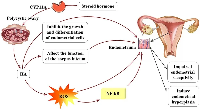
Fig 1: Pathophysiology of endometrial cancer in related to PCOS
2.3 Breast Cancer
There is still no conclusive evidence linking PMOS to breast cancer. PMOS and the overall risk of breast cancer have not consistently shown a strong independent association in current epidemiological evidence [7,13,14].
Nonetheless, a number of PMOS-related traits might be pertinent to the biology of breast cancer. Chronic inflammation, insulin resistance, and obesity are prevalent in PMOS and are independently linked to an increased risk of cancer [8,11,33]. Although the exact role of these anomalies is still unknown, altered sex-steroid signaling and hyperandrogenism have also been suggested as potential biological contributors [20].
Thus, it is important to describe the relationship with breast cancer with caution. Although PMOS and breast carcinogenesis may share endocrine and metabolic pathways, there is currently insufficient data to support PMOS's status as a recognized independent risk factor for breast cancer [7,13,14].
This portion of the original publication had global disability-adjusted life-year data for PMOS; however, since this data does not explicitly support a link between PMOS and breast cancer, it has been omitted from the amended edition [19].
3. PATHOPHYSIOLOGICAL MECHANISMS LINKING PMOS AND CANCER
3.1 Chronic Anovulation and Altered Steroid Signalling
One of the greatest physiologic connections between PMOS and endometrial disease is chronic anovulation. Progesterone exposure becomes insufficient when ovulation is irregular, yet estrogen-mediated endometrial growth may persist [11,15]. In vulnerable women, persistent proliferative signaling can cause endometrial hyperplasia and raise the risk of malignant transformation.
Abnormal gonadotropin signaling further affects the endocrine environment of PMOS. Ovarian theca cells may produce too much androgen as a result of increased pulsatile GnRH activity and abnormal LH secretion [21–24]. The endocrine abnormalities are then maintained by follicular stoppage and ovulation failure.
However, compared to its association with endometrial pathology, the link between persistent anovulation and breast or ovarian cancer is far less clear. Therefore, endometrial carcinogenesis should continue to be the focus of the most compelling mechanistic interpretation.
3.2 Hyperandrogenism and Androgen Signalling
A hallmark endocrine feature of PMOS is hyperandrogenism, which can be brought on by an increase in the synthesis of androgens by the ovaries and/or the adrenal glands [21–24]. Testosterone, androstenedione, dehydroepiandrosterone, and dehydroepiandrosterone sulfate are among the frequently elevated androgens [22–24].
Reduced SHBG concentrations raise circulating free testosterone, whereas greater LH stimulation can improve androgen production by theca cells at the ovarian level [22, 27]. Follicular stoppage, persistent anovulation, and interference with follicular maturation can all be caused by excessive androgen signaling [21–24].
Additionally, androgen receptor signaling may have an impact on transcriptional programs related to cellular proliferation and differentiation. An example of how epigenetic regulation may interact with androgen signaling is the involvement of BRD4 in the transcription of androgen receptors and the remodeling of ovarian tissue in PCOS.
Another significant molecular element is DENND1A. Overexpression of the DENND1A.V2 isoform in ovarian theca cells has been associated with increased expression of steroidogenic enzymes and a PCOS-like hyperandrogenic phenotype [46].
However, it is still unclear how hyperandrogenism directly contributes to the development of cancer in humans. Therefore, rather than being evidence of carcinogenic causation, the existence of androgen-related molecular changes should be interpreted as mechanistic plausibility.
3.3 Insulin Resistance and Metabolic Dysfunction
Insulin resistance is one of the major metabolic abnormalities associated with PMOS. Both lean and obese phenotypes may experience it, though metabolic dysfunction is typically made worse by obesity [25–30]. Defects in post-receptor insulin signaling, including anomalies involving IRS proteins and PI3K/Akt signalling, contribute to reduced glucose uptake [28,29].
By lowering SHBG concentrations and boosting ovarian androgen production, compensatory hyperinsulinemia may increase the availability of free androgen [27,47]. This creates an endocrine–metabolic feedback loop in which insulin resistance aggravates hyperandrogenism and hyperandrogenism contributes to reproductive dysfunction.
From a cancer perspective, hyperinsulinemia may influence cellular proliferation through insulin and IGF signalling. Activation of PI3K/Akt/mTOR and MAPK/ERK pathways can regulate cell growth, metabolism, survival and angiogenesis [15,47,48].
Thus, a key contact between PMOS and carcinogenesis may be the metabolic environment. However, when analyzing relationships, it is important to take into account the fact that obesity, type 2 diabetes, and other metabolic diseases are separate cancer-risk factors.
3.4 Assessment of Insulin Resistance
Using a single clinical measure to describe insulin resistance is challenging. Although the hyperinsulinemic–euglycemic clamp is still a standard technique for evaluating insulin sensitivity experimentally, its intricacy prevents regular clinical application [28, 31].
Although waist circumference and BMI are frequently used anthropometric measures, they are not sufficient to detect insulin resistance in all women with PMOS because metabolic dysfunction can happen without obesity [28, 30].
HOMA-IR, QUICKI, and fasting insulin have been studied as more useful surrogate markers [28, 30–32]. However, some women with impaired glucose tolerance may still have fasting insulin levels within the reference range; in these cases, an oral glucose tolerance test may offer more information [32].
These restrictions are crucial since metabolic phenotype may affect the amount of PMOS-associated cancer risk.
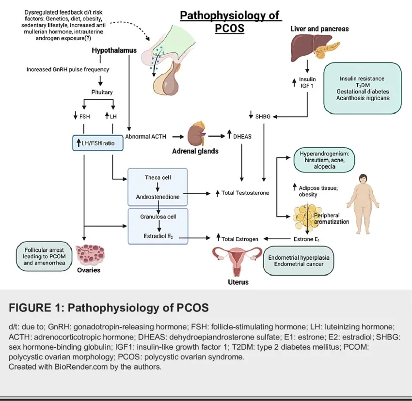
Fig 2: Pathophysiology of PCOS
4. CHRONIC INFLAMMATION AND OXIDATIVE STRESS
4.1 Chronic Low-Grade Inflammation
In PMOS, low-grade chronic inflammation is commonly seen. Women with the syndrome have been found to have elevated levels of inflammatory mediators such as CRP, IL-6, IL-18, and TNF-α [33, 36–39].
This inflammatory condition may be exacerbated by adipose tissue, especially when obesity and adipocyte dysfunction are present. While IL-18 has been linked to insulin resistance and metabolic syndrome, TNF-α can disrupt insulin receptor signaling and cause insulin resistance [37–39].
Pathways like NF-κB and STAT3, which control cytokine production, cell survival, proliferation, and tissue remodeling, can be triggered by persistent inflammatory signaling [16,52]. The biology of cancer is also affected by these pathways.
Nevertheless, inflammation is not unique to PMOS and should not be considered a biomarker for malignancy. Instead, it is a possible biological process by which tissue homeostasis may be affected by metabolic malfunction.
4.2 Oxidative Stress
An imbalance between the generation of reactive oxygen species and antioxidant defense is linked to PMOS [33–35]. Increased ROS production may be caused by hyperandrogenism, insulin resistance, obesity, and mitochondrial dysfunction [40–43].
In addition to harming lipids, proteins, and DNA, oxidative stress can disrupt cellular signaling [33–35]. While antioxidant systems like superoxide dismutase, glutathione, glutathione peroxidase, catalase, and total antioxidant capacity may be affected, markers like malondialdehyde, protein carbonyls, and advanced oxidation protein products have been studied as indicators of oxidative damage [34, 35, 43].
Oxidative stress and chronic inflammation are linked because ROS can activate the NF-κB, AP-1, and MAPK pathways and increase the production of inflammatory cytokines [40–42].
This interaction might offer a mechanistic connection between genomic instability and metabolic dysfunction. However, there is still little concrete proof that oxidative stress causes cancer development in PMOS on its own.
5. GENETIC AND MOLECULAR ALTERATIONS
The diverse phenotype of PMOS is significantly influenced by genetic predisposition. Numerous loci linked to ovarian function, steroidogenesis, gonadotropin signaling, and insulin action have been found by genome-wide association studies [44, 45].
DENND1A, THADA, LHCGR, FSHR, INSR, HMGA2, YAP1, and RAB5B are among the candidate genes identified in PMOS [44,45]. These genes are involved in processes related to ovarian physiology, metabolic control, and reproductive function.
DENND1A is very important for the production of androgens. Experimental research has demonstrated that the DENND1A.V2 variant produces a theca-cell phenotype similar to PCOS, which is marked by elevated steroidogenic activity [46].
An further molecular interaction is insulin receptor signaling. While maintaining pathways implicated in ovarian androgen synthesis, aberrant serine phosphorylation of IRS proteins can disrupt insulin signaling [47].
The mTOR pathway may have an impact on follicular development, autophagy, and granulosa-cell proliferation in reproductive biology [48]. A likely molecular link between PMOS and proliferative signaling is provided by the dysregulation of mTOR, a significant regulator of cell growth and metabolism.
These pathways shouldn't be readily seen as mechanisms unique to cancer, though. Rather of proving a direct malignant change, the majority of existing research indicates their function in PMOS pathogenesis.
6. MICRORNAS AND EPIGENETIC DYSREGULATION
In PMOS, microRNAs offer an extra level of post-transcriptional control. Numerous miRNAs have been studied in connection with insulin signaling, inflammation, granulosa-cell function, and apoptosis, including miR-93, miR-21, miR-222, miR-146a, and miR-320 [49].
Reproductive and metabolic processes may be impacted by altered miRNA expression. For instance, miR-93 has been linked to insulin sensitivity and GLUT4 expression control, whereas miR-21 and miR-146a have been linked to ovarian and inflammatory processes [49].
DNA methylation, chromatin remodeling, histone modification, and non-coding RNA-mediated control are all included in epigenetic regulation [1,50,51]. These processes offer a possible link between metabolic or environmental stressors and genetic vulnerability.
Epigenetic changes linked to prenatal androgen exposure and PCOS-like traits have been shown in experimental research [50,51]. Genes related to steroidogenesis, insulin signaling, inflammation, and reproductive function may be impacted by these changes.
It is important to assess the connection between these epigenetic anomalies and cancer with caution. Evidence that some PMOS-associated epigenetic indicators independently predict malignant transformation is still lacking, despite the fact that epigenetic dysregulation is a known characteristic of carcinogenesis.
Therefore, rather than being proven cancer predictions, epigenetic modifications should now be regarded as prospective biomarkers and candidate processes.
7. ONCOGENIC SIGNALLING PATHWAYS
PMOS-associated metabolic problems and cancer biology may converge on a number of intracellular signaling pathways.
7.1 PI3K/Akt/mTOR
In order to promote cellular growth, protein synthesis, and survival, insulin and IGF signaling can activate PI3K/Akt and mTOR [15,47,48]. Hyperinsulinemia and aberrant insulin signaling may make these pathways more active in PMOS.
In cancer, dysregulation of the PI3K/Akt/mTOR axis is also common. Consequently, its involvement offers a tenable mechanistic link between metabolic disorders and proliferation linked to cancer.
7.2 MAPK/ERK
Insulin, IGF, and other growth-factor signals can activate the MAPK/ERK pathway, which controls cell proliferation and differentiation [15,47]. Despite compromised metabolic signaling in PMOS, modified insulin signaling may maintain or improve mitogenic pathways.
7.3 NF-κB and STAT3
Increased production of inflammatory cytokines and other mediators can arise from the activation of NF-κB and STAT3 by chronic inflammatory signaling [16,52]. Persistent activation of these pathways may support tissue remodeling, inflammation, and cellular survival.
7.4 Wnt/β-catenin
Wnt/β-catenin signaling may interact with hormonal and metabolic pathways and has been linked to ovarian and endometrial biology [16]. Nevertheless, there is still little data explicitly connecting PMOS-associated cancer to Wnt/β-catenin dysregulation.
8. GUT MICROBIOTA AND THE ESTROGEN–MICROBIOME AXIS
The pathogenesis of PMOS has been linked to the gut microbiota. Metabolic dysfunction, inflammation, and hyperandrogenism have all been linked to changes in microbial diversity and composition [5,54].
Systemic inflammatory signaling and intestinal permeability may be impacted by gut dysbiosis. Metabolic inflammation may result from increased translocation of microbial compounds such lipopolysaccharide [54–56].
Another possibly important process is the estrobolome. It refers to microbial genes that are involved in the metabolism of estrogen, especially bacterial β-glucuronidase activity, which might affect enterohepatic recirculation and estrogen deconjugation [56].
Changes in microbiome-mediated estrogen metabolism may potentially affect hormone-responsive tissues since estrogen signaling is important for endometrial biology. However, this process is still a developing theory rather than a proven explanation for the cancer risk linked to PMOS.
Geographical location, drug exposure, nutrition, obesity, and other environmental factors also affect the composition of the microbiome. Therefore, future research must control these variables.
9. INTEGRATED MOLECULAR PERSPECTIVE
One biological route is unlikely to account for the possible connection between PMOS and cancer. Rather, cellular homeostasis may be altered by the interaction of endocrine, metabolic, inflammatory, oxidative, genetic, epigenetic, and microbial disorders [9,16,21].
Endometrial cancer exhibits the strongest mechanistic convergence, with metabolic dysfunction, extended estrogenic stimulation, and chronic anovulation offering a cogent scientific explanation [11,15,16].
Although there is conflicting epidemiological evidence, endocrine and inflammatory pathways are conceivable for ovarian cancer [7,14,17]. The link is considerably less definite in the case of breast cancer [7,13,14].
Because PMOS should not be viewed as a universal cancer-predisposition condition, this tissue-specific interpretation is crucial. Rather, the combination of the PMOS phenotype, metabolic status, reproductive history, tissue-specific hormone responsiveness, and other environmental or genetic variables may determine cancer risk.
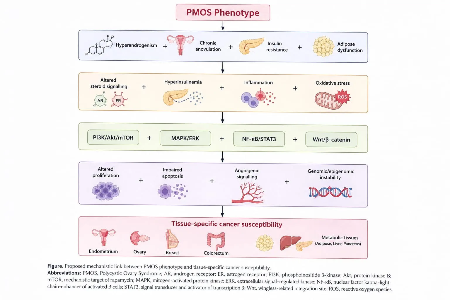
Fig 3: Mechanistic link between PMOS and tissue specific cancer susceptibility
10. CLINICAL IMPLICATIONS AND RISK REDUCTION
10.1 Clinical Risk Assessment
Instead of presuming that every woman with PMOS has the same cancer risk, clinical care should concentrate on identifying and changing known reproductive and metabolic risk factors [57,58].
Menstrual history, reproductive history, body weight, blood pressure, glucose regulation, and other metabolic risk variables should all be included in the clinical evaluation [57,58].
Other known risk factors for endometrial hyperplasia and cancer include long-term untreated amenorrhea, increased body weight, type 2 diabetes, and persistent endometrial thickness [58].
The proper diagnostic assessment should be performed for persistent abnormal uterine bleeding. To assess suspected endometrial pathology, transvaginal ultrasonography and endometrial biopsy may be utilized when clinically warranted [58,59].
Crucially, despite an elevated relative risk, standard endometrial cancer screening is not now advised for asymptomatic women with PMOS because the absolute chance of cancer is very low [58].
10.2 Metabolic Management
Modifying one's lifestyle is still crucial to managing PMOS. Reproductive results and metabolic health may be enhanced by appropriate physical exercise, diet, and weight control [26,57,58].
Because obesity can worsen insulin resistance, inflammation, and the risk of endometrial cancer, weight control is very important.
When taken appropriately, metformin can improve insulin sensitivity and associated endocrine problems in women with metabolic indications [47,57,58].
Metformin, however, shouldn't be marketed as a proven anticancer medication in PMOS. There is further research to be done on any possible anticancer effects.
10.3 Hormonal Management
Because prolonged untreated anovulation might increase endometrial exposure to unopposed estrogen, managing menstrual dysfunction is crucial [11,15,58].
When clinically required, appropriate hormonal therapy can be utilized to protect the endometrium and control menstrual cycles [58].
In women with PMOS, regular progestogen exposure is explicitly acknowledged as a prophylactic measure against endometrial hyperplasia [58].
10.4 Fertility Management
Individualized fertility treatment should be based on clinical phenotype and reproductive objectives.
While other medications, such as clomiphene and gonadotropins, may be taken into consideration based on clinical conditions, letrozole is frequently utilized for ovulation induction [57,58].
Instead of being referred to as cancer-prevention therapy, these treatments should be called fertility-management strategies.
11. PHARMACOLOGICAL AND LIFESTYLE STRATEGIES
|
Intervention |
Principal mechanism |
Potential role in PMOS |
|
Metformin |
Improves insulin sensitivity and reduces hepatic glucose production |
Metabolic dysfunction and selected reproductive indications [47,57,58] |
|
Combined hormonal contraceptives |
Regulate cycles and suppress ovarian androgen activity |
Menstrual irregularity and hyperandrogenic symptoms [58] |
|
Anti-androgens |
Reduce androgen-mediated effects |
Hirsutism/acne in appropriate patients [58] |
|
Letrozole |
Aromatase inhibition and ovulation induction |
First-line fertility treatment in appropriate patients [58] |
|
Clomiphene citrate |
Estrogen-receptor modulation and increased gonadotropin release |
Ovulation induction [58] |
|
GLP-1 receptor agonists |
Improve weight and metabolic parameters |
Selected patients with obesity/metabolic indications [58] |
|
Myo-/D-chiro-inositol |
Modulation of insulin signalling |
Selected metabolic/reproductive applications [58] |
|
Lifestyle intervention |
Improves metabolic fitness and supports weight management |
Fundamental component of PMOS management [26,57,58] |
12. EMERGING BIOMARKERS AND PRECISION RISK STRATIFICATION
Opportunities to find molecular phenotypes linked to various long-term outcomes in PMOS may arise from the growing application of genomics, epigenomics, transcriptomics, metabolomics, and microbiome studies [1,9,44,49–56].
Inflammatory mediators, metabolic signatures, sex-steroid profiles, microRNAs, DNA-methylation patterns, and microbiological signatures are examples of possible potential biomarkers.
However, clinical value is not established by the discovery of a molecular difference. Before a potential biomarker may be included in routine risk assessment, it must show reproducibility, sufficient predictive performance, and clinical benefit.
A precision-risk framework of the future might incorporate:
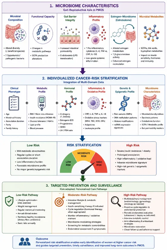
Fig 4: Framework for a gut-reproductive axis and its relationship with individualized cancer risk stratification and targeted prevention.
13. Gaps in Knowledge and Prospects for Further Research
A number of significant issues are still unanswered.
First, it's uncertain if certain PMOS phenotypes are associated with a disproportionately increased risk of cancer. Obesity, extended anovulation, severe hyperandrogenism, and significant insulin resistance may all constitute unique risk profiles that need to be assessed independently [7,15,16].
Second, because obesity, diabetes, infertility, parity, age, and reproductive history may function as confounders or mediators, it is still challenging to ascertain the independent contribution of PMOS [7,13,14].
Third, rather than prospective causal investigations, many postulated biological pathways, such as microRNAs, epigenetic modifications, and changes in the gut microbiota, are mostly supported by experimental or associative data [49–56].
Fourth, due to the biological heterogeneity of ovarian malignancies, ovarian cancer should be investigated based on histology and molecular subtype.
Fifth, future studies should ascertain whether combining clinical and genomic models might enhance prediction in ways that go beyond traditional risk variables.
Thus, it is necessary to have large longitudinal cohorts with consistent PMOS phenotyping, thorough metabolic characterisation, and long-term cancer follow-up. Subgroups with different biological trajectories and cancer susceptibilities may be found by integrating multi-omics data.
14. EMERGING RESEARCH FRAMEWORK
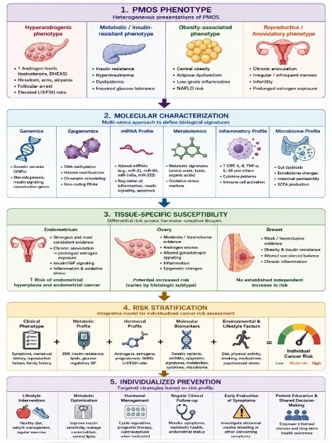
Fig 5: Future precision-risk framework for research work related to PCOS
CONCLUSION
The effects of PMOS, a diverse endocrine-metabolic condition, go beyond problems with reproduction. While relationships with ovarian and breast malignancies are still inconsistent, the strongest epidemiological data points to a link between PMOS and endometrial cancer [7,13,14].
Chronic anovulation, steroid-hormone signaling, hyperandrogenism, insulin resistance, inflammation, oxidative stress, genetic susceptibility, epigenetic regulation, and intracellular signaling are all interrelated abnormalities in the biological relationship between PMOS and carcinogenesis [9,11,15,16,21,33,44,47].
Possible molecular interfaces between metabolic and inflammatory disorders and altered cellular behavior are provided by PI3K/Akt/mTOR, MAPK/ERK, and NF-κB-related signaling [15,16,47,48]. Additional layers of regulation may be provided by gut microbial changes, microRNAs, and epigenetic modifications [1,49–56].
However, biological plausibility and causality are not the same thing. Obesity, diabetes, infertility, and other reproductive or metabolic traits may significantly increase the observed risk; the strength of the link varies depending on the type of cancer [7,13,14].
Clinically speaking, managing metabolic dysfunction, irregular menstruation, and other known risk factors should be the top goal. Although frequent screening of asymptomatic women is not advised, women with PMOS should be made aware of their elevated endometrial risk [58].
High-risk molecular subgroups may be identified by future prospective studies that combine clinical phenotyping with genomes, epigenomics, metabolomics, and microbiome study. In the end, these methods might help women with PMOS receive tailored prevention and more accurate cancer-risk assessment.
REFERENCES


Abarnadevika A., Dhivakar R.*, Salvia Placid S., Sushmitha S., Gokul R., Polyendocrine Metabolic Ovarian Syndrome And Cancer Risk: Integrated Endocrine, Metabolic And Molecular Mechanisms, Int. J. Sci. R. Tech., 2026, 3 (9), 593-608. https://doi.org/10.5281/zenodo.22975758
 10.5281/zenodo.22975758