

We use cookies to ensure our website works properly and to personalise your experience. Cookies policy
Department of Chemistry, Gopikabai Sitaram Gawande College, Umarkhed, Dist. Yavatmal – 445206 (M.S.), India
The action of phenylhydrazine hydrochloride on three 3-benzoyl-6-chlorochroman-4-ones [3-I(a–c)] in DMSO–piperidine has been studied. Rather than simple condensation at the C-4 carbonyl, the reaction proceeds with opening of the pyranone ring and recyclisation to give the 4-benzoyl-3-(5-chloro-2-hydroxyphenyl)-1-phenyl-?2-pyrazolines [5-I(a–c)] in 68–72% yield. The structures follow from a consistent body of spectroscopic evidence. The ¹H NMR spectrum of each product displays the classical ABX pattern of three mutually coupled doublets of doublets (H-4a, ? 4.20, J = 12, 3 Hz; H-4b, ? 5.35, J = 12, 11 Hz; H-5, ? 5.82, J = 11, 3 Hz) in place of the two AB doublets of the flavanone, together with a new, strongly deshielded phenolic O–H singlet at ? 11.20 — direct evidence that the pyranone ring has been opened and the phenol liberated. In the infrared, a broad hydrogen-bonded O–H band appears near 3400 cm?¹ and the flavanone C=O at 1680 cm?¹ is replaced by the C=N stretch of the ?2-pyrazoline ring at 1600 cm?¹. The electronic spectrum shows a large bathochromic shift of about 70 nm (310 ? 380 nm), attributable to the n??* transition of the C=N chromophore conjugated with the ortho-hydroxyphenyl ring, and the molecular ion increases by 90 Da, corresponding to the net addition of phenylhydrazine with loss of water. Knorr’s test is positive for all three products.
Chroman-4-ones (flavanones) and their derived nitrogen heterocycles are of continuing interest because of their broad biological profile (Harborne & Williams, 2000; Cushnie & Lamb, 2005). The 3-aroylchroman-4-ones used in the present work are readily available from o-hydroxyacetophenones through the Baker–Venkataraman transformation followed by Claisen–Schmidt condensation (Baker, 1933; Mahal & Venkataraman, 1934).
The reaction of a carbonyl compound with phenylhydrazine is normally expected to give a phenylhydrazone. When the carbonyl is embedded in a chroman-4-one bearing an acyl group at C-3, however, a more profound change is possible: nucleophilic attack of the hydrazine may be followed by opening of the pyranone ring at the C–O bond and recyclisation onto the C-3 acyl group, converting the six-membered oxygen heterocycle into a five-membered nitrogen heterocycle and liberating a free phenolic hydroxyl (Knorr, 1883; Levai, 2005; Elguero, 1996). Pyrazolines obtained in this way are of pharmacological interest in their own right (Kumar et al., 2009; Ansari et al., 2017; Sahu et al., 2008). In the present paper we take one reaction — the action of phenylhydrazine hydrochloride on the 3-benzoylchroman-4-ones [3-I(a–c)] — and present the spectroscopic evidence which shows that ring-opening followed by recyclisation, rather than simple hydrazone formation, is what actually occurs.
2. EXPERIMENTAL
2.1 Materials and instrumentation
All chemicals were of analytical reagent grade and solvents were dried and distilled before use (Furniss et al., 1989). Reactions were monitored by TLC on silica gel G with benzene as eluent. Melting points were determined in open capillaries and are uncorrected. UV–Visible spectra were recorded over 200–500 nm, IR spectra as KBr pellets over 4000–400 cm⻹, and ¹H NMR spectra at 300 MHz in CDCl₃ with TMS as internal standard (δ in ppm, J in Hz); mass spectra were obtained by electron impact. Assignments follow standard correlation data (Silverstein et al., 2005; Pavia et al., 2015). The starting chroman-4-ones [3-I(a–c)] were prepared as described previously.
2.2 General procedure
The chroman-4-one [3-I(a–c)] (0.01 mol) and phenylhydrazine hydrochloride (0.02 mol, 2.88 g) were placed in a round-bottomed flask with DMSO (20 mL) and piperidine (0.5 mL). The mixture was refluxed on a water bath for 1.5 h and then poured into ice-cold water. The separated solid was filtered, washed with water and crystallised from ethanol–acetic acid (9:1) to give the Δ2-pyrazoline as a yellowish-white crystalline solid. The products give a positive Knorr’s test (blue-green colour with bromine vapour) and dissolve in dilute sodium hydroxide, consistent with the liberated phenolic hydroxyl.
2.3 Physical and analytical data
[5-I(a)] 4-Benzoyl-5-(4-chlorophenyl)-3-(5-chloro-2-hydroxyphenyl)-1-phenyl-Δ2-pyrazoline. From [3-I(a)] (0.01 mol, 3.97 g). Yellowish-white solid; m.p. 180 °C; yield 72%. Knorr’s test positive; soluble in dilute NaOH. Anal. Calcd for C₂₈Hâ‚‚â‚€Clâ‚‚Nâ‚‚Oâ‚‚ (487): C, 69.00; H, 4.14. Found: C, 68.95; H, 4.17. UV (CHCl₃) λmax: 380 nm (n→π*, C=N), 258 nm. IR (KBr): 3400 (br, phenolic O–H), 3060, 1645, 1600 (C=N), 1490, 760 cm⻹. ¹H NMR (300 MHz, CDCl₃): δ 4.20 (dd, J = 12, 3 Hz, 1H, H-4a), 5.35 (dd, J = 12, 11 Hz, 1H, H-4b), 5.82 (dd, J = 11, 3 Hz, 1H, H-5), 7.30 (m, 18H, ArH), 11.20 (s, 1H, Ar–OH). MS (EI): m/z 486 (Mâº), 488 (M+2), 490 (M+4) — two Cl; 409 (M–77); 105 (base peak, PhCOâº); 77.
[5-I(b)] 4-Benzoyl-3-(5-chloro-2-hydroxyphenyl)-1-phenyl-5-(p-tolyl)-Δ2-pyrazoline. From [3-I(b)] (0.01 mol). Yellowish-white solid; m.p. 168 °C; yield 70%. Anal. Calcd for C₂₉H₂₃ClNâ‚‚Oâ‚‚ (466.5): C, 74.59; H, 4.97. Found: C, 74.54; H, 5.00. UV: 382, 258 nm. IR: 3398 (br), 1643, 1598 cm⻹. ¹H NMR: δ 2.37 (s, 3H, Ar–CH₃), 4.22 (dd), 5.38 (dd), 5.80 (dd), 7.30 (m, 18H), 11.18 (s, 1H, Ar–OH). MS: m/z 466 (Mâº), 468 (M+2, one Cl); 105 (base).
[5-I(c)] 4-Benzoyl-3-(5-chloro-2-hydroxyphenyl)-1,5-diphenyl-Δ2-pyrazoline. From [3-I(c)] (0.01 mol). Yellowish-white solid; m.p. 162 °C; yield 68%. Anal. Calcd for C₂₈Hâ‚‚â‚ClNâ‚‚Oâ‚‚ (452.5): C, 74.25; H, 4.67. Found: C, 74.20; H, 4.70. UV: 376, 255 nm. IR: 3395 (br), 1640, 1598 cm⻹. ¹H NMR: δ 4.18 (dd), 5.32 (dd), 5.78 (dd), 7.27 (m, 19H), 11.15 (s, 1H). MS: m/z 452 (Mâº), 454 (M+2); 105 (base).
3. RESULTS AND DISCUSSION
3.1 The reaction
Refluxing the flavanones [3-I(a–c)] with two equivalents of phenylhydrazine hydrochloride in DMSO–piperidine for 1.5 h gives the Δ2-pyrazolines [5-I(a–c)] in 68–72% yield. The reaction is best understood as a three-stage process. Nucleophilic addition of the terminal nitrogen of phenylhydrazine to the C-4 carbonyl of the chromanone gives a carbinolamine which dehydrates to the hydrazone. The nitrogen lone pair then attacks the C-3 benzoyl carbonyl intramolecularly, and this is accompanied by cleavage of the C-2–O bond of the pyranone ring; the ring oxygen departs as a phenolate, which is protonated on work-up to give the free phenolic hydroxyl. Recyclisation onto the benzoyl group closes the five-membered pyrazoline ring, so that the carbon which was C-2 of the flavanone becomes C-5 of the pyrazoline and the carbon which was C-3 becomes C-4. Overall the products correspond to the addition of phenylhydrazine to the flavanone with loss of one molecule of water. Two experimental observations support this course immediately: the products dissolve readily in dilute sodium hydroxide, and they give a positive Knorr’s test, neither of which is true of the starting flavanones.
3.2 ¹H NMR evidence for ring-opening and recyclisation
The ¹H NMR spectra provide the decisive evidence and are shown in Figure 1. The flavanone [3-I(a)] shows the two mutually coupled methine doublets of H-2 (δ 5.38) and H-3 (δ 5.94) with J = 11 Hz, and shows no signal downfield of the aromatic region. In the product these two doublets have gone; in their place is a three-spin ABX pattern of doublets of doublets at δ 4.20 (J = 12, 3 Hz), δ 5.35 (J = 12, 11 Hz) and δ 5.82 (J = 11, 3 Hz), assigned to H-4a, H-4b and H-5 of the pyrazoline ring, respectively. The appearance of a geminal coupling of 12 Hz between the two protons at δ 4.20 and δ 5.35 shows that these are now a diastereotopic methylene pair — a unit which simply does not exist in the flavanone — and this is possible only if a new ring has been formed in which C-4 carries two hydrogens.
Equally telling is the singlet at δ 11.20, integrating for one proton and exchangeable with Dâ‚‚O. A phenolic hydroxyl at this chemical shift can only arise if the pyranone ring has been opened, since in the flavanone that oxygen is tied up in the ring. Its strongly deshielded position is consistent with intramolecular hydrogen bonding to the adjacent pyrazoline nitrogen. Taken together, the ABX system and the liberated phenol establish the ring-opening–recyclisation pathway and exclude simple hydrazone formation, which would have retained the pyranone ring and its two AB doublets.
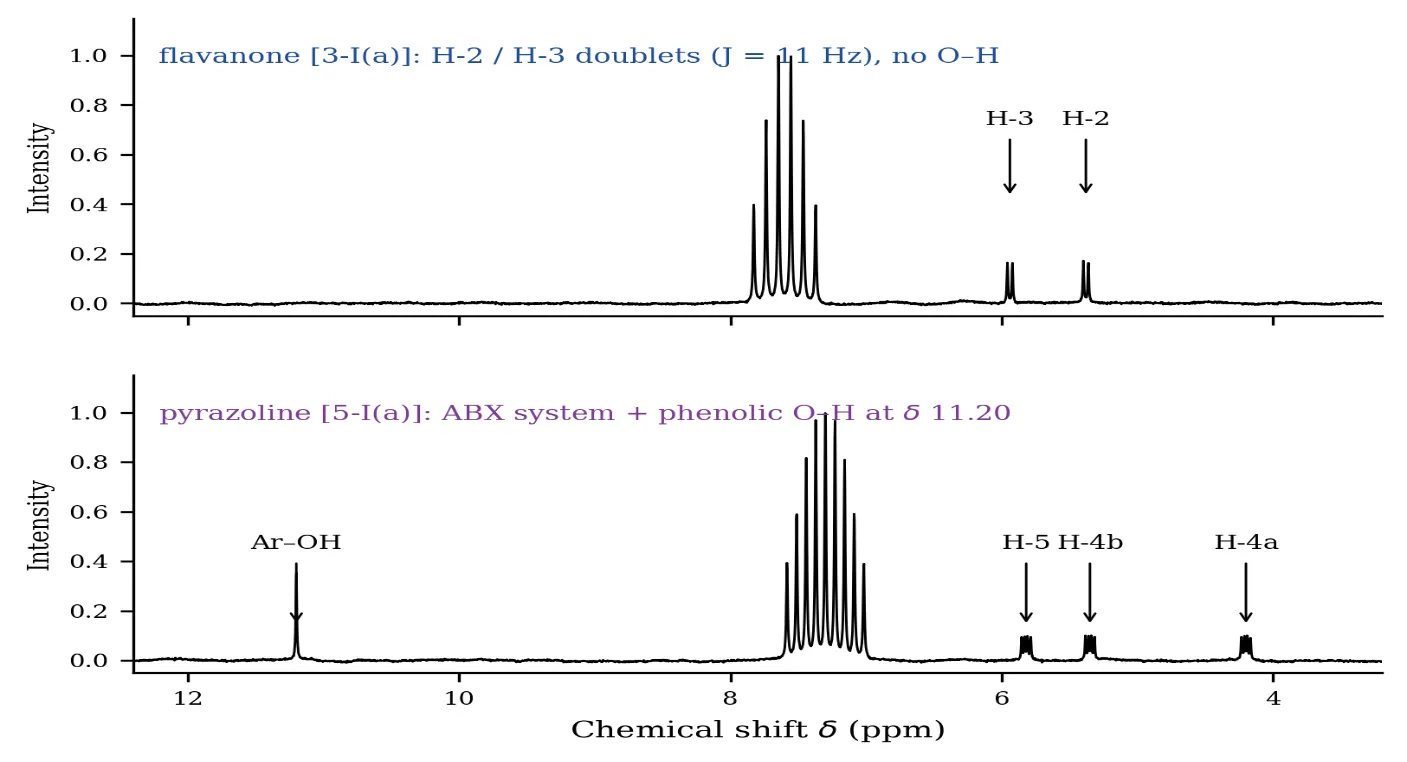
Figure 1. ¹H NMR spectra (300 MHz, CDCl₃) of the flavanone [3-I(a)] and the derived pyrazoline [5-I(a)]. The AB doublets of H-2 and H-3 are replaced by the ABX pattern of H-4a, H-4b and H-5, and a new phenolic O–H appears at δ 11.20.
3.3 Infrared spectra
The IR spectra (Figure 2) confirm the same two structural changes. The flavanone shows a conjugated ketone carbonyl at 1680 cm⻹ and no absorption in the hydroxyl region. In the pyrazoline this carbonyl band is absent and is replaced by a strong band at 1600 cm⻹, assigned to the C=N stretch of the Δ2-pyrazoline ring; at the same time a broad band appears near 3400 cm⻹, characteristic of an intramolecularly hydrogen-bonded phenolic O–H. The breadth of this band is itself evidence of hydrogen bonding and matches the downfield ¹H NMR shift of the same proton. The retained benzoyl carbonyl of the C-4 substituent accounts for the residual absorption near 1645 cm⻹.
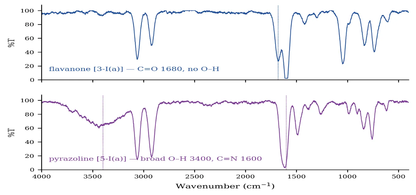
Figure 2. IR spectra (KBr) of [3-I(a)] and [5-I(a)]. The flavanone C=O at 1680 cm⻹ is replaced by the pyrazoline C=N at 1600 cm⻹, and a broad hydrogen-bonded O–H band appears at 3400 cm⻹.
3.4 Electronic spectra
The transformation is accompanied by a large bathochromic shift of the principal absorption band, from 310 nm in the flavanone to 380 nm in the pyrazoline — a displacement of about 70 nm (Figure 3). This is far greater than could be produced by any simple substituent effect and reflects the creation of a genuinely new chromophore: the C=N of the pyrazoline ring is conjugated both with the N-phenyl group and, through C-3, with the ortho-hydroxyphenyl ring liberated in the ring-opening step. The band is assigned to an n→π* transition of this extended azomethine system. Its position (376–382 nm across the three products) is diagnostic, and it is worth noting that it lies well to the red of the corresponding aromatic pyrazoles (≈ 340 nm), so that the electronic spectrum alone distinguishes the Δ2-pyrazoline from its aromatised counterpart.
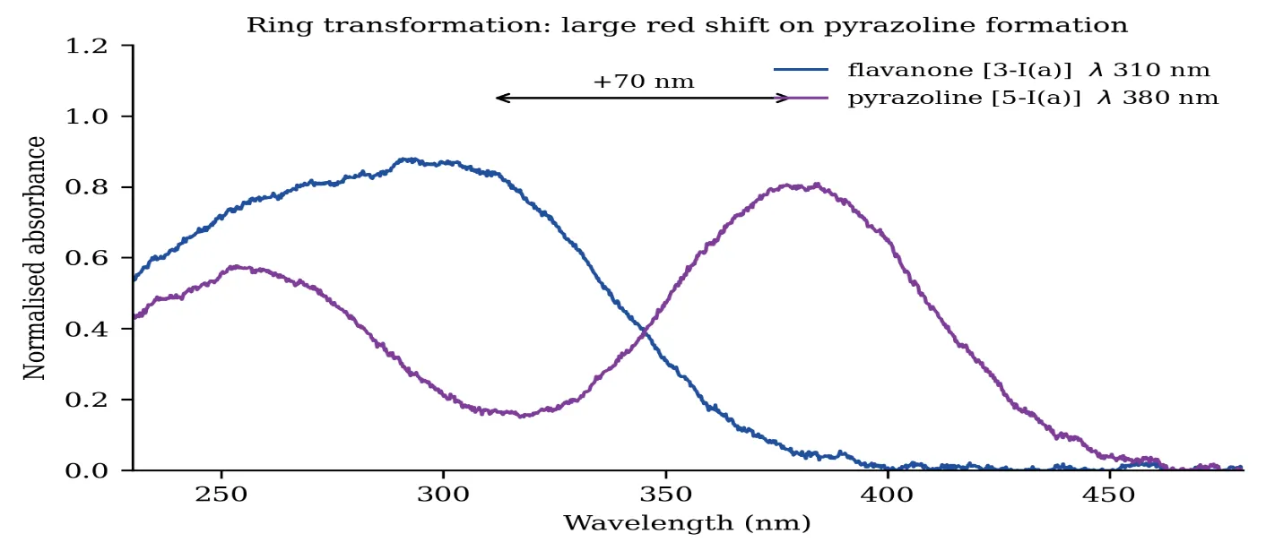
Figure 3. UV–Visible spectra of [3-I(a)] and [5-I(a)] in chloroform, showing the ca. 70 nm red shift on formation of the conjugated C=N chromophore.
3.5 Mass spectra
The mass spectra (Figure 4) confirm the stoichiometry of the transformation. The molecular ion rises from m/z 396 in the flavanone to m/z 486 in the pyrazoline, an increase of exactly 90 Da. This corresponds to the addition of phenylhydrazine (C₆H₈Nâ‚‚, 108) with the loss of water (18), which is precisely what the proposed ring-opening–recyclisation requires. The two-chlorine isotope cluster (M, M+2, M+4 in an approximate 9:6:1 ratio) is preserved intact, showing that neither chlorine is lost in the reaction. The benzoyl cation at m/z 105 remains the base peak, confirming that the C-4 benzoyl substituent survives the transformation, and the fragment at m/z 409 corresponds to loss of the N-phenyl group.
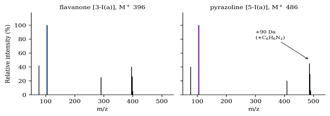
Figure 4. EI mass spectra of [3-I(a)] and [5-I(a)]. The molecular ion increases by 90 Da (net addition of phenylhydrazine minus water) while the two-chlorine isotope cluster is retained.
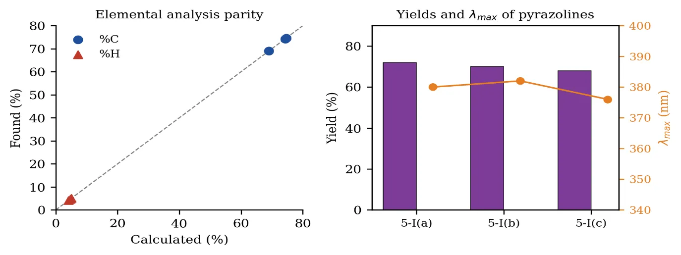
Figure 5. (Left) Parity plot of found versus calculated elemental composition for the three pyrazolines. (Right) Isolated yields and principal absorption maxima.
CONCLUSION
The action of phenylhydrazine hydrochloride on 3-benzoylchroman-4-ones does not stop at hydrazone formation but proceeds with opening of the pyranone ring and recyclisation onto the C-3 benzoyl group, giving 4-benzoyl-3-(2-hydroxyaryl)-1-phenyl-Δ2-pyrazolines in 68–72% yield. Four independent observations establish this course: the replacement of the flavanone AB doublets by a three-spin ABX pattern containing a geminal (12 Hz) methylene coupling; the appearance of a new, strongly deshielded phenolic O–H at δ 11.20, which can only be liberated by ring-opening; the replacement of the C=O band at 1680 cm⻹ by the pyrazoline C=N at 1600 cm⻹ together with a broad hydrogen-bonded O–H at 3400 cm⻹; and an increase of exactly 90 Da in the molecular ion, corresponding to the net addition of phenylhydrazine with loss of water. The large red shift of the principal UV band (about 70 nm) reflects the new, extensively conjugated azomethine chromophore. The products are soluble in dilute alkali and give a positive Knorr’s test, as the liberated phenol requires.
REFERENCES

Arati A. Narwade, Suresh P. Rathod*, Ring Transformation Of 3-Benzoylchroman-4-Ones Into 4-Benzoyl-3-(2-Hydroxyaryl)-Δ2-Pyrazolines: Evidence For Pyranone Ring-Opening Followed By Recyclisation, Int. J. Sci. R. Tech., 2026, 3 (8), 186-191. https://doi.org/10.5281/zenodo.21806081
 10.5281/zenodo.21806081