Rashtriya College Of Pharmacy Hatnoor, Kannad �Chh. Sambhajinagar 431103
Momordica dioica, a member of the Cucurbitaceae family, is a perennial dioecious climber with tuberous roots widely distributed in India and other parts of the Indian subcontinent. The fruits, leaves, and tuberous roots of M. dioica are used as a folk remedy for diabetes mellitus in India. Phytochemical analysis of M. dioica fruits has revealed the presence of alkaloids, steroids, triterpenoids, flavonoids, glycosides, saponins, triterpenes, and other bioactive compounds. The fruit is rich in nutrients, containing high levels of carbohydrates, protein, lipids, fibre, minerals (calcium, iron, potassium, zinc, sodium), and vitamins (carotene, thiamin, riboflavin, niacin). M. dioica exhibits significant antioxidant activities, which may contribute to its potential in preventing and managing oxidative stress-related diseases such as diabetes. Oxidative stress, resulting from an imbalance between free radical generation and antioxidant defences, plays a crucial role in the pathogenesis of diabetes and its complications. M. dioica's antioxidant properties are attributed to its enzymatic and non-enzymatic antioxidant components, including catalase, glutathione system, thioredoxin system, superoxide dismutase, vitamin C, vitamin E, carotenoids, flavonoids, and polyphenols. These antioxidants neutralise reactive oxygen and nitrogen species (RONS), which can cause damage to lipids, DNA, and proteins when present in excessive levels. The beneficial role of M. dioica in diabetes management may be due to its ability to scavenge free radicals, reduce lipid peroxidation, and enhance antioxidant enzyme activities. Further research is needed to elucidate the specific mechanisms by which M. dioica exerts its antioxidant and anti-diabetic effects and to explore its potential as a natural therapeutic agent for diabetes and other oxidative stress-related disorders
�
� � � �
� � � � � � 
� � � �
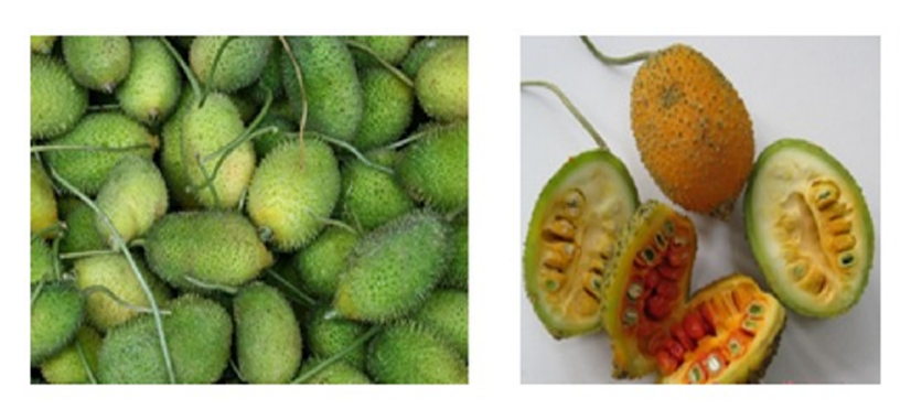
�Fig.1
Cucurbitaceae is� a plant family� commonly known as melons, gourds or cucurbits and includes crops like cucumbers,� squashes (including� pumpkins),� luffas, and� melons� (including� watermelons).� (Sivasudha� et� al 2012) the� family is predominantly distributed around the tropics, where those with edible fruits� were amongst the earliest cultivated plants� in both the old and new� world. Major� genera� under� this� family� are� Trichosanthes� (100 species), Cayaponia (60 species), Momordica (47 species), Gurania (40 species), Sicyos (40 species) and Cucumis (34 species).� This� is� one� of� the� most� genetically� diverse groups� of food� plants� in� the� plant� kingdom.� The� plants belonging� to� this� family� are� forst-sensitive,� drought tolerant, and� intolerant to� wet� and� poorly� drained� soils, production of cucurbits seems to have increased over the time� due to high� demand and consumer.� They� are well known� for� the� bitter� taste� due� to� the� presence� of phytochemical� (alkaloid)� and� have� a� wide� range� of medicinal values.1-3A Momordica� species� is� an� annual or perennial climber that contains about 80 species (Raj et al 1993). This is generally found throughout India, Pakistan, Bangladesh, and also extends from Himalayas to Ceylon. Reported up to an altitude of 1500m in Assam, Garo hills of Meghalaya (Ram et al 2002) and Western Ghats, one of the mega diversity hotspots, hold a rich treasure of diversity in Momordica L, it comprises M. charantiavar. muricata, M.� charantia var.� charantia, M.� dioica and M. sahyadrica (Joseph & Antony, 2008).� The revival of interest in natural drugs started in last decade mainly because of the wide spread belief that green medicine is healthier than� synthetic� products.� Nowadays,� there� is manifold� increase� in� medicinal� plant� based� industries. Due� to� the� increase� in� the� interest� of� medicinal� plants throughout� the� world� which� are� growing� at� a� rate of 7-15% annually, despite the� major advances in the� modern medicine,� the� development� of� new� drugs� from� natural products is still considered important. Medicinal plants as a possible therapeutic measure� has become� a subject of active� scientific� investigations.� The� Momordica� species have been� used in� indigenous medical� systems in� various countries� in� Asia� and� Africa.� Based� on� the� indigenous knowledge,� wild� plant� foods� play� a� vital� role� in� the complex� cultural� system� of� tribal� people� for� reducing various disorders. Research has shown that many edible wild� plants� are� rich� in specific� constituents,� referred� as phytochemical, which may have health promoting effects. So� far� no� review� has� been� covered� from� the� literature encompassing� valuable� attributes� of� M.� dioica� in� all dimensions. Its versatile utility as a nutritious� vegetable, folk medicine and functional food ingredient provoked us to compile a comprehensive� review of this multipurpose fruit� on� the� distribution,� nutritional� attributes� and phytochemical composition and its medicinal properties. Biogeography and Botanical description Based on the both historical literature and recent analysis Momordica� dioica� Roxb is a perennial� dioecious� climber with� tuberous roots.� Taxon Momordica� dioica� Roxb� has been� verified� by� US� Department� of� Agriculture� as member� of� family� Cucurbitaceae,� subfamily cucurbitoideae. Genus Momordica could perhaps refer to sculptured seed orknown as �Kakora� in Gwalior Chambal Division of M.P., is supposed to have originated in� Indo- Malayan� region (Rashid,� 1976� &� Singh,� 1990).� In India, it is distributed widely from Himalayas to Southern peninsula and amongst other parts of Indian subcontinent including Pakistan, Bangladesh, Myanmar and Srilanka, growing wild and mostly cultivated for its fruit which is used as a vegetable (Sastri 1962, Singh et al 2009). The fruit is oval with soft spines; aerial part of the plant dies at the beginning of winter. Plant perennates through sprouting of tubers at the onset of survival of the plant and creates a big production loss.� The species is cultivated by vegetative propagation method from underground tuberous roots.� uneven appearance of fruit which look as if they have been bitten.� The plant commonly known as �Kakora� in Gwalior Chambal Division of M.P., is supposed to have originated in Indo- Malayan region (Rashid, 1976 & Singh, 1990).� In India, it is distributed widely from� Himalayas� to� Southern� peninsula� and amongst� other� parts� of� Indian� subcontinent� including Pakistan,� Bangladesh,� Myanmar� and� Srilanka,� growing wild and mostly cultivated for� its fruit which� is used as a vegetable (Sastri 1962, Singh et al 2009). The fruit is oval with� soft� spines;� aerial� part� of� the� plant� dies� at� the beginning of winter. Plant perennates� through sprouting of tubers at the onset of survival of the plant and creates a� big� production� loss.� The� species� is� cultivated� by vegetative� propagation� method� from� underground tuberous
roots.[1]
Phytochemical and Nutrient Study
The fruit of Momordica dioica contains ashes: 9.1%, crude protein: 5.44%, crude lipid: 3.25%, crude fibre: 22.9%, and carbohydrate: 59.31%. Its fruit has high energy value (288.25?kcal/100?g) in dry weight. Its mineral ranges (mg/100?g dry weight,) are: potassium (4.63), sodium (1.62), calcium (7.37), iron (5.04), and zinc (3.83) [14]. In another investigation, its nutritional value of per 100?g edible fruit is reported to contain 84.1% moisture, 7.7?g carbohydrate, 3.1?g protein, 3.1?g fat, 3.0?g fibre and 1.1?g minerals and small quantities of essential vitamins like carotene, thiamin, riboflavin and niacin [15].Ali and Deokule evaluated some of its micronutrient and secondary metabolites as follows: calcium: 0.5?mg/g, sodium: 1.5?mg/g, potassium: 8.3?mg/g, iron: 0.14?mg/g, zinc: 1.34?mg/g, protein: 19.38%, fat: 4.7%, total phenolic compound: 3.7?mg/g, phytic acid: 2.8?mg/g, and ash value: 6.7% [16]. Moreover, its fruit is recommended as nutritionally rich source of protein and good source of lipid, crude fibre, carbohydrate, iron, calcium, phosphorous. Additionally, it is the highest amount of carotene (162?mg/100?g of edible portion) container amongst the cucurbitaceous vegetables [17�19]. The ash content is reported as 3-4% containing a trace of manganese [20].Tirmizi et al. screened it as a potential source of chromium and zinc [21]. Whereas, Momordica dioica (peeled) contained 0.27?mg/kg of chromium and 4.91?mg/kg of zinc, Momordica dioica (unpeeled) contained 0.26?mg/kg of chromium and 11.0?mg/kg of zinc. The protein content of leaves and dry weight of aerial plant parts remained higher in male as compared to female defruited and monoecious plants [22]. The fruit contains higher amount of ascorbic acid and iodine [23, 24]. The presence of secondary metabolites of fruit including alkaloids, steroids, triterpenoids, and saponins was determined [25]. Among them, four compounds were isolated from ethyl acetate extract and five compounds were isolated from methanol extract consisting of alkaloids and flavonoids with NH and C=O functional groups, respectively. The alkaloids present in seed and root were called momordicin and Momordica foetida, respectively [26]. Phytochemical investigations summarised in Table 1 also showed the presence of lectins, ?-sitosterol, saponin glycosides, triterpenes of ursolic acid, hederagenin, oleanolic acid, ?-spinasterol, stearic acid, gypsogenin, momodicaursenol, and three new compounds named 3?-o-benzoyl-11-oxo-ursolic acid, 3?-o-benzoyl-6-oxo-ursolic acid, and 3-o-?-D-glucuronopyranosyl gypsogenin [2]
Table 1. Nutrient and phytochemical study of Momordica dioica as described in this paper
|
Classification |
Compound |
Extract or preparation |
Reference |
|
Crude protein |
� |
Quantitative analysis showed 5.44% |
[14] |
|
Protein |
� |
Quantitative analysis showed 3.1/100?g |
[15] |
|
� |
Quantitative analysis showed 19.38% |
[16] |
|
|
Crude lipid |
� |
Quantitative analysis showed 3.25% |
[14] |
|
Fat |
� |
Quantitative analysis showed 3.1/100?g |
[15] |
|
� |
Quantitative analysis showed 4.7% |
[16] |
|
|
Crude fibre |
� |
Quantitative analysis showed 22.9% |
[14] |
|
Carbohydrate |
� |
Quantitative analysis showed 59.31% |
[14] |
|
� |
Quantitative analysis showed 7.7/100?g |
[15] |
|
|
Niacin |
� |
Not specified |
[15] |
|
Thiamin |
� |
Not specified |
[15] |
|
Carotene |
� |
Not specified |
[15] |
|
� |
Quantitative analysis showed 162?mg/100?g of edible portion |
||
|
Ascorbic acid |
� |
Not specified |
[24] |
|
Potassium |
� |
Quantitative analysis showed 4.63?mg/100?g dry weight |
[14] |
|
� |
Quantitative analysis showed 8.3?mg/g |
[16] |
|
|
Sodium |
� |
Quantitative analysis showed 1.62?mg/100?g dry weight |
[14] |
|
� |
Quantitative analysis showed 1.5?mg/g |
[16] |
|
|
Calcium |
� |
Quantitative analysis showed 7.37?mg/100?g dry weight |
[14] |
|
� |
Quantitative analysis showed 0.5?mg/g |
[16] |
|
|
Iron |
� |
Quantitative analysis showed 5.04?mg/100?g dry weight |
[14] |
|
� |
Quantitative analysis showed 0.14?mg/g |
[16] |
|
|
Zinc |
� |
Quantitative analysis showed 3.83?mg/100?g dry weight |
[14] |
|
� |
Quantitative analysis showed 1.34?mg/g |
[16] |
|
|
� |
Not specified |
[21] |
|
|
� |
Quantitative analysis showed 4.91?mg/kg (peeled), 11.0?mg/g (unpeeled) |
[22] |
|
|
Manganese |
� |
Not specified |
[20] |
|
Iodine |
� |
Not specified |
[23] |
|
Chromium |
� |
Quantitative analysis showed 0.27?mg/kg (peeled), 0.26?mg/kg (unpeeled) |
[22] |
|
� |
Not specified |
[21] |
|
|
Phytic acid |
� |
Quantitative analysis showed 2.8?mg/g |
[16] |
|
Total phenolic compound |
� |
Quantitative analysis showed 3.7?mg/g |
[16] |
|
Alkaloids |
� |
Identified in ethyl acetate, methanol extract |
[25] |
|
Flavonoid |
� |
Identified in methanol, hexane extract |
[25] |
|
Steroids |
� |
Identified in ethyl acetate, methanol, aqueous extract |
[25] |
|
Saponins |
� |
Identified in methanol, aqueous extract |
[25] |
|
Triterpenoids |
� |
Identified in ethyl acetate, methanol, aqueous extract |
[25] |
|
Alkaloid |
Momordicin |
Identified in seed oil |
[26] |
|
Lectin |
Anti-H-Lectin |
Not specified |
[30] |
|
Alkaloid |
Momordicafoetida |
Not specified |
[26] |
|
Stearic acid |
� |
Identified in methanol extract |
[31] |
|
Steroid |
?-spinasterol octadecanoate |
Identified in methanol extract |
[31] |
|
?-spinasterol-3-O-?-D-glucopyranoside |
Identified in methanol extract |
[31] |
Antioxidants are groups of compounds that neutralise free radicals and reactive oxygen species (ROS) in the cell (Abuajah et al. 2015). Antioxidant activity in food and beverages has become one of the most interesting features in the science community. These antioxidants provide protection against damage caused by free radicals played important roles in the development of many chronic disease including cardiovascular diseases, aging, heart disease, anaemia, cancer, inflammation[3]
The role of antioxidants in preventing oxidative stress-related diseases
Various sets of colonic microbiota cooperate to metabolise different phytochemicals. For instance, ellagic acids are metabolised to urolithins by bacteria like Clostridium spp., Ruminococcaceae, Eubacterium spp., Gordonibacter spp., and Ellagibacter isourolithinifaciens [4]. Daidzeins, isoflavones from soybeans, are instead converted into equals by another community of bacteria such as Streptococcus intermedia�s, Bacteroids ovatus, and Ruminococcus productus [5]. Lignans undergo modifications by diverse bacteria, including Clostridium scindens, Eggerthella lenta, Clostridiales, and Lactonifactor longoviformis [6]. Consequently, phytochemical metabolites can vary among individuals consuming the same food due to the individual differences in colonic microbiota composition. Many diseases heavily impacted by dietary factors entail oxidative damage as an initial occurrence or an early stage in the progression of the disease. Consequently, a significant emphasis in dietary disease prevention has been placed on antioxidant intervention. Over the past decade, a wealth of research, including numerous human intervention studies, has consistently highlighted the pivotal role of antioxidants, particularly phytochemicals, in mitigating the risk of chronic diseases [7].Traditionally, the beneficial role of antioxidants has been associated with curtailing the undesirable and uncontrolled production of reactive oxygen species, leading to a state known as oxidative stress. However, contemporary scientific understanding increasingly recognises that the mechanism of action of antioxidants in vivo may be far more intricate than previously thought.Beyond their role in mitigating oxidative stress, antioxidants and phytochemicals demonstrate multifaceted mechanisms that contribute to disease prevention. These mechanisms include the modulation of inflammatory pathways, the enhancement of cellular repair and regeneration, and interaction with signalling cascades involved in cell growth and apoptosis. Moreover, antioxidants exhibit the potential to influence epigenetic processes, altering gene expression patterns associated with disease susceptibility. Recent studies have illuminated the intricate interplay between antioxidants and the gut microbiota, revealing a symbiotic relationship. Antioxidants, particularly those derived from plant-based sources, can impact the amount and diversity of the gut microbiota, which, in turn, contribute to overall health and disease prevention. Additionally, antioxidants demonstrate neuroprotective effects and may play a crucial role in preserving cognitive function and preventing neurodegenerative diseases.
As research progresses, the understanding of the comprehensive impact of antioxidants and phytochemical on human health continues to evolve. Insights into their nuanced mechanisms of action open avenues for targeted interventions, personalised nutrition strategies, and the development of novel therapeutic approaches for a wide array of diseases influenced by dietary factors.
Phytochemical Constituents of� Momordica dioica
�Following the methods of qualitative analysis reveals the presence of phytochemical in root extracts of M. dioica prepared in petroleum ether and acetone, used for variety of ethnic medicinal uses. The alkaloids, terpenoids, phenols, carbohydrates and steroids are present in both petroleum ether and acetone extract of M. dioica. The flavonoids and saponins are present only in the acetone extract of M. dioica root and absent in petroleum ether extract, while the cardiac glycosides and tannins are present only in the petroleum ether extract of M. dioica root and are absent in acetone.[7]
�Following the methods of quantitative analysis of root extract reveals that the total alkaloid content estimated were 3.43 mg/g and 1.89 mg/g in petroleum ether and acetone respectively. 2.67 mg/g of flavonoids were present in acetonic root extract. The total phenolic content present in petroleum ether and acetone root extract were 2.83 and 1.69 mg/g respectively. The total tannins estimated in petroleum ether root extract was 3.13 mg/g, while the total saponins estimated in[8]
Table-2
|
Sr.no. |
Phytochemical constituents [mg/g] |
Petroleum ether (mg/g |
Acetone (mg/g) |
|
1 |
Alkaloids |
3.43 |
1.89 |
|
2 |
Flavonoids |
-- |
2.67 |
|
3 |
Phenols |
2.83 |
1.69 |
|
4 |
Phenols |
2.83 |
1.69 |
|
5 |
Saponin |
-- |
2.12 |
Analytical Methods Used in Phytochemical Identification
Steps Involved In Plant Collection
2.1.Collection of Plants Plants under consideration may be collected either from wild forests or from herbariums. When plants are collected from wild, there is a risk that they have been incorrectly identified. The major advantage of wildlife plants is that they will not contain any pesticides. After the plants are collected from wild or from herbarium they have to be processed for cleaning in order to prevent the deterioration of phytochemical present in plants.
2.2.Cleaning of Plants After plants collection they have to be cleaned properly. The cleaning process may involve the following steps. Cleaning, washing, peeling or stripping leaves from stems. Cleaning has to be done by hands in order to get better results.
2.3.Drying The main purpose of drying is to remove the water content from plants so that the plants can be stored. Plants have to be dried immediately as soon as the plants collection or this will lead to spoilage of plant materials. The drying consists of two methods. Drying can be done either by natural process or by artificial process.
2.3.1. Natural Process Natural process includes sun- drying. Sometimes plants are placed on drying frames or on stands, to be air-dried in barns or sheds. But this may take few weeks for complete drying. The time depends on temperature and humidity.
2.3.2. Artificial Drying Artificial drying can be done with the help of artificial driers. This process will reduce the drying time to several hours or minutes. The common method that is followed in drying medicinal plants is warm-air drying. In this process plants are placed in the plates of drier on which warm air is blown. This method is mainly applicable to fragile flower and leaves and this requires large number of workers since loading and unloading of plants has to be done manually. 2.4.Powdering After complete drying of plants they have to be powdered well for further analysis [9]
METHODS OF EXTRACTION
3.1. Plant Tissue Homogenisation Plant tissue homogenisation in solvent has been widely used by researchers. Dried or wet, fresh plant parts are grinder in a blender to fine particles, put in a certain quantity of solvent and shaken vigorously for 5 - 10 min or left for 24 h after which the extract is filtered. The filtrate then may be dried under reduced pressure and re-dissolved in the solvent to determine the concentration. Some researchers however centrifuged the filtrate for clarification of the extract.
3.2� Serial Exhaustive Extraction It is another common method of extraction which involves successive extraction with solvents of increasing polarity from a non-polar (hexane) to a more polar solvent (methanol) to ensure that a wide polarity range of compounds could be extracted. Some researchers employ Soxhlet extraction of dried plant material using organic solvent. This method cannot be used for thermolabile compounds as prolonged heating may lead to degradation of compounds.
3.3. Soxhlet Extraction Soxhlet extraction is only required where the desired compound has a limited solubility in a solvent, and the impurity is insoluble in that solvent. If the desired compound has a high solubility in a solvent then a simple filtration can be used to separate the compound from the insoluble substance. The advantage of this system is that instead of many portions of warm solvent being passed through the sample, just one batch of solvent is recycled. This method cannot be used for thermolabile compounds as prolonged heating may lead to degradation of compounds
3.4. Maceration In maceration (for fluid extract), whole or coarsely powdered plant- drug is kept in contact with the solvent in a stoppered container for a defined period with frequent agitation until soluble matter is dissolved. This method is best suitable for use in case of the thermolabile drugs
�3.5. Decoction This method is used for the extraction of the water soluble and heat stable constituents from crude drug by boiling it in water for 15 minutes, cooling, straining and passing sufficient cold water through the drug to produce the required volume.
3.6. Infusion It is a dilute solution of the readily soluble components of the crude drugs. Fresh infusions are prepared by macerating the solids for a short period of time with either cold or boiling water.
�3.7. Digestion This is a kind of maceration in which gentle heat is applied during the maceration extraction process. It is used when moderately elevated temperature is not objectionable and the solvent efficiency of the menstrual is increased thereby.
�3.8. Percolation This is the procedure used most frequently to extract active ingredients in the preparation of tinctures and fluid extracts. A percolator (a narrow, cone-shaped vessel open at both ends) is generally used. The solid ingredients are moistened with an appropriate amount of the specified menstrual and allowed to stand for approximately 4 h in a well closed container, after which the mass is packed and the top of the percolator is closed. Additional men-strum is added to form a shallow layer above the mass, and the mixture is allowed to macerate in the closed percolator for 24 h. The outlet of the percolator then is opened and the liquid contained therein is allowed to drip slowly. Additional men-strum is added as required, until the percolate measures about three-quarters of the required volume of the finished product. The Marc is then pressed and the expressed liquid is added to the percolate. Sufficient men-strum is added to produce the required volume, and the mixed liquid is clarified by filtration or by standing followed by decanting.
3.9. Sonication
The procedure involves the use of ultrasound with frequencies ranging from 20 kHz to 2000 kHz; this increases the permeability of cell walls and produces cavitation. Although the process is useful in some cases, like extraction of rauwolfia a root, its large-scale application is limited due to the higher costs. One disadvantage of the procedure is the occasional but known deleterious effect of ultrasound energy (more than 20 kHz) on the active constituents of medicinal plants through formation of free radicals and consequently undesirable changes in the drug molecules[12]
4.Qualitative And Quantitative Analysis Of Phytochemical
4.1.Preliminary Qualitative Analysis
1. Test for Alkaloids a. Mayer� s test
To a few ml of plant sample extract, two drops of Mayer?s reagent are added along the sides of test tube. Appearance of white creamy precipitate indicates the presence of alkaloids.[6] b. Wagner�s test A few drops of Wagner?s reagent are added to few ml of plant extract along the sides of test tube. A reddish- Brown precipitate confirms the test as positive.
2. Test for Amino acids
�The extract (100 mg) is dissolved in 10 ml of distilled water and filtered through Whatmann No. 1 filter paper and the filtrate is subjected to test for Amino acids. a. Ninhydrin test Two drops of ninhydrin solution (10 mg of ninhydrin in 200 ml of acetone) are added to 2 ml of aqueous filtrate. Appearance of purple colour indicates the presence of amino acids.
3. Test for Carbohydrates
�Molisch� s test To 2 ml of plant sample extract, two drops of alcoholic solution of ?- naphthol are added. The mixture is shaken well and few drops of concentrated sulphuric acid is added slowly along the sides of test tube. A violet ring indicates the presence of carbohydrates. b. Benedict� s test To 0.5 ml of filtrate, 0.5 ml of Benedict?s reagent is added. The mixture is heated on a boiling water bath for 2 minutes. A characteristic coloured precipitate indicates the p
Bharathi LK, Munshi AD, Chandrashekaran S, Behera TK, Das AB, John KJ. Cytotaxonomical analysis of Momordica L. (Cucurbitaceae) species of Indian occurrence. Journal of Genetics. 2011;90

Bhende Kailas, Waghmare S.U., Chavan Umesh, Phytochemical Profile And Antioxidant Activities Of Momordica Dioica, Int. J. Sci. R. Tech., 2025, 2 (1), 123-140. https://doi.org/10.5281/zenodo.14616593
 10.5281/zenodo.14616593
