

We use cookies to ensure our website works properly and to personalise your experience. Cookies policy
Delight College of Pharmacy, Koregaon Bhima, Pune, Maharashtra, India - 412216.
Parkinson’s disease is a progressive neurological disorder associated with degeneration of the dopaminergic neurons, particularly in the substantia nigra, and reduced dopamine signaling. Levodopa remains as a central symptomatic treatment, but convectional delivery can be complicated by variable gastro intestinal therapeutic molecules across the Blood Brain Barrier. Intranasal administration is being investigated as a non- invasive route that may support transport from the nasal cavity toward the central nervous system through olfactory and trigeminal pathways. Nanoparticles may protect encapsulated molecules, alter release, improve contact with nasal mucosa, and influence tissue distribution. It describes key formulation development stages and evaluation domains: particle size and distribution, surface change, morphology, drug content and entrapment, brain targeting and local safety. Nanoparticle based drug delivery has consequently been investigated as a means of modifying the pharmacokinetic and pharmacodynamic behavior of L-DOPA and improving its delivery to the central nervous system.
Review of Parkinson’s Disease --
Parkinson’s disease (PD) is a chronic and progressive neurodegenerative disorder characterized primarily by the degeneration of dopaminergic neurons in the substantia nigra pars compacta of the midbrain. This neuronal degeneration results in a reduction in dopamine levels within the striatum, leading to impaired motor control. Parkinson’s disease was first described by James Parkinson in 1817 and is now recognized as one of the most common neurodegenerative disorders, particularly among older adults. Its development involves a complex interaction of genetic susceptibility, environmental factors, and cellular mechanisms associated with neuronal damage.1,2
The major pathological characteristics of Parkinson’s disease include the progressive loss of dopaminergic neurons and the accumulation of misfolded alpha-synuclein protein in the form of intracellular inclusions known as Lewy bodies. Several mechanisms, including mitochondrial dysfunction, oxidative stress, impaired protein degradation, and neuroinflammation, contribute to the progression of neuronal damage. Although the exact cause of Parkinson’s disease remains incompletely understood, these pathological changes play important roles in its development and progression. [1,3]
Conventional pharmacological treatment primarily aims to alleviate symptoms by restoring or enhancing dopaminergic activity. Levodopa remains a major therapeutic agent; however, conventional drug delivery may be associated with fluctuations in therapeutic response, systemic adverse effects, and difficulties in maintaining consistent symptom control as the disease progresses. Furthermore, the blood–brain barrier (BBB) presents a significant challenge for delivering certain therapeutic agents to the central nervous system. These limitations have encouraged research into alternative drug delivery strategies that may improve drug transport to the brain. [2,3]
Pathophysiology of Parkinson’s Disease –
The principal pathological feature of Parkinson’s disease is the progressive degeneration of dopaminergic neurons in the substantia nigra pars compacta. Under normal physiological conditions, dopamine regulates communication between the substantia nigra and striatum, helping maintain smooth, coordinated movements. As dopaminergic neurons degenerate, dopamine availability decreases, disrupting the direct and indirect pathways of the basal ganglia. These changes interfere with the initiation and execution of voluntary movements, contributing to bradykinesia, rigidity, and other motor manifestations of the disease. [1,22]
Another important pathological characteristic of PD is the abnormal accumulation of α-synuclein, a presynaptic neuronal protein. Misfolded α-synuclein can aggregate and contribute to the formation of Lewy bodies and Lewy neurites, which are pathological features associated with Parkinson’s disease. Abnormal protein aggregation may interfere with synaptic function, cellular transport, protein degradation, and neuronal survival. However, the relationship between α-synuclein aggregation and neuronal degeneration is complex, and the precise mechanisms responsible for disease progression are still being investigated. [1,22]
The current management of Parkinson’s disease primarily aims to control symptoms and maintain patients’ functional ability and quality of life. Levodopa, usually administered with a peripheral dopa-decarboxylase inhibitor such as carbidopa or benserazide, is a major treatment for motor symptoms. Other pharmacological options include dopamine agonists such as pramipexole, ropinirole, and rotigotine, as well as monoamine oxidase-B (MAO-B) inhibitors such as rasagiline and selegiline. Depending on the patient’s condition, treatment may also include rehabilitation, exercise, speech and occupational therapy, and device-aided or surgical interventions.[23]
CURRENT TREATMENT APPROACHES
Parkinson’s disease (PD) is primarily managed using pharmacological therapies that aim to improve dopaminergic neurotransmission and alleviate motor symptoms. Levodopa, dopamine agonists, and monoamine oxidase-B (MAO-B) inhibitors are important therapeutic options. The selection of treatment depends on symptom severity, patient characteristics, disease progression, and the occurrence of adverse effects. [4,5]
1.Dopamine Precursor:
Levodopa (L-DOPA) is considered the most effective pharmacological treatment for the motor symptoms of Parkinson’s disease. It acts as a precursor of dopamine and can cross the blood–brain barrier (BBB) through amino acid transport systems. Once inside the brain, levodopa is converted into dopamine by the enzyme aromatic L-amino acid decarboxylase, helping compensate for dopamine deficiency in the striatum. Levodopa is generally administered with a peripheral dopa-decarboxylase inhibitor, such as carbidopa, to reduce its peripheral metabolism and improve the availability of levodopa for transport into the brain. [5,6]
Despite its effectiveness, long-term levodopa treatment may be associated with motor complications, including wearing-off phenomena, fluctuations in motor response, and levodopa-induced dyskinesia. Nausea, orthostatic hypotension, and other adverse effects may also occur. Furthermore, delayed gastric emptying, dietary amino acid competition, and variability in gastrointestinal absorption can contribute to unpredictable drug responses in some patients. These limitations have encouraged the investigation of alternative drug delivery approaches that may improve the consistency of drug delivery to the brain. [5]
2. Dopamine Agonists:
Dopamine agonists, including pramipexole, ropinirole, and rotigotine, act by directly stimulating dopamine receptors in the brain. Unlike levodopa, these agents do not require conversion into dopamine to produce their pharmacological effects. They may be used as monotherapy in selected patients or in combination with levodopa to improve symptom control. Their use may help manage motor symptoms and reduce certain levodopa-related motor fluctuations. However, dopamine agonists are associated with adverse effects such as nausea, dizziness, orthostatic hypotension, daytime sleepiness, hallucinations, and impulse-control disorders. The risk of these effects varies according to the drug, dose, age, and clinical condition of the patient. Consequently, treatment requires individualized selection and monitoring. These limitations support continued research into drug delivery systems that may improve therapeutic targeting while minimizing unwanted systemic exposure. [4,5]
3. Monoamine Oxidase-B (MAO-B) Inhibitors:
Monoamine oxidase-B inhibitors, including selegiline, rasagiline, and safinamide, are used in the management of Parkinson’s disease. These agents inhibit the MAO-B enzyme, which is involved in dopamine metabolism in the brain. By reducing dopamine breakdown, MAO-B inhibitors help prolong dopaminergic activity and improve motor symptoms. They may be prescribed alone in selected cases or as adjunctive therapy with levodopa, particularly in patients experiencing motor fluctuations. [4,5]
CHALLENGES IN BRAIN DRUG DELIVERY:
Effective drug delivery to the brain remains a major challenge in the treatment of Parkinson’s disease. Although several pharmacological agents are available, their therapeutic performance can be limited by the protective properties of the blood–brain barrier, inadequate brain exposure, systemic adverse effects, and limitations associated with conventional dosage forms. These challenges have encouraged the development of alternative delivery systems capable of improving drug transport to the central nervous system. [6,7]
1.Blood–Brain Barrier (BBB) –
The blood–brain barrier is a highly selective physiological interface between the systemic circulation and the central nervous system. It is primarily formed by brain microvascular endothelial cells connected by tight junctions, together with supporting pericytes, astrocytes, and associated structures of the neurovascular unit. The BBB maintains brain homeostasis by regulating the movement of substances from the blood into brain tissue while restricting the entry of potentially harmful compounds.Although the BBB protects the brain, it also restricts the delivery of many therapeutic agents. Large molecules, hydrophilic compounds, and drugs that are substrates of active efflux transporters may show limited penetration into the central nervous system. Consequently, achieving therapeutically effective concentrations at the intended site of action can be difficult. The development of advanced delivery systems, including nanoparticles and intranasal formulations, is being investigated as a strategy to overcome some of these barriers. [6,7]
2. Limited Brain Penetration of Drugs --
Many therapeutic agents exhibit limited brain penetration because of their physicochemical properties, molecular size, polarity, and interactions with BBB transport mechanisms. Consequently, the concentration of a drug reaching the brain may be insufficient to produce the desired therapeutic response, even when adequate drug concentrations are achieved in the systemic circulation. In addition, peripheral metabolism and drug efflux mechanisms may further reduce the amount of drug available to brain tissue. In Parkinson’s disease, inadequate and variable brain exposure can complicate symptom management. Nanoparticle-based drug delivery systems are being investigated for their potential to protect drugs from degradation, modify drug release, and improve delivery to specific tissues. [6,7]
3. Systemic Adverse Effects --
Systemic adverse effects are another important limitation of conventional drug delivery. Following oral or other systemic administration, therapeutic agents may distribute to tissues outside the brain and interact with peripheral receptors or metabolic pathways. Such exposure may contribute to adverse effects and can limit the doses that patients tolerate. For dopaminergic treatments, adverse effects may include nausea, orthostatic hypotension, dizziness, sleep disturbances, and dyskinesia, depending on the medication and treatment regimen. [4,5]
Advanced drug delivery systems aim to improve the distribution of therapeutic agents and potentially reduce unnecessary systemic exposure. Intranasal delivery is particularly interesting because it may facilitate drug transport toward the brain without relying exclusively on systemic circulation. [6,7]
INTRA NASAL DRUG DELIVERY FOR PARKINSON’S DISEASE:
Intranasal drug delivery has emerged as a promising approach for delivering therapeutic agents to the central nervous system (CNS). Conventional drug administration is often associated with limited brain penetration because of the blood–brain barrier (BBB), systemic metabolism, and other physiological barriers. Intranasal administration offers the possibility of transporting drugs from the nasal cavity to the brain through the olfactory and trigeminal pathways. Nanoparticle-based formulations are of particular interest because they can potentially improve drug stability, modify drug release, and enhance retention at the nasal mucosa. [8,9,10]
1. Anatomy and Physiology of the Nasal Cavity --
The nasal cavity is a specialized anatomical structure that performs important functions in respiration, air filtration, humidification, and olfaction. It is lined by nasal mucosa and can be divided into three major regions: the nasal vestibule, respiratory region, and olfactory region. These regions differ in their epithelial structure, physiological functions, vascularization, and suitability for drug absorption. The respiratory region occupies most of the nasal cavity and is lined with ciliated pseudostratified columnar epithelium containing goblet cells. Its rich vascular supply supports the absorption of drugs into the systemic circulation. Mucus secreted by goblet cells traps foreign particles, while coordinated ciliary movement transports mucus towards the nasopharynx. [9,10]
2. Mechanism of Nose-to-Brain Drug Transport --
The mechanism of nose-to-brain transport can be explained through the following processes:
1. Deposition in the nasal cavity: Following administration, the formulation is deposited on the nasal mucosa. The extent of deposition depends on the delivery device, particle characteristics, administration technique, and anatomical conditions.
2. Interaction with the nasal mucosa: The drug is released from its formulation and interacts with the nasal epithelium. Mucoadhesive polymers may increase the residence time of a formulation, potentially improving drug absorption before muccociliary clearance occurs.
3. Transport across the nasal epithelium: Drug molecules may cross the epithelium through transcellular transport, paracellular movement, or cellular uptake mechanisms. The relative contribution of these mechanisms depends on the physicochemical properties of the drug and the formulation.
4. Transport towards the CNS: Following absorption, a fraction of the drug may travel along olfactory or trigeminal pathways through intracellular axonal transport or extracellular movement through perineural spaces.
5. Distribution within brain tissues: After reaching the olfactory bulb, brainstem, cerebrospinal fluid (CSF), or associated compartments, the drug may distribute to other CNS regions. However, the extent and pattern of distribution vary considerably between formulations and therapeutic agents.[8,10,11,12]
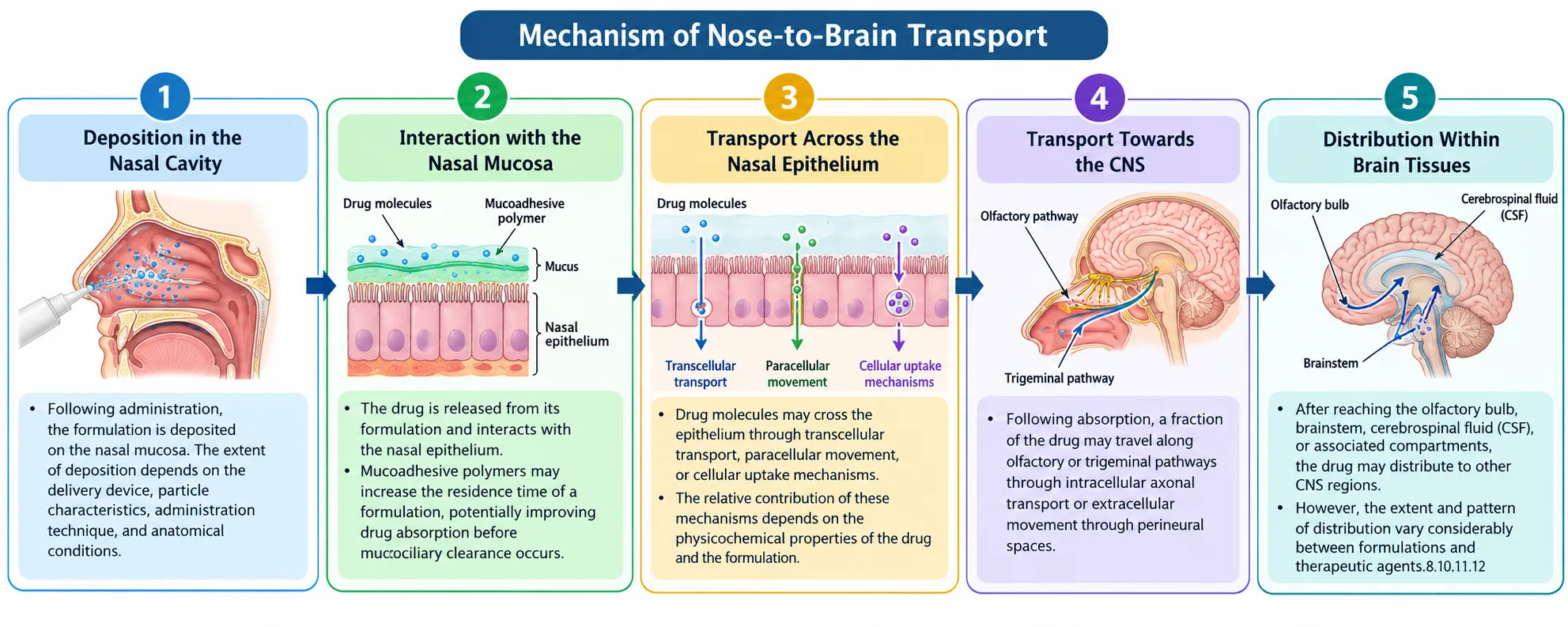
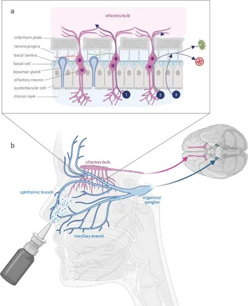
Figure 2: Mechanism of Nose to Brain Delivery
3. Advantages of Intranasal Delivery --
1. Potential for Nose-to-Brain Delivery: The olfactory and trigeminal pathways provide anatomical routes that may facilitate drug transport towards the CNS.
2. Non-Invasive Administration: Intranasal administration avoids the need for injections in many applications and may improve patient convenience.
3. Avoidance of Gastrointestinal Absorption: Intranasally administered drugs do not need to undergo gastrointestinal absorption before entering the body.
4. Reduced First-Pass Metabolism: Drugs absorbed through the nasal mucosa avoid the initial hepatic first-pass effect associated with oral administration, although systemic metabolism may still occur.
5. Potential for Rapid Drug Absorption: The nasal mucosa has a rich blood supply, allowing relatively rapid absorption of suitable drugs. The onset of action depends on the drug and formulation.
6. Potential Reduction in Systemic Exposure: Direct nose-to-brain transport may reduce the amount of drug required to circulate systemically to achieve a desired brain exposure. This benefit must be demonstrated experimentally. [8,9,10]
4. Limitations of Intranasal Delivery –
1. Mucociliary Clearance:
The nasal mucosa contains ciliated epithelial cells that continuously transport mucus towards the nasopharynx. This protective mechanism removes foreign particles and can rapidly eliminate intranasally administered drugs before sufficient absorption occurs. Consequently, nasal residence time is an important factor in formulation development. [9,10]
2. Enzymatic Degradation:
Enzymes present in nasal secretions and epithelial tissues may degrade susceptible drugs, particularly peptides and proteins. This degradation can reduce the quantity of intact therapeutic agent available for absorption. Nanoparticle encapsulation may protect some drugs from degradation, depending on the carrier and formulation characteristics. [8,10]
3. Limited Administration Volume:
The nasal cavity can accommodate only a limited volume of formulation. Excessive administration may cause leakage, swallowing, or drainage towards the nasopharynx. This limitation can restrict the dose that can be delivered through the intranasal route. [9,10]
4. Variability in Nasal Absorption:
Nasal absorption can vary because of differences in nasal anatomy, mucus production, mucosal condition, congestion, and administration technique. Such variability may result in inconsistent drug exposure and can complicate dose optimization. [9,10]
5. Nasal Irritation and Toxicity:
Certain drugs, solvents, surfactants, and absorption-enhancing excipients may irritate or damage the nasal epithelium. Repeated administration requires particular attention to local tolerability, mucosal integrity, and potential long-term toxicity. Appropriate in vitro, ex vivo, and in vivo safety assessments are therefore necessary. [9,10]
NANOPARTICLES AS DRUG DELIVERY CARRIER:
Nanoparticles are colloidal particulate systems generally ranging from approximately 1 to 1000 nm in size, although the commonly used definition of nanomaterials often emphasizes dimensions between 1 and 100 nm. They can be prepared from biodegradable polymers, lipids, or other suitable materials to encapsulate, incorporate, or adsorb therapeutic agents. Nanoparticles have attracted considerable attention in pharmaceutical research because their physicochemical properties can be tailored to improve drug solubility, stability, release characteristics, and delivery to specific tissues. [13,14]
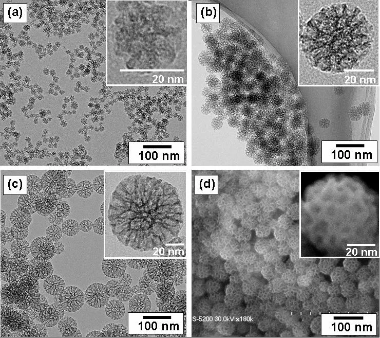
Figure 3: Nanoparticles [31]
Types of Nanoparticles --
1. Polymeric Nanoparticles:
Polymeric nanoparticles are nanoscale drug delivery systems prepared using natural or synthetic polymers. Commonly investigated polymers include poly (lactic-co-glycolic acid) (PLGA), polylactic acid (PLA), and chitosan. Depending on their internal structure, polymeric nanoparticles may be classified as nanospheres, in which the drug is distributed throughout a polymeric matrix, or Nanocapsules, in which the drug is contained within a core surrounded by a polymeric shell. Polymeric nanoparticles are being investigated for the delivery of dopaminergic and neuroprotective agents for Parkinson’s disease. [14,16]
2. Solid Lipid Nanoparticles (SLNs):
Solid lipid nanoparticles are colloidal drug delivery systems composed of lipids that remain solid at room and body temperatures. They generally contain a solid lipid matrix stabilized by surfactants in an aqueous dispersion. Examples of lipids used in SLN formulations include glyceryl monostearate, stearic acid, and other physiologically suitable solid lipids. SLNs can incorporate lipophilic drugs and, depending on their structure and formulation, certain hydrophilic drugs. They offer potential advantages such as biodegradability, protection of encapsulated drugs, and modification of drug-release profiles. [13,15]
3. Nanostructured Lipid Carriers (NLCs):
Nanostructured lipid carriers are second-generation lipid-based nanoparticles developed to address certain limitations of SLNs. They contain a mixture of solid and liquid lipids stabilized by surfactants. The incorporation of liquid lipid disrupts the orderly arrangement of the solid lipid matrix, creating a less organized structure that may accommodate more drug and reduce drug expulsion during storage. For Parkinson’s disease, NLCs represent a potential carrier for dopaminergic and neuroprotective agents. Their application in intranasal drug delivery may help improve drug stability and brain exposure. [13,15]
4. Liposomes:
Liposomes are spherical vesicular drug delivery systems composed of one or more phospholipid bilayers surrounding an aqueous compartment. Their amphiphilic structure allows them to incorporate hydrophilic drugs within the aqueous core and lipophilic drugs within the lipid bilayer. Cholesterol is commonly included to modify membrane rigidity, stability, and permeability. In Parkinson’s disease, liposomes are being explored as carriers for therapeutic and neuroprotective compounds. [15,17,31]
5. Nanoemulsions:
Nanoemulsions are dispersions of oil and water stabilized by surfactants or combinations of surfactants and co-surfactants. They commonly contain nanoscale oil droplets dispersed in an aqueous phase or water droplets dispersed in an oil phase. Unlike thermodynamically stable microemulsions, nanoemulsions are kinetically stable systems that require suitable formulation and processing conditions to maintain their physical stability. In Parkinson’s disease research, Nano Emulsions have been explored as potential carriers for drugs and neuroprotective compounds. They may improve the formulation of poorly soluble agents and potentially facilitate nose-to-brain delivery. [15,17]
6. Chitosan-Based Nanoparticles:
Chitosan is a naturally derived, biodegradable polysaccharide obtained through the deacetylation of chitin. It contains amino groups that can become positively charged under suitable acidic conditions. Chitosan-based nanoparticles can be prepared using techniques such as ionic gelation and can also be used as coatings for other nanocarriers. Chitosan-based nanoparticles have been investigated for the intranasal delivery of antiparkinsonian drugs, including pramipexole.16,18
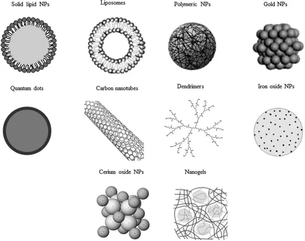
Figure 4: Types of Nanoparticles
Composition of Nanoparticle Formulations –
The major components are described below.
1. Active Pharmaceutical Ingredient (API): The drug or therapeutic agent intended for delivery to the brain. Examples investigated for Parkinson’s disease include levodopa-related formulations, pramipexole, ropinirole, and neuroprotective compounds. The selected drug influences carrier choice and formulation design.
2. Polymer or Lipid Matrix: This component forms the structural framework of the nanoparticle. Polymeric systems may contain PLGA or chitosan, whereas lipid-based systems may contain solid lipids, liquid lipids, or phospholipids. The matrix affects drug incorporation, stability, and release.
3. Surfactants and Stabilizers: These substances help stabilize the dispersion and control particle formation. Suitable surfactants can reduce interfacial tension and help prevent aggregation. Their concentration must be optimized to avoid undesirable effects on nasal tissues.
4. Mucoadhesive Agents: Materials such as chitosan may increase the interaction of the formulation with nasal mucus and prolong residence time. However, Mucoadhesion alone does not guarantee improved brain delivery.
5. Cross-Linking Agents: Certain nanoparticle systems, including chitosan nanoparticles, may require suitable cross-linking agents to form and stabilize their structure. The type and concentration of cross-linker influence particle characteristics and drug release.
6. Aqueous Phase and Solvents: Water, buffers, or suitable processing solvents may be used during preparation. Residual solvents, pH, and other formulation characteristics must be controlled to ensure product quality and nasal compatibility. [14,15,18]
Advantages of Nanoparticles --
The major potential advantages include:
1. Improved Drug Stability: Encapsulation may protect certain therapeutic agents from enzymatic degradation and other environmental influences.
2. Controlled Drug Release: Appropriate carrier materials can modify drug-release kinetics and potentially prolong drug availability.
3. Improved Solubilization: Lipid-based and other suitable nanoparticle systems may improve the formulation of poorly water-soluble drugs.
4. Enhanced Nasal Residence: Mucoadhesive materials, particularly chitosan, may prolong contact with nasal mucosa and reduce premature clearance.
5. Potential for Enhanced Brain Delivery: Certain formulations may facilitate transport towards the CNS through olfactory and trigeminal pathways. [13,15,18]
Role of Nanoparticles in Parkinson’s Disease Treatment --
Parkinson’s disease is characterized by the progressive degeneration of dopaminergic neurons, particularly in the substantia nigra pars compacta, resulting in reduced dopamine availability in the striatum. Although pharmacological treatments can improve symptoms, drug delivery to the brain remains a challenge for several therapeutic agents. Nanoparticle-based delivery systems are therefore being investigated as potential platforms for improving the delivery of dopaminergic and neuroprotective compounds. [13,15,17]
1. Delivery of Dopaminergic Drugs:
Nanoparticles have been investigated for the delivery of antiparkinsonian drugs such as ropinirole, rotigotine, rasagiline, and pramipexole. Suitable formulations may improve drug stability, modify release characteristics, and potentially enhance brain exposure following intranasal administration. [13,16]
2. Potential for Improved Brain Targeting:
The olfactory and trigeminal pathways provide potential routes for transporting intranasally administered drugs towards the CNS. Nanoparticles can be designed to modify interactions with the nasal mucosa and potentially support transport through these pathways.Their performance depends on particle size, surface charge, drug release, mucoadhesive properties, and nasal deposition. Brain-targeting studies should therefore assess brain and plasma drug concentrations and, where appropriate, calculate relevant drug-targeting parameters. [13-15]
3. Delivery of Neuroprotective Agents:
Oxidative stress, mitochondrial dysfunction, protein aggregation, and neuroinflammation are among the mechanisms associated with Parkinson’s disease pathology. Consequently, researchers have investigated compounds with antioxidant or other neuroprotective properties as potential therapeutic approaches.Nanoparticle formulations may help protect selected compounds from degradation and improve their delivery characteristics. Lipid-based nanoparticles, polymeric nanoparticles, and other nanocarriers have been studied for the delivery of experimental neuroprotective agents. [15,17]
DRUGS UESD IN NANOPARTICLE-BASED INTRANASAL DELIVERY:
Nanoparticle-based intranasal drug delivery has emerged as a promising research approach for improving the delivery of anti-Parkinsonian drugs to the brain. The intranasal route may facilitate nose-to-brain transport through the olfactory and trigeminal pathways, potentially reducing some limitations associated with conventional systemic administration. Nanocarriers, including polymeric nanoparticles, solid lipid nanoparticles (SLNs), and nanostructured lipid carriers (NLCs), have been investigated to improve drug stability, nasal residence time, brain exposure, and controlled release. [6,19]
1. Levodopa --
Levodopa (L-DOPA) is a dopamine precursor and remains a principal pharmacological treatment for the motor symptoms of Parkinson’s disease. It crosses the blood–brain barrier and is converted into dopamine in the brain. However, its clinical use may be complicated by peripheral metabolism, fluctuations in therapeutic response, and motor complications associated with long-term treatment.
Nanoparticle-based formulations have been investigated to protect levodopa from premature metabolism and improve its delivery to the central nervous system. Polymeric and lipid-based nanoparticles may provide controlled drug release and offer opportunities for targeted delivery. Intranasal administration is also being investigated as a potential alternative route for increasing brain exposure while reducing reliance on systemic transport. Nevertheless, the performance of a particular formulation depends on its composition, particle characteristics, drug loading, and administration route. Further preclinical and clinical research is required to establish its safety and therapeutic benefits. [6,19]
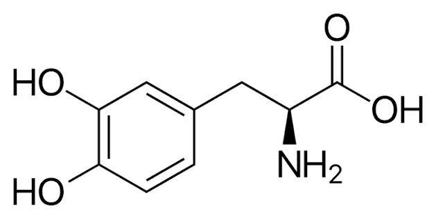
Figure 4: Structure of Levodopa
2.Rotigotine:
Rotigotine is a dopamine agonist that stimulates dopamine receptors and helps manage the motor symptoms of Parkinson’s disease. Its application in nanoparticle-based intranasal delivery has attracted interest because of the potential to transport the drug directly from the nasal cavity to the brain. Intranasal administration produced greater brain accumulation than conventional intravenous administration in the experimental model. [20]
3. Pramipexole:
Pramipexole is a dopamine Dâ‚‚/D₃ receptor agonist used in the management of Parkinson’s disease. Its therapeutic action involves stimulation of dopamine receptors, helping compensate for reduced dopaminergic activity in the brain. [21]
4.Rasagiline:
Rasagiline is a selective monoamine oxidase-B (MAO-B) inhibitor used in Parkinson’s disease. It reduces the breakdown of dopamine in the brain, helping maintain dopaminergic activity. Rasagiline mesylate has been investigated in lipid-based nanoparticle formulations intended for intranasal administration. Solid lipid nanoparticles may offer advantages such as protection of the incorporated drug, controlled release, and the potential to improve delivery through the nasal mucosa. Lipid-based carriers are particularly relevant to nose-to-brain research because their physicochemical characteristics can be adjusted to influence drug loading and release. [6,8]
MATERIALS AND METHODS
Levodopa formulations are available in conventional oral dosage forms, and nanoparticle-based intranasal delivery for Parkinson’s disease. The principal formulation study describes levodopa-loaded chitosan and PLGA nanoparticles for intranasal delivery have been proved to be efficient in treatment of Parkinson's Disease.
Materials reported in the selected nanoparticle study
The reported materials included L-Dopa as the active pharmaceutical ingredient; low-molecular-weight chitosan (50–60 kDa) as a biodegradable, mucoadhesive polymer; poly (lactic-co-glycolic acid) (PLGA, 50:50) as a biodegradable polymer matrix; sodium tripolyphosphate (TPP) as the ionic cross-linking agent for chitosan; Tween 80 as a stabilizer/resuspending agent; polyvinyl alcohol (PVA) as an emulsion stabilizer; acetic acid to dissolve chitosan; dichloromethane as the organic solvent for PLGA; and deionised water as the aqueous phase. The selection and purpose of each material are summarized in Table 1 [25].
Table 1: Materials Used in Reported Levodopa Nanoparticle Formulations and Their Purpose
| Material | Role in formulation | Why it is used |
|---|---|---|
| Levodopa (L-Dopa) | Active pharmaceutical ingredient | Dopamine precursor used to improve motor symptoms of Parkinson’s disease. |
| Chitosan | Polymeric carrier | Biodegradable and mucoadhesive; may increase nasal residence time. |
| PLGA (50:50) | Polymeric carrier | Biodegradable matrix that can entrap drug and influence release. |
| Sodium tripolyphosphate (TPP) | Cross-linking agent | Ionically cross-links chitosan to form nanoparticles. |
| Tween 80 | Stabilizer/resuspending agent | Helps reduce aggregation and stabilize the dispersion. |
| Polyvinyl alcohol (PVA) | Emulsion stabilizer | Stabilizes the emulsion during PLGA nanoparticle preparation. |
| Acetic acid | Solvent for chitosan | Helps dissolve chitosan before ionic gelation. |
| Dichloromethane | Organic solvent for PLGA | Dissolves PLGA during preparation; must be removed from the final product. |
| Deionised water | Aqueous phase/vehicle | Used to prepare aqueous solutions and emulsions. |
Conventional levodopa dosage forms
Levodopa is available in oral dosage forms including immediate-release tablets and modified/extended-release tablets or capsules. Combination products commonly contain carbidopa or benserazide, which inhibit peripheral conversion of levodopa to dopamine and help more levodopa become available for transport into the brain32. Tablets are convenient and allow dose adjustment, although gastrointestinal absorption and clinical response can vary between patients5,26.
Figure 5: Generic Illustrative Image of Tablets and a Medicine Container [32]
Literature-based preparation methods for levodopa-loaded nanoparticles
Chitosan nanoparticles were reported to be prepared by ionic gelation. Chitosan was dissolved in dilute acetic acid, Tween 80 was added, and L-Dopa was incorporated. An aqueous TPP solution was then added to initiate ionic cross-linking and nanoparticle formation. The resulting dispersion was centrifuged, and the nanoparticle fraction was collected and lyophilized for analysis. The study compared four chitosan concentrations (0.1–0.4% w/v), with L-Dopa at 0.1% w/v, Tween 80 at 0.5% w/v, and TPP at 0.1% w/v [25].
PLGA nanoparticles were reported to be prepared using a modified water-in-oil-in-water (W/O/W) solvent-evaporation method. L-Dopa was dissolved in water, PLGA was dissolved in an organic solvent, and the phases were homogenized to form a primary emulsion. An aqueous PVA solution was then added to form a double emulsion. The organic solvent was removed under reduced pressure, after which nanoparticles were collected by centrifugation and lyophilized. The study compared three PLGA concentrations (1.25, 2.50, and 5.00 mg/mL), with L-Dopa at 1 mg/mL and PVA at 0.2% w/v [25].
Evaluation of nanoparticle formulations
The selected study evaluated particle size and polydispersity, zeta potential, morphology using electron microscopy, drug entrapment/content, solid-state properties using X-ray diffraction, chemical interactions using FTIR, and pharmacokinetic performance after intranasal administration in an animal model [25].
FORMULATION STUDY OF LEVODOPA NANOPARTICLES FOR THE TREATMENT OF PARKINSON’S DISEASE:
Levodopa (L-Dopa) is a precursor of dopamine and is widely used in the treatment of Parkinson’s disease (PD). The disease is associated with the degeneration of dopaminergic neurons in the substantia nigra, resulting in reduced dopamine levels in the brain. Although oral Levodopa is an established treatment, its therapeutic effectiveness may be affected by peripheral metabolism, fluctuations in drug availability and adverse effects associated with long-term treatment.
Nanoparticle-based drug delivery systems have been investigated to improve Levodopa delivery to the central nervous system (CNS). Polymeric nanoparticles can encapsulate the drug and potentially protect it from premature degradation. Intranasal administration is particularly interesting because it may facilitate drug transport towards the brain through the olfactory and trigeminal pathways, partially bypassing the blood–brain barrier (BBB). However, the effectiveness of this approach depends on the formulation and requires further investigation. [24]
Selection of Nanocarrier --
Polymeric nanoparticles prepared using chitosan and poly (lactic-co-glycolic acid) (PLGA) have been investigated for intranasal Levodopa delivery.
Chitosan nanoparticles: Chitosan possesses mucoadhesive properties that may increase contact between the formulation and nasal mucosa and support drug absorption.
PLGA nanoparticles: PLGA is a biodegradable polymer that can encapsulate drugs and provide a matrix for controlled drug release.
In a study published in 2022, researchers prepared Levodopa-loaded chitosan and PLGA nanoparticles and evaluated their physicochemical properties and pharmacokinetic performance following intranasal administration.[19]
Methodology for preparation of Levodopa-Loaded Nanoparticles --
1.Ionic Gelation Method:
Chitosan nanoparticles containing Levodopa were prepared using the ionic gelation method. Chitosan was combined with the drug, and sodium tripolyphosphate (TPP) was used as a cross-linking agent. Ionic interactions between chitosan and TPP facilitated nanoparticle formation.
Four chitosan formulations were developed and evaluated to identify a formulation with suitable particle size, surface charge and drug entrapment.
2. Modified Solvent Evaporation Method:
PLGA nanoparticles were prepared using a modified water-in-oil-in-water (W/O/W) solvent evaporation technique. This method involves the formation of primary and secondary emulsions, followed by removal of the organic solvent to produce polymeric nanoparticles.
Three PLGA formulations were developed and characterized. These methods demonstrate how different polymeric carriers can be investigated for intranasal delivery of Levodopa. [6]
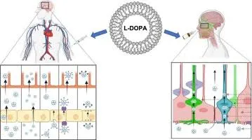
Figure 6: Levodopa Loaded Nanoparticles Acts on Intra Nasal Route
Advantages --
1.Potential to improve Levodopa absorption following intranasal administration.
2.Chitosan may increase nasal mucosal residence time because of its mucoadhesive properties.
3.Polymeric carriers may protect the drug and influence its release profile.
4.Intranasal administration may offer an alternative to conventional oral delivery.
Limitations --
1.Nasal mucociliary clearance may remove the formulation before sufficient absorption occurs.
2.Nanoparticle stability, drug loading and reproducibility require careful optimization.
3.Nasal mucosal safety and long-term tolerability need further evaluation.
4.The findings from animal studies cannot be directly extrapolated to humans.[24]
EVALUATION PARAMETERS OF NANOPARTICLE FORMULATIONS: [25- 27]
The evaluation of nanoparticle formulations is essential to determine their physicochemical properties, drug-loading capacity, stability, drug-release behaviour and suitability for intranasal administration. For Parkinson’s disease, these parameters help assess whether a nanoparticle formulation has the potential to improve drug absorption and delivery towards the brain. The major valuation parameters include particle size, polydispersity index, zeta potential, entrapment efficiency, morphology, in-vitro drug release, nasal mucosal compatibility and in-vivo performance.
Table 2: Evaluation Parameters
| Evaluation Parameter | Method commonly used | Purpose |
|---|---|---|
| Particle size | DLS or suitable particle sizing technique | Determines average particle size |
| PDI | DLS | Assesses particle size distribution |
| Zeta Potential | Electrophoretic light scattering | Evaluates surface change |
| Drug assay after separating free drug | Determines the proportion of drug entrapped | |
| Drug loading | Drug content analysis | Determines drug content relative to nanoparticle mass |
| Surface Morphology | SEM/TEM | Examines particle shape and structure |
| FTIR | Infrared Spectroscopy | Investigates functional groups and potential interactions |
| In-vitro drug release | Suitable dissolution or release study | Determines drug release behavior |
| Mucoadhesion | Mucin-binding or residence time tests | Evaluates interaction with nasal mucus |
| Nasal compatibility | Cell based or tissue-based studies | Assesses potential irritation or toxicity |
| In-vivo pharmacokinetics | Drug concentration- time analysis | Determines systemic drug exposure |
| Stability | Storage studies | Evaluates changes in formulation quality |
| Brain Distribution | Brain-tissue drug analysis | Investigates drug delivery to the brain |
CHALLENGES AND LIMITATIONS:
1.Mucociliary Clearance –
The nasal cavity possesses a natural mucociliary clearance mechanism that removes mucus, foreign particles and deposited substances. This mechanism can rapidly eliminate intranasally administered nanoparticles before sufficient drug absorption occurs, thereby reducing nasal residence time and therapeutic availability. Mucoadhesive polymers, such as chitosan, may improve retention; however, excessive interaction with mucus may also restrict nanoparticle movement towards the nasal epithelium. [6]
2. Limited Nasal Absorption and Blood–Brain Barrier Challenges --
Although intranasal administration may facilitate drug transport towards the brain through olfactory and trigeminal pathways, not all administered drugs reach the central nervous system. Drug absorption depends on physicochemical properties, nasal permeability, formulation characteristics and the location of deposition within the nasal cavity. Furthermore, bypassing the blood–brain barrier (BBB) does not guarantee that a sufficient therapeutic concentration will reach the specific brain regions affected by Parkinson’s disease.[28]
3. Enzymatic Degradation --
Enzymes present in the nasal mucosa may degrade susceptible therapeutic molecules before they are absorbed. This can reduce the amount of active drug available for transport. Nanoparticles may protect certain drugs from premature degradation, but the extent of protection depends on the carrier composition, drug location within the carrier and release characteristics. Therefore, formulation-specific studies are necessary to establish the protective effect of each nanocarrier. [29]
5. Nasal Toxicity and Long-Term Safety --
The nasal mucosa is a sensitive biological barrier, and repeated administration of nanoparticles or formulation excipients may cause irritation, inflammation or epithelial damage. Certain surface modifications and penetration-enhancing excipients may also affect mucosal integrity. Therefore, nasal histopathology, mucosal compatibility, ciliary function and long-term tolerability should be evaluated before clinical application. The safety of a formulation cannot be established solely from its physicochemical properties or short-term animal studies. [30]
CONCLUSION:
Parkinson’s disease (PD) is a progressive neurodegenerative disorder characterized by the degeneration of dopaminergic neurons in the substantia nigra, resulting in dopamine deficiency and impaired motor function. Although conventional pharmacological treatments, particularly Levodopa, provide significant symptomatic relief, their therapeutic effectiveness may be limited by peripheral metabolism, fluctuations in drug availability and difficulties in achieving consistent drug concentrations in the brain. Therefore, novel drug delivery approaches are being investigated to improve therapeutic outcomes and patient quality of life. Nanoparticle-based intranasal drug delivery has emerged as a promising research approach for delivering therapeutic agents towards the central nervous system. This strategy combines the advantages of nanotechnology with the accessibility of the nasal route. Intranasally administered drugs may reach the brain through the olfactory and trigeminal pathways, potentially reducing dependence on systemic circulation and partially bypassing the blood–brain barrier (BBB). However, the extent of brain delivery depends on the drug’s physicochemical properties, formulation characteristics, nasal deposition and transport mechanisms. [6,29]
Different nanocarrier systems, including polymeric nanoparticles, solid lipid nanoparticles (SLNs), nanostructured lipid carriers (NLCs), liposomes and Nanoemulsions, have been investigated for intranasal drug delivery. Polymeric nanoparticles prepared using chitosan or poly (lactic-co-glycolic acid) (PLGA) are of particular interest because their composition can be modified to influence drug encapsulation, release characteristics, stability and interactions with nasal mucosa. Chitosan may also improve formulation residence time because of its mucoadhesive properties. Nevertheless, the suitability of each carrier depends on the drug, formulation composition and intended therapeutic application. [25]
Levodopa-loaded polymeric nanoparticles provide an example of this approach. Ahmad et al. developed Levodopa-loaded chitosan and PLGA nanoparticles using ionic gelation and a modified water-in-oil-in-water (W/O/W) solvent evaporation method, respectively. The formulations were characterized for physicochemical properties and evaluated for their potential in intranasal delivery. The optimized chitosan formulation demonstrated an entrapment efficiency of 82.38 ± 1.63% and increased systemic drug exposure following intranasal administration in an animal model. These findings support further investigation of polymeric nanoparticles for Levodopa delivery; however, increased systemic exposure alone does not establish enhanced direct brain delivery or clinical efficacy in humans.Despite these promising developments, several challenges remain, including mucociliary clearance, enzymatic degradation, variable nasal absorption, formulation instability, potential nasal irritation, dose limitations and difficulties in large-scale manufacturing. Furthermore, much of the evidence for nanoparticle-based intranasal treatment of Parkinson’s disease remains preclinical. Differences between animal models and humans necessitate further investigations into nasal tolerability, pharmacokinetics, brain distribution, long-term safety and therapeutic effectiveness. Standardized formulation and evaluation protocols are also needed to improve reproducibility and facilitate clinical translation.[29]
Nanoparticle-based intranasal delivery represents a promising research strategy for improving drug delivery towards the brain in Parkinson’s disease, but it is not yet an established replacement for conventional treatment. Future research should focus on optimizing drug–carrier combinations, improving nasal retention without damaging the mucosa, achieving reproducible formulation characteristics and confirming brain-targeting efficiency through well-designed preclinical and clinical studies. These developments may help determine whether intranasal nanocarriers can provide meaningful therapeutic advantages for patients with Parkinson’s disease.[25]
REFERENCES



Tanvi B. Palve, Vishal S. Madankar, Sampat D. Navale, A Review on Nano Particle-Based Intra Nasal Drug Delivery for the Treatment of Parkinson’s Disease, Int. J. Sci. R. Tech., 2026, 3 (11), 365-380. https://doi.org/10.5281/zenodo.23976004
 10.5281/zenodo.23976004