
We use cookies to ensure our website works properly and to personalise your experience. Cookies policy
Department of Orthopaedic Physiotherapy, R.V.S College of Physiotherapy, Coimbatore, The Tamil Nadu Dr M.G.R. Medical University, Chennai, Tamil Nadu, India
A study on the effectiveness of muscle energy technique versus static stretching exercise on pain, craniovertebral angle, neck disability in subjects with upper cross syndrome. Objectives :Research to ascertain the effectiveness of muscle energy technique versus static stretching exercise on pain, craniovertebral angle, neck disability in subjects with upper cross syndrome. Design :Pre and post test of experimental study design is adopted. Setting :the site of the study was R.V.S College of Physiotherapy out patient department , Sulur , Coimbatore. Result : on comparing the mean post treatment GMFM -88 scores of both groups, Group A (muscle energy technique and strengthening exercise ) exhibited. Conclusion :Muscle energy technique and strengthening exercise are more effective in the management of pain, craniovertebral angle and neck disability among subjects with forward head posture due to upper cross syndrome.
Upper Crossed Syndrome (UCS) is characterized by tightness of the upper trapezius, pectoralis major, and levator scapulae muscles, along with weakness of the rhomboids, serratus anterior, middle and lower trapezius, deep cervical flexors (rectus capitis anterior, rectus capitis lateralis, longus capitis, and longus colli), and scalene muscles. Janda termed this condition "Upper Crossed Syndrome" because the pattern of shortened and weakened muscles forms a crossed arrangement in the upper body (Mohanty, 2015).The prevalence of UCS in the general population ranges from 28% to 67% annually. Certain occupational groups demonstrate a higher prevalence, including office workers (32.43%), drivers (24.32%), housewives (27.03%), teachers (16.22%), and medical students (37.1%) (Azam, 2022).
The cervical vertebral column consists of seven vertebrae and is divided into two distinct regions: the upper cervical spine (craniovertebral region) and the lower cervical spine. The craniovertebral region includes the occipital condyles and the first two cervical vertebrae, C1 (atlas) and C2 (axis). The lower cervical spine comprises vertebrae C3 to C7. Vertebrae C3 to C6 share similar structural characteristics and are therefore considered typical cervical vertebrae (Chaurasia, 2004).Most compressive forces acting on the cervical spine are transmitted through the vertebral bodiesand intervertebral discs, while approximately one-third of these forces pass through the zygapophyseal joints. Compressive loads are relatively low during upright standing and sitting postures but increase significantly at the end ranges of cervical flexion and extension. Cervical dysfunction may result from several factors, including degenerative changes, traumatic injuries, medical conditions such as rheumatoid arthritis, spinal stenosis, cervical radiculopathy, tumors, and lifestyle-related factors such as poor posture, repetitive movements, and stress (Cynthia, 2017).
In UCS, tightness of the upper trapezius and levator scapulae on the posterior aspect of the body crosses with tightness of the pectoralis major and minor muscles anteriorly. Simultaneously, weakness of the deep cervical flexors crosses with weakness of the middle and lower trapezius muscles. This characteristic pattern of muscle imbalance contributes to joint dysfunction, particularly at the atlanto-occipital joint, C4–C5 segment, cervicothoracic junction, glenohumeral joint, and T4–T5 segment. Janda observed that these regions correspond to transitional zones where adjacent vertebrae differ in morphology, making them more susceptible to mechanical stress (Janda, 1988).UCS is commonly associated with postural abnormalities such as forward head posture, protracted scapulae, medially rotated humerus, increased thoracic kyphosis, and cervical extension. These postural deviations place excessive stress on the cervicocranial junction, C4–C5 and T4 segments, and the shoulder complex due to altered glenohumeral joint mechanics. Excessive loading of the T4 segment may occasionally produce chest pain resembling angina pectoris. Alteration in the orientation of the glenoid fossa can lead toscapular rotation and abduction, requiring increased activity of the levator scapulae and upper trapezius to stabilize the humeral head (Ashok, 2021).
These biomechanical alterations may contribute to impingement syndromes and cervical or upper thoracic pain. Muscles subjected to repetitive overuse tend to become shortened and tight, a phenomenon known as adaptive shortening. Conversely, opposing muscles are exposed to prolonged stretching during sustained postures, resulting in lengthening and weakness, referred to as stretch weakness (Thacker, 2011).
Upper Crossed Syndrome is characterized by several postural abnormalities, including forward head posture, increased cervical lordosis, thoracic hyperkyphosis, rounded shoulders, and scapular winging. These abnormalities disrupt normal scapular movement patterns, adversely affecting shoulder and neck stability and mobility. Sustained poor posture increases joint loading, accelerates degenerative changes, contributes to muscle imbalance, and may result in pain. If left untreated, these impairments can lead to altered movement patterns and progressive joint dysfunction (Mincheol, 2023).
Various treatment approaches have been proposed for the management of UCS, including stretching exercises, strengthening exercises, muscle energy techniques (MET), myofascial release, postural correction exercises, electrical stimulation, and deep cervical flexor activation exercises. Recent therapeutic trends have also demonstrated benefits from corrective exercise games, kinesio taping, scapular stabilization exercises, and proprioceptive neuromuscular facilitation (PNF) techniques (Ashok, 2021).
Muscle Energy Techniques are manual therapy procedures derived from osteopathic medicine and are designed to lengthen muscles and fascia while improving joint mobility. These techniques involve voluntary muscle contractions performed by the patient against a precisely controlled resistance applied by the therapist. Typically, the patient maintains a gentle contraction for 3–5 seconds, followed by relaxation. The procedure is repeated three to five times. When applied appropriately, MET is considered safe and effective for treating joint restrictions associated with musculoskeletal disorders at various stages of healing (Leon, 2006).
Stretching refers to therapeutic interventions aimed at increasing soft tissue extensibility to improve flexibility and range of motion (ROM). Stretching is indicated when ROM is restricted due to adhesions, contractures, scar tissue formation, muscle shortening, or soft tissue tightness. It may also be incorporated into fitness and sports conditioning programs to prevent musculoskeletal injuries and improve functional performance (Kisner, 2017).
Strengthening exercises are physical activities designed to enhance the force-generating capacity of muscles by requiring them to work against resistance. Resistance may be provided by body weight, free weights, resistance bands, or exercise machines. Common types of strengthening exercises include isometric, isotonic, and isokinetic exercises (Kisner, 2017).
The Numerical Pain Rating Scale (NPRS) is one of the most commonly used tools for assessing pain intensity due to its simplicity, validity, and reliability. Patients are asked to rate their pain on a scale from 0 to 10, where 0 indicates no pain and 10 represents the worst imaginable pain (Maide, 2021).
The Neck Disability Index (NDI) is the most widely used region-specific questionnaire for assessing neck-related disability. It has demonstrated strong reliability and validity. The questionnaire consists of 10 items, each scored from 0 to 5, with higher scores indicating greater self-reported disability (Bryan, 2011).
Kinovea is a free two-dimensional motion analysis software used to measure kinematic parameters. It enables video analysis with or without markers, although the use of passive markers may improve measurement reliability. Kinovea has been utilized in sports science, clinical practice, and research settings to assess movement patterns such as running and vertical jumping. One notable feature of the software is its ability to measure object movement while accounting for camera perspective, making it a cost-effective tool for biomechanical assessment (Puig, 2019).
METHODOLOGY
Study setting
The study was conducted in outpatient department of RVS College of Physiotherapy, Sulur, Coimbatore.
Study design and study duration
The study design was a pre and post -test experimental study conducted over a period of six months
Inclusion criteria
• Age between 20-35 year
• Craniovertebral angle greater less than 48
• Both male and female
• Subjects willing to participate
• Neck pain for more than 4 weeks
Exclusion criteria
• Any congenital conditions
• Recent surgery
• Recent fracture
• Rheumatoid arthritis,
• Ankylosing spondylitis,
• Fibromyalgia
• Congenital spinal deformities.
• Osteoporosis
• Tumors
• Pregnancy
• Cervical radiculopathy
Orientation to the subject
Before the collection of data, all the subjects were explained about the study. The investigation had given a detailed orientation about various test procedures. The concern and full cooperation of each participant was collected after a complete explanation of condition and demonstration of procedure.
Procedure
Measurement procedure
Pain was measured by using numerical pain rating scale is asked and results are noticed before the treatment.30 subjects with forward head posture where taken and each subjects is given separate neck disability form to assess.Lateral view of subjects neck is videographed and used to measure craniovertebral angle using kinovea software. Verbal explanation and demonstration were provided, patient was made comfortable and relaxed, then score is recorded.
Treatment procedure
|
Serial no |
Exercise |
position |
Procedure |
|
1 |
upper trapezius (shoulder shrugs)

Figure 2: upper trapezius. |
Patient position: Standing or sitting Position Therapist position: standing. |
Subject was asked to hold weight in hands by sides and lift shoulders straight up toward your ears. Hold for 2 seconds, then slowly lower. 3 sets of 10–15 reps.
|
|
2 |
Levator scapulae
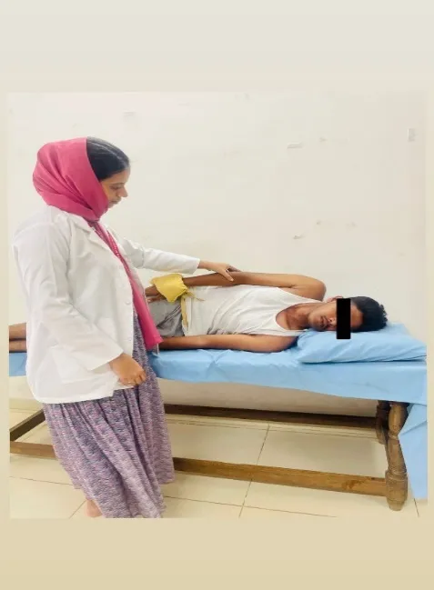
Figure 3: levator scapulae |
Patient position: side lying. Therapist position: standing near to the side to be treated |
Subject was asked to lie on the side with working arm on top. subject was asked to hold a light dumbbell or no weight at first. Instruct to keeping the arm straight and lift the shoulder (scapula) toward the ear, hold, then lower. neck compensation must be discouraged 3 sets of 10-12 repetitions |
|
3 |
Strengthening exercise for pectoralis major
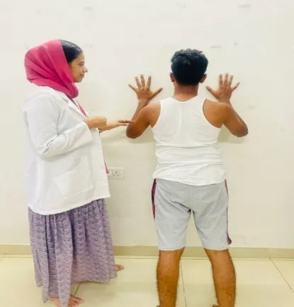
Figure 4: pectoralis major |
Patient position: standing. Therapist position : Standing near the side to be treated.
|
Subject was asked to stand facing a wall with hands at chest height and perform a push-up movement against the wall. Keep elbows at 45° and shoulder blades retracted. 3 sets of 10-12 repetitions |
Table 1: Strengthening exercise
Group B: Static stretching
|
Serial no |
Exercise |
position |
procedure |
|
1 |
Upper trapezius

Figure 8: upper trapezius |
Patient position: sitting or standing. Therapist position : standing near the side to be treated. |
Patient was asked to keep shoulders relaxed and spine neutral. Patient was asked to hold the bottom of chair with the hand on the side to be stretched, or let the arm hang relaxed and weighted down. Head was side-bended away and tilted head towards the opposite shoulder (ear to shoulder.) gently pull the head further into the stretch. Do not pull force full Hold for 20–30 seconds, breathing slowly Repeat on the other side 2-4 times per side. 3-5 days per week |
|
|
Levator scapula
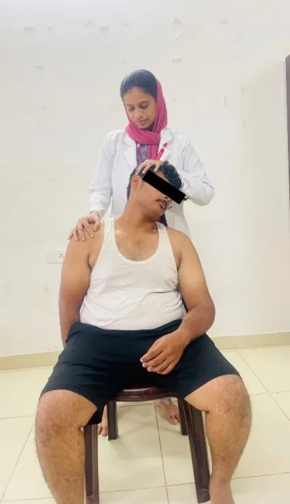
Figure 9: levator scapulae |
Patient position: sitting or standing. Therapist position:standing next to the position to be treated.
|
Patient was asked to keep shoulders relaxed, spine neutral and place the arm behind your back, or gently depress the shoulder with hand or a strap. Ask the patient to turn and tilt the head away Rotate head about 45° to the opposite side and bring chin downward toward your armpit on that side.This creates a diagonal stretch that targets the levator scapulae specifically. opposite hand was used (e.g., left hand if stretching right side) to gently pull your head downward, deepening the stretch. Hold the stretch for 20–30 seconds 2-4 times per side. 3-5 days per week |
|
3 |
Pectoralis major.
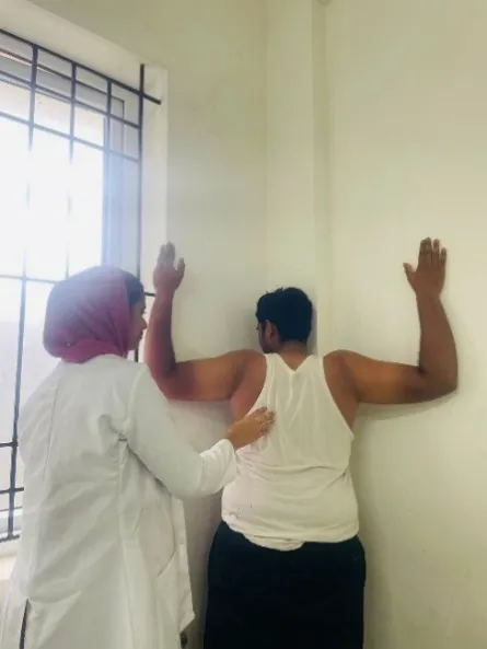
Figure 10: pectoralis major |
Patient and therapist in standing position |
Corner Stretch: Stand facing the corner of a room. Place both forearms on the walls with elbows just below shoulder height. Lean chest toward the corner until a stretch was felt. Hold for 30 seconds. 2-4 times per side. 3-5 days per week |
Table 3: Static stretching exercise
DATA ANALYSIS AND RESULTS
|
Variables |
Groups |
Test |
Mean |
Mean difference |
Standard deviation |
Paired ‘t’ value |
|
Pain |
Group A |
Pre test |
7.133 |
4.6 |
0.5069 |
35.6 |
|
Post test |
2.533 |
|||||
|
Group B |
Pre test |
7.733 |
2.8 |
0.414 |
26.19 |
|
|
Post test |
4.933 |
|||||
|
Neck disability |
Group A |
Pre test |
45.33 |
12.33 |
1.17 |
41.047 |
|
Post test |
33 |
|||||
|
Group B |
Pre test |
37.93 |
11.133 |
1.29 |
30.99 |
|
|
Post test |
26.8 |
|||||
|
Craniovertebral angle |
Group A |
Pre test |
40.96 |
7.97 |
0.3651 |
84.54 |
|
Post test |
48.93 |
|||||
|
Group B |
Pre test |
41.507 |
7.64 |
0.3853 |
76.86 |
|
|
Post test |
49.194 |
0.005 level of significance
The table shows the mean value, mean difference, standard deviation and paired ‘t’ test value between pre test and post test scores of pin, neck disability, craniovertbral angle among group A and group B.
|
Variables |
Groups |
Mean |
Mean difference |
Standard deviation |
Unpaired ‘t’ value |
|
Pain |
Group A |
4.6 |
1.8 |
0.4628 |
10.68 |
|
Group B |
2.8 |
||||
|
Neck disability |
Group A |
12.33 |
1.197 |
0.641 |
5.4199 |
|
Group B |
11.133 |
||||
|
Craniovertebral angle |
Group A |
7.97 |
0.33 |
0.2802 |
3.1562 |
|
Group B |
7.64 |
0.005 level of significance
The table shows the mean value, mean difference, standard deviation and unpaired ‘t’ test value between Group A and Group B scores of pain, neck disability and craniovertebral angle.
DISCUSSION
The study aimed to evaluate the effectiveness of Muscle Energy Technique (MET) versus static stretching exercises on pain, craniovertebral angle (CVA), and neck disability in individuals with Forward Head Posture (FHP) caused by Upper Crossed Syndrome (UCS).Pain was measured using the Numerical Pain Rating Scale (NPRS), neck disability by the Neck Disability Index (NDI), and posture (CVA) through Kinovea software before and after treatment.
Results showed a significant improvement in pain, neck disability, and CVA in both groups, but MET was more effective than static stretching.
Supporting evidence from Shwetha et al. (2024)—a randomized controlled trial with 30 participants—showed similar findings: both interventions improved pain and function, but MET produced greater effects.
CVA was described as a key indicator of head and neck alignment, influenced by muscle imbalance and posture. Improvement in CVA after exercises indicates better posture (≥50° considered normal), reduced imbalance, and improved proprioception.
Exercises reduced neck disability by Strengthening weak cervical and scapular muscles, correcting posture, increasing flexibility, and enhancing neuromuscular control and stability.
Pain relief may occur due to gate control mechanisms and endorphin release. The amount of exercise is crucial for effectiveness, though increasing intensity beyond a point does not enhance outcomes.
CONCLUSION
A comparative study was conducted to find the effectiveness of muscle energy technique versus static stretching exercise on pain, craniovertebral angle and neck disability in subjects with forward head posture due to upper cross syndrome.30 subjects with upper cross syndrome were included in the study and randomly divided into two equal groups.
Group A- Muscle energy technique and strengthening exercise
Group B – static stretching and strengthening exercises
Pain, craniovertebral angle, and neck disability were assessed before and after intervention by numerical pain rating scale, kinovea software and neck disability index.
From statistical results, it is concluded that the muscle energy technique and strengthening exercise are more effective in the management of pain, craniovertebral angle and neck disability among subjects with forward head posture due to upper cross syndrome.
Limitation:
Suggestions:
REFERENCES


Fairusa*, Divya J. Pawani*, Franklin Shaju M. K., A Study On The Effectiveness Of Muscle Energy Technique Versus Static Stretching Exercise On Pain, Craniovertebral Angle And Neck Disability In Subjects With Upper Cross Syndrome, Int. J. Sci. R. Tech., 2026, 3 (6), 1626-1634. https://doi.org/10.5281/zenodo.20936006
 10.5281/zenodo.20936006