

We use cookies to ensure our website works properly and to personalise your experience. Cookies policy
Department of Orthodontics and Dentofacial Orthopedics, Hitech Dental College and Hospital, Bhubaneswar, Odisha, India.
Three-dimensional (3D) bioprinting has emerged as an enabling technology in regenerative medicine with the potential to redefine the future of orthodontic treatment. By integrating living cells, biomaterials, and bioactive molecules through layer-by-layer fabrication, bioprinting enables the development of patient-specific constructs that closely replicate native craniofacial tissues. In orthodontics, this technology offers potential solutions for challenges such as alveolar bone deficiency, periodontal tissue loss, root resorption, craniofacial defects, and compromised tissue healing following orthodontic interventions. Recent advances in stem cell biology, bioactive hydrogels, tissue engineering, growth factor delivery, and digital manufacturing have expanded the scope of regenerative orthodontics beyond conventional biomechanical tooth movement. The incorporation of artificial intelligence, computer-aided design, cone-beam computed tomography, and additive manufacturing further supports precision treatment planning and customized regenerative therapies. Despite encouraging experimental outcomes, routine clinical implementation remains limited by challenges related to vascularization, scaffold maturation, manufacturing standardization, regulatory approval, long-term safety, and economic feasibility. Continued interdisciplinary collaboration among orthodontists, bioengineers, material scientists, and regulatory authorities is essential to translate laboratory innovations into predictable clinical applications. This article reviews the biological principles, technological advancements, clinical applications, translational challenges, and future perspectives of bioprinting, highlighting current evidence, existing limitations, and research priorities toward clinical translation.
Orthodontics has traditionally focused on correcting malocclusion through the application of controlled mechanical forces that induce remodeling of the periodontal ligament and alveolar bone. Although advances in biomechanics, appliance systems, and digital technologies have significantly improved treatment precision, the biological response of supporting tissues ultimately governs the efficiency, stability, and safety of orthodontic tooth movement. Consequently, successful treatment extends beyond achieving ideal occlusion and increasingly emphasizes the preservation of periodontal health, maintenance of alveolar bone integrity, and minimization of adverse effects such as root resorption, gingival recession, and bone dehiscence. ¹, ²
The growing understanding of craniofacial biology has shifted orthodontics toward a more biologically driven specialty, where tissue preservation and regeneration are becoming integral components of treatment planning. This evolution has coincided with recent advances in regenerative medicine, a multidisciplinary field that combines cellular biology, biomaterials science, engineering, and molecular therapeutics to restore the structure and function of damaged tissues. Within dentistry, regenerative approaches have already demonstrated potential applications in periodontal regeneration, implant dentistry, endodontics, and craniofacial reconstruction, encouraging similar innovations in orthodontic practice. ³, â´
Among emerging regenerative technologies, three-dimensional (3D) bioprinting has attracted increasing interest because of its ability to fabricate living, patient-specific tissue constructs with high spatial precision. Unlike conventional additive manufacturing, which produces inert structures from synthetic materials, bioprinting integrates living cells, bioactive molecules, and biodegradable biomaterials into organized three-dimensional architectures that closely mimic the native extracellular matrix. This technology enables simultaneous control over scaffold geometry, cellular distribution, and biological composition, thereby creating constructs capable of supporting tissue repair, regeneration, and functional integration after implantation. âµ, â
The relevance of bioprinting to orthodontics lies in the biological complexity of orthodontic tooth movement. Mechanical loading initiates a cascade of cellular and molecular events involving osteoblasts, osteoclasts, fibroblasts, endothelial cells, immune cells, and extracellular matrix components. These coordinated processes regulate bone resorption on the pressure side and bone formation on the tension side, allowing controlled dental movement within the alveolar process. However, compromised tissue quality resulting from periodontal disease, trauma, congenital anomalies, systemic disorders, or previous surgical interventions may impair this biological response and limit treatment outcomes. In such situations, regenerative strategies have the potential to complement conventional biomechanics by restoring the supporting tissues before, during, or after orthodontic therapy. â·, â¸
Recent developments in stem cell biology and tissue engineering have substantially expanded the therapeutic possibilities of bioprinting. Mesenchymal stem cells derived from bone marrow, periodontal ligament, dental pulp, gingiva, and other oral tissues exhibit remarkable proliferative and multilineage differentiation capacities, making them attractive candidates for craniofacial regeneration. When incorporated into biocompatible hydrogel-based bioinks, these cells can be precisely positioned within customized scaffolds that promote cell survival, vascularization, and tissue maturation. Simultaneously, advances in printable biomaterials have enabled the fabrication of constructs with controlled mechanical properties, degradation rates, and biological functionality that more closely replicate native craniofacial tissues. â¹, ¹â°
Parallel progress in digital dentistry has further accelerated the clinical potential of bioprinting. High-resolution cone-beam computed tomography, intraoral optical scanning, facial imaging, computer-aided design and manufacturing (CAD/CAM), and artificial intelligence now permit highly accurate digital reconstruction of craniofacial anatomy. These technologies facilitate the design of individualized scaffolds that precisely match patient-specific defects while optimizing structural integrity and biological performance. Such convergence of digital planning and regenerative manufacturing represents an important step toward precision orthodontics, where treatment is tailored according to each patient's anatomical, biological, and functional characteristics rather than relying solely on standardized therapeutic approaches. ¹¹, ¹²
The literature included in this narrative review was identified through searches of PubMed, Scopus, Web of Science, and Google Scholar using combinations of the keywords '3D bioprinting', 'orthodontics', 'tissue engineering', 'craniofacial regeneration', 'bioinks', 'stem cells', and 'regenerative medicine'. Preference was given to English-language articles, systematic reviews, landmark studies, and recent publications (2015–2026) relevant to orthodontic applications.
Despite encouraging experimental progress, the translation of bioprinting from laboratory research to routine orthodontic practice remains limited. Major challenges include maintaining long-term cellular viability, achieving adequate vascularization within engineered constructs, reproducing the hierarchical architecture of periodontal tissues, and ensuring predictable integration with host tissues. Additional barriers, including manufacturing standardization, regulatory approval, cost-effectiveness, and long-term clinical validation, must also be addressed before widespread clinical adoption can be achieved.
Nevertheless, rapid advances in biomaterials, stem cell biology, biofabrication technologies, digital dentistry, and artificial intelligence are expanding the possibilities of regenerative orthodontics. Rather than replacing conventional biomechanical principles, bioprinting is increasingly viewed as a complementary strategy capable of restoring the biological integrity of the dentoalveolar complex while facilitating personalized treatment approaches. This review aims to critically evaluate current advances in orthodontic bioprinting, summarize available biological and technological evidence, discuss translational challenges, and identify future research priorities.
2. Orthodontic Background
Orthodontic tooth movement (OTM) is a biologically regulated process initiated by controlled mechanical forces that induce remodeling of the periodontal ligament (PDL) and alveolar bone. Rather than being a purely mechanical phenomenon, OTM depends on coordinated cellular and molecular responses involving osteoclasts, osteoblasts, fibroblasts, endothelial cells, and inflammatory mediators, which collectively regulate bone resorption on the pressure side and bone formation on the tension side. ¹³, ¹â´
Although contemporary biomechanical principles have significantly improved treatment efficiency, successful orthodontic outcomes remain constrained by the biological response of the supporting tissues. Excessive or prolonged forces, reduced alveolar bone volume, periodontal inflammation, systemic diseases, and individual variations in bone metabolism may impair tissue remodeling, resulting in delayed tooth movement, root resorption, gingival recession, or compromised periodontal stability. ¹âµ, ¹â¶ These limitations underscore that biomechanics alone cannot overcome deficiencies in tissue quality or regenerative capacity.
Recent advances in regenerative biology have shifted the focus from merely moving teeth to preserving and restoring the dentoalveolar complex. Strategies incorporating stem cells, biomaterials, and tissue engineering have demonstrated the potential to regenerate alveolar bone and periodontal tissues while creating a more favorable biological environment for orthodontic treatment. Within this evolving paradigm, bioprinting represents a potential technology capable of fabricating patient-specific living constructs that may enhance tissue regeneration, improve treatment predictability, and broaden therapeutic options for patients with complex craniofacial or periodontal defects. ¹â·, ¹â¸
3. Evolution of Bioprinting
Bioprinting evolved from conventional three-dimensional (3D) printing, a technology initially developed for rapid prototyping in engineering and industrial manufacturing. The incorporation of living cells, biomaterials, and bioactive molecules transformed additive manufacturing into a biofabrication platform capable of producing biologically active tissue constructs. This advancement marked a major shift in regenerative medicine by enabling the fabrication of tissues that more closely replicate the structural and functional characteristics of native organs. ¹â¹, ²â°
Over the past two decades, advances in printing technologies, bioink formulations, scaffold design, and computer-aided design have significantly improved the precision, reproducibility, and biological performance of bioprinted constructs. The integration of patient-specific imaging with digital workflows has further enabled the fabrication of customized tissue constructs tailored to individual anatomical defects, supporting the transition toward precision regenerative medicine. ²¹, ²²
In dentistry, bioprinting has progressed from experimental investigations of dental pulp and periodontal regeneration to applications involving alveolar bone and craniofacial tissue engineering. Although orthodontic practice continues to rely primarily on biomechanical principles, emerging evidence suggests that bioprinting may overcome biological limitations by facilitating patient-specific regeneration of the dentoalveolar complex. Consequently, bioprinting is increasingly recognized as a regenerative platform with the potential to complement conventional orthodontic therapy and improve long-term clinical outcomes. ²³, ²â´
4. Principles of Bioprinting
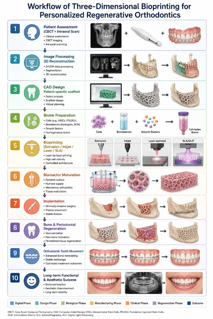
Bioprinting is founded on the precise spatial deposition of living cells, biomaterials, and bioactive molecules to fabricate three-dimensional constructs that replicate the architecture and biological function of native tissues. The workflow typically begins with patient-specific imaging using cone-beam computed tomography (CBCT), computed tomography (CT), or magnetic resonance imaging (MRI), followed by computer-aided design (CAD) to generate a digital model that guides layer-by-layer fabrication. This integration of imaging and digital design enables the production of customized constructs with high anatomical accuracy, an essential prerequisite for personalized regenerative therapies. ²âµ, ²â¶ The complete workflow is illustrated in Figure 1.
The success of bioprinting depends on the coordinated interaction of three fundamental components: viable cells, bioinks, and the printing platform. Cells must withstand the mechanical stresses generated during printing while retaining their proliferative and differentiation potential. Likewise, bioinks should exhibit appropriate rheological properties to ensure print fidelity without compromising cell viability. Following fabrication, printed constructs require a maturation phase during which extracellular matrix deposition, tissue organization, and functional integration are promoted under controlled biological conditions. ²â·, ²â¸
Beyond precise fabrication, successful tissue regeneration requires recreation of the native cellular microenvironment. Scaffold architecture, pore interconnectivity, mechanical stiffness, nutrient diffusion, and vascularization collectively influence cell behavior and tissue maturation. In orthodontic applications, these factors are particularly important because regenerated tissues must integrate with the periodontal ligament and alveolar bone while withstanding continuous biomechanical loading. Consequently, the effectiveness of bioprinting depends not only on manufacturing precision but also on its ability to reproduce the biological complexity of the dentoalveolar environment. ²â¹, ³â°
5. Bioprinting Technologies
Bioprinting technologies differ in their printing mechanisms, resolution, cell compatibility, and suitability for specific tissue types. Consequently, selecting an appropriate bioprinting platform requires balancing structural fidelity, printing speed, biomaterial properties, and post-printing cell viability. In craniofacial tissue engineering, extrusion-, inkjet-, laser-assisted-, and light-based bioprinting are the most extensively investigated techniques, each offering distinct advantages and limitations. ³¹, ³² A comparison of major bioprinting technologies is presented in Table 1.
Extrusion-based bioprinting is the most widely used technique for craniofacial applications because it can print high-viscosity bioinks containing large numbers of viable cells. The method is technically simple, scalable, and capable of producing mechanically stable constructs suitable for alveolar bone and periodontal regeneration. However, the relatively high shear stress generated during extrusion and its lower printing resolution may compromise cell viability and limit the fabrication of intricate periodontal microarchitectures. ³³, ³â´
Inkjet bioprinting deposits bioinks as microscopic droplets, providing rapid fabrication, low cost, and high cell viability. Nevertheless, its dependence on low-viscosity bioinks restricts the production of mechanically robust, load-bearing constructs, reducing its applicability for regenerating large craniofacial defects. Laser-assisted bioprinting achieves exceptional spatial resolution without nozzle-related clogging or shear stress, making it suitable for precise cell patterning. Despite these advantages, its high cost, complex instrumentation, and limited availability continue to hinder widespread clinical adoption. ³âµ, ³â
Light-based techniques, including stereolithography (SLA) and digital light processing (DLP), fabricate constructs through photopolymerization of photosensitive bioinks, producing excellent structural accuracy and smooth surface characteristics. These methods are particularly attractive for fabricating anatomically complex craniofacial scaffolds, although the availability of suitable photocrosslinkable biomaterials remains a limiting factor. Overall, no single bioprinting technology satisfies all clinical requirements. Current research therefore focuses on hybrid bioprinting platforms that combine the strengths of multiple techniques to improve construct fidelity, biological function, and clinical applicability in personalized regenerative orthodontics. ³â·, ³â¸
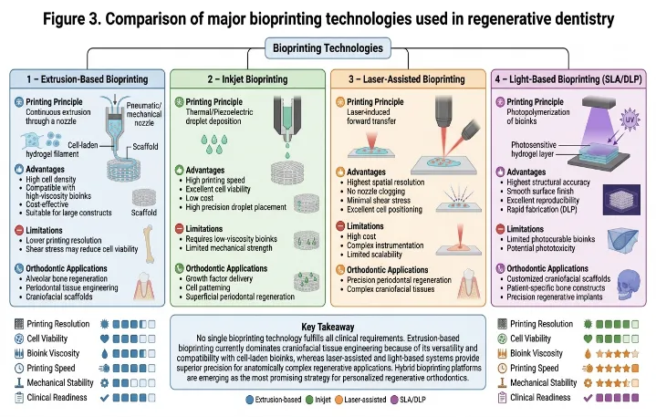
Current evidence indicates that extrusion-based bioprinting is the most extensively investigated platform for craniofacial tissue engineering because of its compatibility with cell-laden, high-viscosity bioinks and its ability to fabricate mechanically stable constructs. In contrast, inkjet and laser-assisted systems provide superior printing precision but remain constrained by bioink viscosity, manufacturing complexity, or cost. Although hybrid bioprinting strategies have demonstrated encouraging improvements in construct fidelity and biological performance, available evidence is derived predominantly from laboratory investigations and animal studies. Consequently, comparative clinical studies are required to determine the optimal bioprinting platform for specific orthodontic applications. The major bioprinting technologies are compared in Figure 3.
6. Bioinks and Biomaterials
Bioinks are the functional core of bioprinting, comprising living cells suspended within biocompatible biomaterials that provide mechanical support and a favorable microenvironment for tissue regeneration. An ideal bioink should combine excellent printability with high cell viability, appropriate mechanical strength, controlled biodegradation, and the ability to promote cell adhesion, proliferation, and lineage-specific differentiation. Because no single material fulfills all these requirements, bioink selection remains one of the most critical determinants of successful tissue fabrication. ³â¹, â´â°
Natural biomaterials, including collagen, gelatin, alginate, fibrin, hyaluronic acid, and gelatin methacrylate (GelMA), closely mimic the native extracellular matrix and support cellular attachment and biological activity. However, their limited mechanical strength restricts their use in load-bearing applications. In contrast, synthetic polymers such as polyethylene glycol (PEG), polycaprolactone (PCL), and polylactic acid (PLA) provide superior mechanical stability and controlled degradation but lack intrinsic bioactivity. Consequently, composite bioinks integrating natural and synthetic materials have emerged as a potential approach to balance biological performance with structural integrity. â´¹, â´²
Recent advances have shifted bioink development beyond printability toward enhanced biological functionality. Hydrogels remain the most widely used matrices because of their extracellular matrix-like properties, while decellularized extracellular matrix (dECM)-based bioinks, self-healing hydrogels, and stimuli-responsive biomaterials provide tissue-specific biochemical cues that improve cell survival, proliferation, and differentiation. These innovations have enabled more biomimetic tissue constructs with improved regenerative potential. â´³, â´â´
For orthodontic applications, bioinks are increasingly reinforced with osteoconductive materials such as hydroxyapatite, β-tricalcium phosphate, and bioactive glass to improve bone regeneration, osseointegration, and mechanical stability. â´âµ, â´â¶ In addition, nanoparticle-reinforced and ion-functionalized biomaterials have demonstrated enhanced osteogenic differentiation, angiogenesis, and extracellular matrix mineralization in preclinical studies, supporting their potential for craniofacial and periodontal tissue engineering. â´â·, â´â¸ However, variability in bioink composition, crosslinking strategies, and printing parameters continues to limit standardization and direct comparison among studies. Future research should focus on developing multifunctional bioinks capable of promoting vascularization, controlled growth factor delivery, and predictable clinical translation in personalized regenerative orthodontics. The characteristics of commonly used bioinks are summarized in Table 2.
7. Stem Cells
Stem cells constitute the biological foundation of bioprinting because they provide the capacity for self-renewal, proliferation, and differentiation required for tissue regeneration. Among the various cell populations investigated, mesenchymal stem cells (MSCs) remain the preferred choice for craniofacial tissue engineering owing to their osteogenic potential, immunomodulatory properties, ease of isolation, and favorable safety profile. Their incorporation into bioprinted constructs has significantly improved the regeneration of mineralized and soft tissues, making them indispensable for regenerative dentistry. â´â¹, âµâ°
The oral cavity offers an accessible and clinically relevant source of stem cells, including periodontal ligament stem cells (PDLSCs), dental pulp stem cells (DPSCs), stem cells from the apical papilla (SCAP), and gingival mesenchymal stem cells. PDLSCs are particularly attractive for orthodontic applications because of their ability to regenerate cementum, periodontal ligament, and alveolar bone, while DPSCs and SCAP contribute to mineralized tissue formation and craniofacial repair. Their ease of harvesting, high proliferative capacity, and reduced ethical concerns further support their translational potential. âµ¹, âµ²
Recent advances in induced pluripotent stem cell (iPSC) technology have expanded opportunities for patient-specific regenerative therapies by generating pluripotent cells without the ethical concerns associated with embryonic stem cells. However, challenges related to genomic instability, tumorigenicity, manufacturing complexity, and regulatory approval currently limit their clinical application. Consequently, adult dental-derived stem cells remain the most practical and clinically relevant cell source for orthodontic bioprinting. âµ³, âµâ´
Successful regeneration depends not only on stem cell selection but also on the interaction between cells, biomaterials, and biochemical signals within the engineered microenvironment. Mechanical stimulation, scaffold architecture, and bioactive cues collectively regulate stem cell survival and lineage commitment after implantation. Optimizing these interactions is expected to enhance periodontal regeneration, alveolar bone remodeling, and the long-term success of bioprinted therapies in orthodontics. The major stem cell sources relevant to orthodontic bioprinting are summarized in Table 3. âµâµ, âµâ
Although preclinical investigations have demonstrated encouraging regenerative outcomes using dental-derived mesenchymal stem cells, variations in cell isolation methods, scaffold composition, and experimental models continue to limit direct comparison among studies. Well-designed clinical trials are therefore required to establish standardized protocols and confirm their long-term safety and efficacy in orthodontic applications.
8. Tissue Engineering
Tissue engineering provides the biological and engineering framework that underpins bioprinting by integrating three essential components: regenerative cells, biomaterial scaffolds, and bioactive signaling molecules. The coordinated interaction of this tissue engineering triad aims to recreate the native microenvironment required for tissue repair, enabling controlled cell proliferation, differentiation, extracellular matrix deposition, and functional tissue regeneration. âµâ·, âµâ¸ The tissue engineering triad is illustrated in Figure 2.
Scaffolds serve as temporary three-dimensional templates that support cell attachment and guide new tissue formation until they are gradually replaced by native extracellular matrix. An ideal scaffold should possess interconnected porosity, appropriate mechanical strength, biocompatibility, controlled biodegradation, and adequate permeability to facilitate nutrient diffusion and vascular ingrowth. Recent advances in scaffold fabrication, including multiphasic and gradient scaffolds, have improved the regeneration of complex craniofacial tissues by more closely replicating the structural organization of bone and periodontal tissues. âµâ¹, â¶â°
Despite these advances, successful tissue regeneration remains limited by inadequate vascularization, insufficient mechanical integration, and the difficulty of reproducing the hierarchical architecture of native tissues. Bioreactor-based maturation systems and dynamic culture conditions have therefore been developed to improve tissue organization before implantation and enhance the functional performance of engineered constructs. ⶹ, ⶲ
In orthodontics, these principles provide the biological basis for regenerating alveolar bone, periodontal ligament, and other supporting tissues compromised by trauma, periodontal disease, congenital defects, or orthodontic treatment. When integrated with bioprinting, tissue engineering enables the fabrication of patient-specific regenerative constructs, representing a significant step toward biologically driven and personalized orthodontic care.
Contemporary evidence indicates that tissue engineering strategies integrating stem cells, biomimetic scaffolds, and bioactive signaling molecules produce superior regenerative outcomes compared with scaffold-only approaches. Multiphasic scaffolds that more closely replicate the hierarchical organization of periodontal and osseous tissues have demonstrated enhanced cellular organization, vascular infiltration, and tissue-specific differentiation in preclinical studies. Nevertheless, inadequate vascularization, inconsistent scaffold degradation, and limited long-term in vivo validation remain the principal barriers to clinical translation. Future research should prioritize standardized scaffold fabrication, functional vascularization, and well-designed clinical trials to establish predictable regenerative protocols for orthodontic applications.
9. Bone Biology
Bone remodeling is a dynamic process that maintains skeletal homeostasis through the coordinated activities of osteoclast-mediated bone resorption and osteoblast-mediated bone formation. During orthodontic tooth movement, mechanical forces alter the local microenvironment of the periodontal ligament, initiating a cascade of biochemical signals that regulate alveolar bone remodeling. The magnitude and duration of these forces determine the balance between bone resorption and deposition, thereby influencing the rate, efficiency, and stability of tooth movement. ⶳ, â¶â´
Osteocytes function as the primary mechanosensors within bone, converting mechanical stimuli into biochemical signals that regulate cellular activity. Among the molecular pathways involved, the receptor activator of nuclear factor-κB (RANK), RANK ligand (RANKL), and osteoprotegerin (OPG) signaling axis plays a pivotal role in controlling osteoclast differentiation and maintaining bone homeostasis. Disruption of this balance may contribute to adverse effects such as delayed tooth movement, alveolar bone loss, and external apical root resorption during orthodontic treatment. â¶âµ, â¶â
An understanding of bone biology is fundamental to the design of bioprinted constructs intended for craniofacial regeneration. Biomaterials that promote osteogenesis, angiogenesis, and mineralization while providing adequate mechanical support are essential for successful integration with the host bone. Recent advances in nanostructured scaffolds and bioactive ion-functionalized biomaterials have demonstrated enhanced cellular attachment, vascularization, and matrix mineralization, supporting their application in alveolar bone regeneration and personalized regenerative orthodontics. â¶â·, â¶â¸
10. Growth Factors and Signaling
Growth factors regulate the cellular events that govern tissue repair, angiogenesis, and extracellular matrix remodeling, making them indispensable components of tissue-engineered and bioprinted constructs. Rather than acting independently, these molecules function through interconnected signaling networks that coordinate stem cell recruitment, proliferation, differentiation, and tissue maturation. Controlled incorporation of growth factors into bioinks and scaffolds has therefore emerged as an effective strategy to enhance the regenerative potential of bioprinted tissues. â¶â¹, â·â°
Among the key osteogenic mediators, bone morphogenetic proteins (BMPs) stimulate osteoblast differentiation and bone formation, whereas vascular endothelial growth factor (VEGF) promotes angiogenesis, ensuring adequate oxygen and nutrient delivery to regenerating tissues. Platelet-derived growth factor (PDGF) facilitates cell migration and proliferation, while transforming growth factor-β (TGF-β) regulates extracellular matrix synthesis and tissue remodeling. Fibroblast growth factors (FGFs) further contribute to wound healing, angiogenesis, and cellular proliferation, collectively creating a favorable microenvironment for tissue regeneration. â·¹, â·²
In addition to soluble growth factors, intracellular signaling pathways such as Wnt/β-catenin, Notch, and Hedgehog regulate osteogenesis, stem cell differentiation, and skeletal development. Advances in biomaterial engineering have enabled localized and sustained delivery of these bioactive molecules through bioprinted scaffolds, minimizing systemic exposure while enhancing biological efficacy. Such controlled signaling strategies may improve alveolar bone regeneration, periodontal healing, and integration of engineered tissues, thereby supporting the development of personalized regenerative therapies in orthodontics. â·³, â·â´ The principal growth factors involved in tissue regeneration are summarized in Table 4.
11. Orthodontic Applications
Although bioprinting remains in the early stages of clinical translation, its potential applications in orthodontics are rapidly expanding. Unlike conventional orthodontic treatment, which primarily relies on biomechanical manipulation of teeth, bioprinting offers the possibility of biologically reconstructing the supporting dentoalveolar tissues. This regenerative approach may improve treatment outcomes in patients with alveolar bone deficiencies, periodontal defects, craniofacial anomalies, and compromised healing capacity, thereby extending the scope of orthodontic therapy beyond tooth movement alone. â·âµ, â·â
One of the most potential applications is alveolar bone regeneration. Insufficient alveolar bone volume frequently limits orthodontic tooth movement and may compromise treatment stability. Patient-specific bioprinted scaffolds incorporating osteogenic cells and bioactive biomaterials have demonstrated favourable preclinical findings in promoting new bone formation while maintaining the anatomical contour of the alveolar ridge. Such constructs may reduce dependence on conventional bone grafting procedures and facilitate orthodontic treatment in patients with localized bone defects. â·â·, â·â¸
Bioprinting also shows considerable potential for periodontal regeneration, particularly in reconstructing the complex architecture of the periodontal ligament, cementum, and alveolar bone. Multiphasic scaffolds capable of spatially organizing different cell populations and biomaterials may improve regeneration of the periodontal attachment apparatus, thereby enhancing periodontal support before, during, or after orthodontic treatment. This approach may be particularly beneficial for adult patients with reduced periodontal support or a history of periodontal disease. â·â¹, â¸â°
Another important application is the management of craniofacial defects, including alveolar clefts and post-traumatic bone loss. Integration of cone-beam computed tomography with computer-aided design enables fabrication of patient-specific constructs that closely match complex anatomical defects, potentially improving graft adaptation and reducing surgical morbidity. Emerging research also suggests future applications in temporomandibular joint regeneration and bioengineered dental tissues, although these approaches remain largely experimental. ⸹, ⸲
Despite these advances, evidence supporting orthodontic applications of bioprinting is derived predominantly from laboratory investigations and preclinical animal studies. Well-designed clinical trials are still required to determine long-term safety, functional integration, treatment predictability, and cost-effectiveness before these technologies can be incorporated into routine orthodontic practice. Nevertheless, current evidence indicates that bioprinting is expected to complement conventional biomechanics by introducing biologically guided regenerative strategies that support personalized orthodontic care. Current and future orthodontic applications are summarized in Table 5.
12. Digital Dentistry and Artificial Intelligence
Digital dentistry has become an integral component of modern bioprinting workflows by enabling accurate visualization, planning, and fabrication of patient-specific regenerative constructs. High-resolution cone-beam computed tomography (CBCT), intraoral scanners, and computer-aided design/computer-aided manufacturing (CAD/CAM) technologies facilitate precise reconstruction of craniofacial anatomy, allowing customized scaffold design that closely conforms to individual defects. Such image-guided workflows improve structural accuracy while supporting the development of personalized regenerative therapies. ⸳, â¸â´
Artificial intelligence (AI) further enhances bioprinting by optimizing scaffold design, predicting bioink performance, and refining printing parameters to improve construct quality and reproducibility. Machine learning algorithms can analyze large biological and imaging datasets to identify optimal scaffold architectures, simulate tissue maturation, and predict regenerative outcomes. Integration of AI with finite element analysis also enables biomechanical simulation of orthodontic tooth movement, providing valuable insights into scaffold behavior and tissue adaptation under functional loading. â¸âµ, â¸â
The convergence of digital imaging, AI, and bioprinting represents a milestone toward precision orthodontics. By integrating patient-specific anatomical, biomechanical, and biological information into a single digital workflow, these technologies have the potential to improve treatment planning, reduce manufacturing variability, and facilitate the clinical translation of personalized regenerative strategies. Nevertheless, widespread implementation will require standardized algorithms, robust clinical validation, and seamless integration into existing digital orthodontic workflows.
13. Clinical Translation, Regulatory and Ethical Considerations
Despite significant advances in bioprinting, its translation into routine orthodontic practice remains limited. Most evidence is derived from in vitro investigations and preclinical animal studies, with relatively few technologies progressing to clinical evaluation. The successful implementation of bioprinted constructs requires reproducible manufacturing, long-term cell viability, adequate vascularization, predictable host integration, and sustained functional performance under physiological loading. In addition, standardization of bioinks, printing parameters, sterilization methods, and quality control protocols is essential to ensure product consistency and clinical reliability. â¸â·, â¸â¸
The regulatory pathway for bioprinted products is particularly complex because these constructs combine living cells, biomaterials, and bioactive molecules, placing them at the intersection of medical devices, biologics, and tissue-engineered products. Consequently, comprehensive assessment of biocompatibility, biodegradation, immunogenicity, mechanical stability, and long-term safety is required before regulatory approval can be granted. The absence of globally harmonized regulatory frameworks further complicates the clinical translation of bioprinting technologies. Major barriers to clinical implementation are summarized in Table 6.
Ethical considerations extend beyond stem cell procurement to include informed consent, patient privacy, equitable access to advanced therapies, and responsible management of patient-derived biological materials. As artificial intelligence becomes increasingly integrated into digital planning and manufacturing workflows, additional concerns regarding algorithm transparency, data security, and clinical accountability must also be addressed. Collaborative efforts among clinicians, bioengineers, regulatory agencies, and industry partners will be essential to establish standardized protocols and facilitate the safe and responsible adoption of bioprinting in personalized regenerative orthodontics.
14. Future Perspectives
The future of bioprinting in orthodontics will be driven by continued advances in biomaterials, stem cell biology, and precision regenerative medicine. Emerging innovations such as four-dimensional (4D) bioprinting, in which printed constructs undergo controlled structural or functional changes in response to biological or mechanical stimuli, may overcome current limitations related to tissue maturation and adaptation. Similarly, smart bioinks capable of controlled release of growth factors, antimicrobial agents, and immunomodulatory molecules are expected to improve tissue regeneration while minimizing postoperative complications.
Future progress will increasingly depend on integrating advanced computational tools with patient-specific bioprinting workflows to optimize scaffold design and improve the predictability of regenerative outcomes. Research should also prioritize vascularized bioprinting, in situ bioprinting, gene-editing technologies, and organ-on-a-chip platforms to better replicate the biological complexity of craniofacial tissues.
Equally important will be the establishment of standardized manufacturing protocols, multicenter clinical trials, and harmonized regulatory frameworks to facilitate safe clinical translation. Continued collaboration among orthodontists, bioengineers, biomaterial scientists, cell biologists, and regulatory authorities will be essential to transform bioprinting from an experimental technology into a clinically reliable component of regenerative orthodontic care.
Table 1. Comparison of major bioprinting technologies and their relevance to orthodontics
| Technology | Printing principle | Major advantages | Limitations | Potential orthodontic applications |
|---|---|---|---|---|
| Extrusion-based bioprinting | Continuous extrusion of cell-laden bioinks through pneumatic or mechanical pressure | High cell density, compatible with viscous bioinks, suitable for large tissue constructs, cost-effective | Lower printing resolution, shear stress may reduce cell viability | Alveolar bone regeneration, periodontal tissue engineering, craniofacial scaffold fabrication |
| Inkjet bioprinting | Deposition of microscopic bioink droplets using thermal or piezoelectric actuation | High printing speed, low cost, excellent cell viability, precise droplet placement | Limited to low-viscosity bioinks, poor mechanical strength for large constructs | Cell patterning, growth factor delivery, superficial periodontal regeneration |
| Laser-assisted bioprinting | Laser-induced transfer of bioinks without nozzle contact | High spatial resolution, minimal shear stress, accurate cell positioning | Expensive equipment, technically demanding, limited scalability | Precision periodontal regeneration, complex craniofacial tissue engineering |
| Stereolithography (SLA) | Layer-by-layer photopolymerization of photosensitive bioinks | Excellent structural accuracy, smooth surface finish, high reproducibility | Restricted to photocurable biomaterials, potential phototoxicity | Customized craniofacial scaffolds, patient-specific bone constructs |
| Digital Light Processing (DLP) | Projection of patterned light to polymerize an entire layer simultaneously | Faster fabrication than SLA, high resolution, improved printing efficiency | Limited bioink compatibility, specialized instrumentation | Personalized regenerative scaffolds and precision craniofacial reconstruction |
| Hybrid bioprinting | Combination of multiple printing techniques within a single construct | Combines high resolution with mechanical strength and biological functionality | Increased manufacturing complexity and cost | Multitissue regeneration, periodontal complex engineering, personalized orthodontic constructs |
Legend
This table summarizes the working principles, principal advantages, inherent limitations, and potential orthodontic applications of the major bioprinting technologies currently used in regenerative dentistry. Extrusion-, inkjet-, laser-assisted-, and light-based bioprinting techniques differ in printing resolution, cell compatibility, bioink requirements, and suitability for craniofacial tissue engineering, highlighting the need to select printing platforms according to specific clinical applications.
Table 2. Bioinks and biomaterials used in craniofacial and orthodontic bioprinting
| Biomaterial/Bioink | Classification | Key biological properties | Limitations | Orthodontic applications |
|---|---|---|---|---|
| Collagen | Natural polymer | Excellent biocompatibility, promotes cell adhesion, reproduce native extracellular matrix | Poor mechanical strength, rapid degradation | Periodontal ligament regeneration, soft tissue engineering |
| Gelatin methacrylate (GelMA) | Modified natural hydrogel | High printability, photocrosslinkable, supports stem cell proliferation and differentiation | Requires photoinitiators, moderate mechanical stability | Periodontal regeneration, alveolar bone engineering |
| Alginate | Natural polysaccharide | Mild gelation conditions, excellent printability, maintains high cell viability | Limited cell adhesion, relatively low bioactivity | Cell encapsulation and scaffold fabrication |
| Hyaluronic acid | Natural glycosaminoglycan | Promotes cell migration, angiogenesis, and extracellular matrix formation | Weak mechanical properties | Soft tissue regeneration and periodontal repair |
| Fibrin | Natural protein hydrogel | Excellent biocompatibility, supports angiogenesis and wound healing | Rapid degradation, low structural stability | Periodontal and gingival tissue regeneration |
| Polyethylene glycol (PEG) | Synthetic polymer | Tunable mechanical properties, controlled degradation, reproducible fabrication | Bioinert unless modified with bioactive molecules | Composite bioinks and customized scaffold fabrication |
| Polycaprolactone (PCL) | Synthetic biodegradable polymer | High mechanical strength, slow degradation, excellent structural support | Limited intrinsic bioactivity | Load-bearing craniofacial scaffolds and alveolar bone regeneration |
| Polylactic acid (PLA) | Synthetic biodegradable polymer | Good mechanical stability and printability | Brittle, limited cellular interaction | Customized bone scaffolds and supportive frameworks |
| Hydroxyapatite (HA) | Bioactive ceramic | Excellent osteoconductivity, promotes mineralization and bone formation | Brittle when used alone | Alveolar bone augmentation and craniofacial reconstruction |
| β-Tricalcium phosphate (β-TCP) | Bioactive ceramic | Osteoconductive, biodegradable, stimulates bone remodeling | Lower mechanical strength than cortical bone | Bone defect repair and alveolar ridge regeneration |
| Bioactive glass | Bioactive ceramic | Enhances osteogenesis, angiogenesis, and mineral deposition | Difficult to use as a standalone scaffold | Bone regeneration and composite bioink reinforcement |
| Decellularized extracellular matrix (dECM) | Natural tissue-derived bioink | Tissue-specific biochemical cues, promotes cell differentiation and tissue maturation | Batch variability and complex preparation | Personalized periodontal and craniofacial tissue engineering |
| Composite bioinks | Hybrid (natural + synthetic) | Combine biological activity with improved mechanical strength and printability | Increased formulation complexity | Patient-specific multiphasic constructs for periodontal and alveolar regeneration |
Legend
This table compares the biological characteristics, mechanical properties, limitations, and orthodontic applications of commonly used natural polymers, synthetic polymers, bioactive ceramics, and composite bioinks employed in craniofacial tissue engineering. The information highlights how biomaterial selection influences printability, cell viability, scaffold stability, and regenerative outcomes.
Table 3. Stem cell sources and their regenerative potential in orthodontics
| Stem cell source | Tissue of origin | Biological characteristics | Regenerative potential | Potential orthodontic applications |
|---|---|---|---|---|
| Bone marrow-derived mesenchymal stem cells (BM-MSCs) | Bone marrow | High osteogenic potential, immunomodulatory properties, multilineage differentiation | Bone and cartilage regeneration | Alveolar bone augmentation and craniofacial reconstruction |
| Periodontal ligament stem cells (PDLSCs) | Periodontal ligament | Differentiate into cementoblasts, osteoblasts, and fibroblasts; maintain periodontal homeostasis | Periodontal ligament, cementum, and alveolar bone regeneration | Periodontal regeneration, enhancement of orthodontic tooth movement, root surface repair |
| Dental pulp stem cells (DPSCs) | Dental pulp | High proliferative capacity, angiogenic and neurogenic potential | Mineralized tissue and pulp regeneration | Alveolar bone repair and dentin–pulp complex regeneration |
| Stem cells from the apical papilla (SCAP) | Apical papilla of developing teeth | Rapid proliferation and strong odontogenic potential | Root development and mineralized tissue formation | Craniofacial regeneration and regenerative endodontic–orthodontic applications |
| Gingival mesenchymal stem cells (GMSCs) | Gingival connective tissue | Easily accessible, rapid proliferation, immunomodulatory activity | Soft tissue and periodontal regeneration | Gingival augmentation and periodontal tissue repair |
| Adipose-derived stem cells (ADSCs) | Adipose tissue | Abundant source, high proliferative capacity, multilineage differentiation | Bone, cartilage, and soft tissue regeneration | Craniofacial tissue engineering and scaffold-based regeneration |
| Induced pluripotent stem cells (iPSCs) | Reprogrammed somatic cells | Pluripotent, patient-specific, unlimited expansion potential | Regeneration of multiple craniofacial tissues | Personalized regenerative therapies and disease modeling (experimental) |
| Embryonic stem cells (ESCs) | Inner cell mass of blastocyst | Unlimited self-renewal and pluripotency | Broad tissue regenerative potential | Primarily experimental because of ethical and regulatory concerns |
Legend
This table summarizes the major stem cell populations investigated for craniofacial tissue engineering, including their tissue sources, biological characteristics, regenerative capacity, and potential orthodontic applications. Dental-derived mesenchymal stem cells, particularly periodontal ligament stem cells and dental pulp stem cells, represent the most clinically relevant cell populations for regenerative orthodontics.
Table 4. Major growth factors and signaling pathways involved in bioprinting and regenerative orthodontics
| Growth factor/Signaling pathway | Primary biological function | Role in tissue regeneration | Potential orthodontic application |
|---|---|---|---|
| Bone morphogenetic proteins (BMP-2, BMP-7) | Promote osteoblast differentiation and osteogenesis | Stimulate bone formation and mineralization | Alveolar bone regeneration, bone defect reconstruction, accelerated healing |
| Vascular endothelial growth factor (VEGF) | Stimulates angiogenesis and vascular permeability | Enhances vascularization of engineered tissues, improving nutrient and oxygen supply | Improved survival of bioprinted constructs and regeneration of periodontal and alveolar tissues |
| Platelet-derived growth factor (PDGF) | Regulates cell migration, proliferation, and chemotaxis | Promotes recruitment of mesenchymal stem cells and periodontal fibroblasts | Periodontal regeneration, wound healing, and enhanced tissue remodeling |
| Transforming growth factor-β (TGF-β) | Controls extracellular matrix synthesis and cell differentiation | Regulates collagen deposition and tissue remodeling | Regeneration of periodontal ligament and connective tissues |
| Fibroblast growth factors (FGFs) | Stimulate cell proliferation, angiogenesis, and wound healing | Promote soft tissue repair and vascular development | Gingival regeneration and periodontal healing |
| RANK/RANKL/OPG pathway | Regulates osteoclast differentiation and bone remodeling | Maintains balance between bone resorption and bone formation | Biological modulation of orthodontic tooth movement and alveolar bone remodeling |
| Wnt/β-catenin signaling | Regulates osteogenesis and stem cell differentiation | Enhances bone regeneration and mineralization | Promotion of alveolar bone formation and regenerative orthodontics |
| Notch signaling | Controls cell proliferation and lineage commitment | Maintains stem cell homeostasis and tissue organization | Regulation of periodontal ligament regeneration |
| Hedgehog signaling | Regulates embryonic development and skeletal morphogenesis | Supports craniofacial tissue formation and osteogenic differentiation | Craniofacial regeneration and bone tissue engineering |
| Controlled growth factor delivery systems | Sustained and localized release of bioactive molecules | Improves biological efficacy while minimizing systemic exposure | Bioactive bioprinted scaffolds for personalized periodontal and alveolar regeneration |
Legend
This table outlines the principal growth factors and molecular signaling pathways that regulate osteogenesis, angiogenesis, extracellular matrix remodeling, and stem cell differentiation during tissue regeneration. Their potential applications in periodontal regeneration, alveolar bone repair, and orthodontic tissue engineering are also summarized.
Table 5. Current and emerging applications of bioprinting in orthodontics
| Orthodontic application | Clinical indication | Bioprinting strategy | Potential clinical benefits | Current level of evidence |
|---|---|---|---|---|
| Alveolar bone regeneration | Alveolar bone deficiency, localized bone defects | Cell-laden osteoconductive scaffolds incorporating MSCs and bioactive biomaterials | Enhanced bone volume, improved orthodontic tooth movement, reduced need for autogenous bone grafts | Preclinical |
| Periodontal tissue regeneration | Periodontal attachment loss, gingival recession | Multiphasic bioprinted constructs containing PDLSCs, cementoblasts, and osteogenic cells | Regeneration of periodontal ligament, cementum, and alveolar bone | Preclinical |
| Alveolar cleft reconstruction | Cleft lip and palate patients | Patient-specific bioprinted bone grafts designed using CBCT and CAD/CAM | Improved graft adaptation, reduced donor-site morbidity, enhanced orthodontic rehabilitation | Early preclinical |
| Craniofacial bone reconstruction | Traumatic defects, tumor resection, congenital anomalies | Customized bioactive scaffolds reinforced with osteogenic cells | Anatomically accurate reconstruction with improved tissue integration | Preclinical |
| Temporomandibular joint (TMJ) regeneration | Condylar defects and TMJ degeneration | Osteochondral bioprinted constructs incorporating cartilage and bone-forming cells | Restoration of joint structure and function | Experimental |
| Personalized regenerative scaffolds | Patient-specific dentoalveolar defects | AI-assisted scaffold design with customized bioprinting | Improved anatomical fit and individualized treatment planning | Experimental |
| Bioactive drug-delivery scaffolds | Patients requiring enhanced tissue healing | Bioprinted constructs with controlled release of growth factors or therapeutic agents | Localized biological stimulation with reduced systemic exposure | Experimental |
| Future bioengineered periodontal complex | Severe periodontal destruction | Integrated regeneration of periodontal ligament, cementum, and alveolar bone | Biological restoration of the dentoalveolar attachment apparatus | Conceptual/Experimental |
Legend
This table summarizes the present and potential future applications of bioprinting in orthodontics, including alveolar bone regeneration, periodontal tissue engineering, craniofacial reconstruction, temporomandibular joint regeneration, customized scaffolds, and biologically active drug-delivery systems. The current level of scientific evidence is indicated for each application.
Table 6. Current challenges and future directions in orthodontic bioprinting
| Challenge | Current limitation | Potential solution | Future research priority |
|---|---|---|---|
| Vascularization | Limited nutrient diffusion and poor survival of large bioprinted constructs | Prevascularized scaffolds, angiogenic bioinks, endothelial cell incorporation | Development of functional vascular networks for clinically relevant tissue constructs |
| Cell viability and maturation | Reduced cell survival due to printing-induced mechanical stress and insufficient tissue maturation | Optimization of printing parameters, advanced bioinks, dynamic bioreactor culture | Long-term functional maturation and improved post-implantation survival |
| Biomaterial optimization | Difficulty balancing printability, mechanical strength, and bioactivity | Multifunctional composite bioinks and smart biomaterials | Bioinks with controlled biodegradation, immunomodulation, and sustained bioactive molecule release |
| Mechanical stability | Inadequate load-bearing capacity of regenerated tissues | Hybrid scaffolds reinforced with ceramics or biodegradable polymers | Biomimetic scaffolds with improved biomechanical performance |
| Clinical translation | Predominance of in vitro and animal studies with limited human evidence | Well-designed multicenter clinical trials and standardized treatment protocols | Generation of high-level clinical evidence supporting orthodontic applications |
| Manufacturing standardization | Variability in bioinks, printing parameters, and quality control | Standardized manufacturing protocols and Good Manufacturing Practice (GMP) compliance | Reproducible large-scale production of bioprinted constructs |
| Regulatory approval | Lack of harmonized international regulations for cell-based bioprinted products | Collaboration among regulatory agencies, researchers, and industry | Development of globally accepted regulatory pathways for clinical implementation |
| Cost and accessibility | High equipment costs and technical expertise required | Automation, scalable manufacturing, and cost-effective printing technologies | Improved affordability and broader clinical adoption |
| Digital integration | Limited interoperability among imaging, CAD/CAM, AI, and bioprinting systems | Unified digital workflows and AI-assisted treatment planning | Fully integrated precision regenerative orthodontic platforms |
| Future innovations | Early-stage development of emerging technologies | 4D bioprinting, in situ bioprinting, digital twins, gene editing, and organ-on-a-chip systems | Personalized, biologically responsive regenerative therapies for routine orthodontic practice |
Legend
This table highlights the major scientific, technical, regulatory, and clinical challenges limiting the routine implementation of bioprinting in orthodontics and outlines emerging strategies and research priorities aimed at facilitating successful clinical translation.
Figure 1. Workflow of three-dimensional bioprinting for personalized regenerative orthodontics.
Legend
Schematic illustration of the complete bioprinting workflow, beginning with patient assessment using cone-beam computed tomography (CBCT) and intraoral scanning, followed by image processing, computer-aided design (CAD), bioink preparation, three-dimensional bioprinting, construct maturation, surgical implantation, tissue regeneration, and subsequent orthodontic treatment. The workflow demonstrates the integration of digital technologies, tissue engineering, and regenerative medicine to enable personalized orthodontic care.
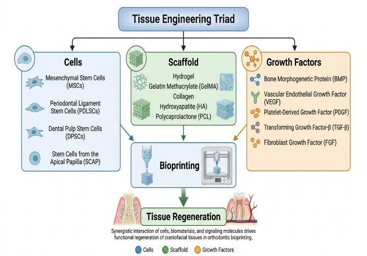
Figure 2. Tissue engineering triad used in orthodontic bioprinting.
Legend
Illustration of the tissue engineering triad consisting of regenerative cells, biomaterial scaffolds, and bioactive growth factors that collectively support tissue regeneration. Mesenchymal stem cells (MSCs), periodontal ligament stem cells (PDLSCs), dental pulp stem cells (DPSCs), and stem cells from the apical papilla (SCAP) are incorporated within biocompatible scaffolds such as hydrogels, gelatin methacrylate (GelMA), collagen, hydroxyapatite (HA), and polycaprolactone (PCL), together with osteogenic and angiogenic growth factors to promote regeneration of periodontal and craniofacial tissues.
Figure 3. Comparison of major bioprinting technologies used in regenerative dentistry.
Legend
Comparison of extrusion-based, inkjet, laser-assisted, and light-based (stereolithography/digital light processing) bioprinting technologies with respect to their printing mechanisms, advantages, limitations, and potential orthodontic applications. The figure highlights the complementary characteristics of each platform and the emerging role of hybrid bioprinting strategies for personalized regenerative orthodontics.
CONCLUSION
Bioprinting represents a significant advancement in regenerative medicine with the potential to redefine the biological foundation of orthodontic treatment. By integrating tissue engineering, stem cell biology, biomaterials, growth factor delivery, and digital technologies, it offers opportunities to regenerate alveolar bone, periodontal tissues, and other craniofacial structures that are critical for successful orthodontic outcomes. Although current applications remain predominantly at the experimental and preclinical stages, continuous progress in biofabrication technologies has substantially expanded the prospects for personalized regenerative orthodontics.
The successful clinical translation of bioprinting will depend on overcoming challenges related to vascularization, construct maturation, long-term functionality, manufacturing standardization, regulatory approval, and economic feasibility. Equally important is the generation of high-quality clinical evidence to establish the safety, efficacy, and long-term predictability of bioprinted therapies within routine orthodontic practice.
Rather than replacing conventional biomechanics, bioprinting is likely to complement existing orthodontic treatment by providing biologically guided regenerative solutions tailored to individual patient needs. Continued interdisciplinary collaboration among orthodontists, bioengineers, biomaterial scientists, cell biologists, and regulatory authorities will be essential to accelerate clinical translation. As these scientific and technological advances converge, bioprinting is expected to play an increasingly important role in the evolution of precision orthodontics, supporting a transition from conventional tooth movement toward biologically integrated, patient-specific regenerative care.
16. Declarations
16.1 Ethical approval
Not required for narrative review.
16.2 Conflict of Interest
The authors declare no conflict of interest.
16.3 Acknowledgements
None.
16.4 Data Availability
Not applicable.
16.5 Funding
No funding received.
16.6 AI-Assisted Writing Disclosure
An AI-assisted writing tool was utilized to improve the technical clarity, language quality, and structural organization of the manuscript during the editorial revision phase. All scientific content, literature interpretation, critical analysis, and final editorial decisions were performed exclusively by the authors, who take full responsibility for the accuracy and integrity of the work.
REFERENCES


Dr. Subash Chandra Nayak, Dr. Anshuman Mishra, Bioprinting in Orthodontics: From Tissue Engineering to Personalized Regenerative Medicine, Int. J. Sci. R. Tech., 2026, 3 (10), 1307-1328. https://doi.org/10.5281/zenodo.23683498
 10.5281/zenodo.23683498