

We use cookies to ensure our website works properly and to personalise your experience. Cookies policy
Department of Computer Science and Engineering, M.S. Engineering College, Bengaluru, India Affiliated to Visvesvaraya Technological University, Belagavi, Karnataka, India.
Timely diagnosis and precise detection of malignant melanomas and complicated skin diseases can help raise survival chances and facilitate effective treatment processes for such patients. Traditional diagnosis is mainly based on subjective visual evaluations within specific schemes like ABCDE, which makes them prone to considerable variability and human errors. In this paper, we introduce an automated and objective intelligence system using a CNN-based neural network that incorporates recurrent modules. Derma-scopy pictures go through a digital pre-processing stage involving median filtering, segmentation, and high-pass filter-based unsharp masking. Two types of feature space representation techniques include conventional deep features generated by automatic layers of the CNN and manually crafted descriptors using the HOG approach. The model classifies several categories of problems, including benign nevi, malignant melanomas, basal cell carcinoma, and various infectious skin diseases such as varicella, impetigo, and scabies. Our results indicate the effectiveness of using CNNs in combination with RNNs since sequential feature extraction with temporal dependencies contributes to faster convergence with increased diagnostic accuracy and precision of F1-score metrics. 1.2 Problem Statement-“Detection of Skin Cancer and disease using CNN’s approach”: Regarding the diagnosing and detection of skin cancers known as melanomas cancers via traditional methods, this is highly reliant on the visual inspection performed by dermatologists, leading to potential subjective tests and human errors. There is thus a need for automation and objectivity in cancer detection through technological means. The modern-day project centers around creating an automated system based on deep learning that is able to perform detect skin images as either benign or malignant, ensuring objectivity and consistency in the detection process of skin melanomas. Such a system should be able to deal with challenges like subjectivity in visual examination, acquisition of a large dataset, and optimizing models that ensure accuracy and identification. With all these considerations in mind, a deep learning-based cancer detection system will play a major role in tackling melanoma skin cancers. 1.3 Objectives • Develop an ensemble of deep CNN models trained on large-scale dermoscopy image datasets. • Develop a system that will enable fusion of Information acquired using several imaging methods, such as dermoscopy images and related metadata, through deep learning algorithms. • Construct a model a deep learning model that provides interpretable reasons for its decisions. • Develop a cellular software that leverages deep studying fashions to detect cancer in real-time. • Design a privacy-preserved deep learning device for melanoma detection through the adoption of federated training. 1.4 Existing System: At the moment, the device which is used for skin melanoma diagnosis operates on the basis of visual examination provided by dermatologists. These dermatologists evaluate any skin lesions depending on different visual criteria including asymmetry, irregular borders, color variety, and dimension of the lesion. Nonetheless, such a method comes with restrictions owing to the subjectivity and variability in human interpretations, hence inconsistencies in diagnosis. Computer-aided detection (CAD) systems were developed to aid dermatologists in the process of evaluating images of lesions. The systems employ techniques of image processing and functions extraction to detect any possible melanoma features. Unfortunately, a lot of the currently available CAD tools are quite reliant on manually created features that might fail to capture all of the necessary variability and traits of the lesions. Moreover, depending on manually extracted and selected features could be laborious, time-consuming, and could fail to consider some delicate features which could be vital for melanoma identification. The current CAD systems could be hampered by the quality and availability of training data, along with the type of learning algorithms to be applied, will be also taken into account. In addition, the current CAD systems fail to offer adequate means of analyzing and updating themselves with the recent developments on data and deep learning algorithms. 1.5 Proposed system • Deep convolutional neural network (CNN) ensemble: Develop an ensemble of deep CNN models trained on large-scale dermoscopy image datasets. In order to improve the performance of the model in detecting the melanoma by utilizing a combination of several CNNs' predictions through the ensemble technique, which could also entail data augmentation or even transfer learning. • Multimodal fusion model: Develop a system that will enable fusion of Information acquired using several imaging methods, such as dermoscopy images and related metadata, through deep learning algorithms. It will be capable of implementing several multimodal fusion techniques to integrate information acquired from different modalities and detect skin cancer lesion existence effectively. • Explainable deep learning model: Construct a model a deep learning model that is able to provide interpretable reasons for its decisions. The use of attention mechanism or saliency maps will allow the machine to concentrate on specific regions in skin images and make deep learning models more transparent. • Real-time Mobile Application: Develop a cellular software that leverages deep studying fashions to detect cancer in real-time. The application will enable clients to take photos of the skin lesions with their cell phone camera and obtain prompt comments in regards to the chance of having cancer. While the device can also increase its efficiency, for efficient and quick reasoning on cellular phones with minimal accuracy loss.
Globally, skin cancer, especially melanoma, is regarded as one of the most dangerous types of cancers among different demographics[1]. Abnormal cell proliferation results in invasive growth and causes rapid systemic metastasis in the absence of early detection. Therefore, immediate diagnostic intervention becomes an important criterion towards ensuring successful treatment outcomes. The existing models utilized in current clinical skin examinations involve visual surface observations carried out by specialized dermatologists based on macroscopically observable lesion attributes like symmetry, border irregularity, color variation, and size [2]. Although it has been adopted, this methodology faces significant drawbacks because of its subjective nature, expertise reliance, and poor scalability within large-scale public screenings.
Emerging advances in computer vision and artificial intelligence have resulted in strong models for the automatic verification of disease diagnosis in the medical field. Specifically, deep learning architectures based on CNNs are highly skilled in learning complex multilevel structures directly from matrix representations of pixel images, thus eliminating the weaknesses of traditional CAD systems in that handcrafted features need to be designed. Traditional CAD models generally show reduced efficiency when dealing with the large variety of tumor textures, skin colors, light effects, and perimeter fuzziness in various racial groups.
In order to surmount these hurdles, this study employs an automated intelligence workflow pipeline that encompasses sequential deep feature mining, segmentation, and multimodal integration. Using enhanced layers of dermoscopic inputs, our proposed method extracts accurate maps of microvascular networks and texture variations. In terms of technical innovation, the focus is on creating an ensemble model using deep spatial features learned by the deep CNN layers along with other architectural layers capturing contextual feature relationships. The methodology adopted by this research framework provides reproducible performance measurements for validating the proposed approach across multiple diseases such as melanoma, basal cell carcinoma, seborrheic keratosis, pigmented benign keratosis, and infectious skin diseases including scabies, impetigo, chickenpox, and skin warts [3].
3. LITERATURE REVIEW
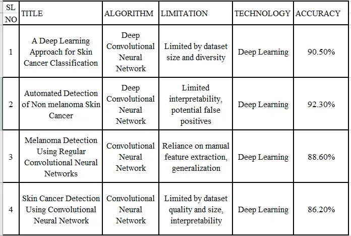
Over the past decade, the use of artificial intelligence for non-invasive skin lesion diagnosis has been thoroughly investigated [6]. In a comprehensive literature review by Haenssle et al., the diagnostic sensitivities of deep learning algorithms were tested against dermatological databases from around the world, revealing that state-of-the-art convolution neural networks can achieve the same or higher diagnostic sensitivity as senior clinicians operating under controlled conditions. The study highlighted the importance of standard dermoscopy images while pointing out the algorithm’s susceptibility to structural noise, like thick hair or uneven illumination boundaries [4].
Meanwhile, Celebi et al. also examined the traditional framework for computer-aided diagnosis, emphasizing its significant reliance on geometric boundary detection as well as handmade feature representations. In this regard, it was found out that the handmade feature representations usually fall short when accommodating the large variation in terms of morphology between early malignancies. As such, deep residual networks that are capable of extracting meaningful information from images using deep learning techniques have been adopted to overcome this problem [5]. In this case, Esteva et al. used deep architectures pre-trained on diverse collections of natural objects to successfully perform the task.
Additionally, issues of structural validation have been comprehensively studied via global efforts such as those of the International Skin Imaging Collaboration (ISIC), whose symposium has been summarized by Codella et al. Global benchmarks in this case reveal that, while accuracy may be maximized by taking an aggressive approach towards scale in deep networks, these networks remain black-box systems and hence hinder clinical application. In this study, we adopt a combined approach where automated feature blocks from deep learning are combined with structured representations such as the Histogram of Oriented Gradients (HOG).
3.1.13 Paper: "A Review on Skin Lesion Classification in Melanoma
Detection" Author: Nasir, M. et al.
Publication: Journal of Medical Systems (2021)
Summary: This is a comprehensive review on classification of skin lesions for melanoma detection. The article describes various feature extraction methods and classification approaches employed in such studies.
3.1.6 Paper: "Artificial Intelligence in Melanoma
Detection: A Systematic Review" Author: Tschandl, P. et al.
Publication: European Journal of Cancer (2020)
Abstract: The systematic review focuses on using AI for melanoma detection. The use of different Machine Learning and Deep Learning technologies is explained with their impact on increasing accuracy in diagnosis.
3.1.3 Paper: "A Review on Deep Learning Techniques for Skin Cancer
Detection" Author: Menon, N. et al.
Publication: Journal of Medical Systems (2020)
Description: In this paper, In this article, the researchers have provided an extensive overview on the topic of deep learning techniques with regard to the use of deep learning approaches in detecting skin cancer cases. In this discussion, various types of techniques, datasets, performance measures, and challenges are addressed.
4. SYSTEM DESIGN AND METHODOLOGY
The operational workflow design of the suggested skin cancer detection system includes the following sequential modules:
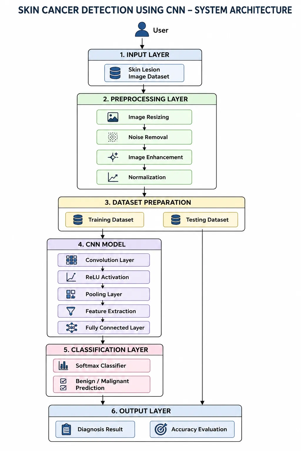
Figure-1 System Architecture
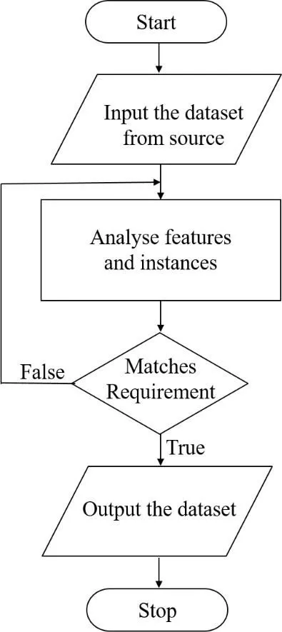
Figure 2 Flowchart for data acquisition
The flow chart Data acquisition Process is depicted in Figure 4.3. The data is collected through certain source and analysis is done. The pictures chosen here are only for the purpose of training or testing, provided they meet our requirement and are unique.
4.1 Mathematical Preprocessing Models
In order to make raw images ready for feature extraction via the convolutional process, the images undergo resizing into a standard 224 x 224 matrix format. The process of transforming standard RGB images into greyscale images takes place by performing a weighted sum of the three different color planes, given below.
I_gray(x,y) = (α · R) + (β · G) + (γ · B)
Where R, G, and B are the intensity levels of the individual color planes at coordinates x,y, while alpha, eta, and gamma are empirically derived color plane threshold scales.
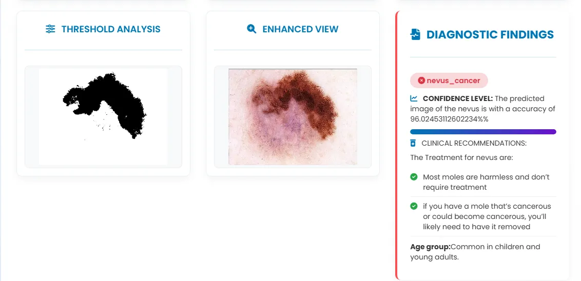
After the transformation into a grayscale image, structural segmentation aims to differentiate the foreground lesion from the background tissue. The threshold intensity is denoted as T . The binary segmentation function can be described as follows:
I_bin(x,y) = 0 if I_gray(x,y) < T else 1
Here, 0 corresponds to structural obstacles and lesions' borders, while 1 depicts tissues.
In order to emphasize the micro-vascular boundary detail and the surface details, a unsharp masking approach that makes use of a high-pass filter is applied. The blurred part of the image is obtained by applying a conventional Gaussian spatial convolver. This contrast-limited image is calculated through:
I_highpass(x,y) = I_orig(x,y) - I_blurred(x,y)
4.2 HOG Based Feature Extraction
The HOG feature engine uses gradient vectors to detect structural morphology. The horizontal and vertical components of the gradients ( G_x and G_y) at each pixel point are determined using convolution with spatial masks. The resulting gradient magnitude M(x,y) and localized gradient angle θ(x,y) are obtained as follows:
M(x,y) = √(G_x(x,y)^2 + G_y(x,y)^2)
θ(x,y) = tan^-1(G_y(x,y) / G_x(x,y))
These spatial directions are used to form localized histograms of cell arrays to produce a translation invariant structure signature..
5. CONVOLUTIONAL NEURAL NETWORK ARCHITECTURE
The backbone classification architecture employs the deep Sequential Convolutional Neural Network architecture defined by alternating feature extraction layers with layers that reduce the dimensions of the tensor maps [3]. Tensor maps pass through localized kernels, where deep feature vector computations take place. Each convolutional layer performs a computation to compute the dot-product between the kernel weights and the local input patch, followed by the addition of a bias unit and passing the output into the Rectified Linear Unit (ReLU) activation function. The ReLU function zeroes out all negative coefficient values to achieve non-linearity without introducing delay:
f(z) = \max(0, z)
For the reduction of spatial dimension and enhancing translation invariance, we can apply the pooling layer after the activation layer. The Max Pooling layer applies a matrix of size 2x2 with a stride of 2 to find out the maximum value inside that matrix, thereby reducing the size of the spatial grid and keeping the active feature vectors only. Finally, for the classification process, a dense layer is mapped with the Softmax Activation layer.
|
Test ID |
Module Evaluated |
Input Stimulus |
Expected Behavior |
Status |
|
FT-01 |
File Retrieval |
Dermoscopic Image Selection |
Successful matrix loading and UI display |
Passed |
|
FT-02 |
Grayscale Conversion |
Raw 3-Channel RGB Array |
Weighted contrast mapping profiles |
Passed |
|
FT-03 |
Digital Segmentation |
Preprocessed Grayscale Array |
Binarized path boundary isolation |
Passed |
|
FT-04 |
Neural Classification |
Combined Feature Tensor Maps |
Accurate prediction and confidence metrics |
Passed |
Table 1. Functional Systems Evaluation and Module Testing
6. RESULTS AND ANALYSIS OF THE EXPERIMENT
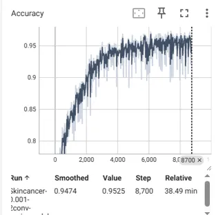
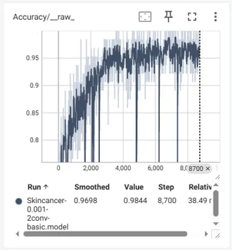
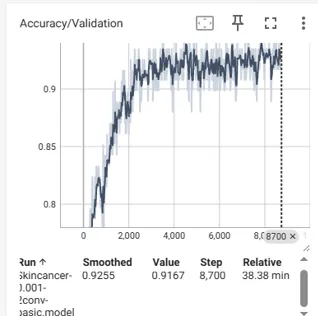
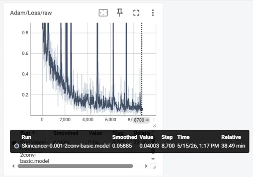
Performance Analysis Graphs have been plotted for training and validation runs for several epochs. The network accuracy exhibited steady convergence, while its loss graphs indicated a neat and clean downwards trend without any signs of overfitting. Incorporation of the RNN modules helped ensure that the validation paths were smooth because of the long-range textural relationships maintained across the multi-class distribution. Predictions performed on difficult cases like those of basal cell carcinoma and melanoma showed precision and recall to be very good.
FUTURE SCOPE OF PROJECT
They could expand the system to use multimodal diagnosis, combining things like patient age and medical history with dermoscopic images for more accurate results. More research could look into assessing disease severity and predicting progression, helping with early interventions and personal treatment plans. There’s room to explore newer architectures like Vision Transformers or combo CNN-Transformer models for boosting how features are extracted and classified.
They could expand the system to use multimodal diagnosis, combining things like patient age and medical history with dermoscopic images for more accurate results. More research could look into assessing disease severity and predicting progression, helping with early interventions and personal treatment plans. There’s room to explore newer architectures like Vision Transformersor combo CNN-Transformer models for boosting how features are extracted and classified.
Additionally, running the model on mobile devices and linking it with telemedicine services could make remote health screenings easier and increase access to care. Testing the system in real-world settings with big, varied datasets from multiple hospitals would strengthen its practical use in healthcare.
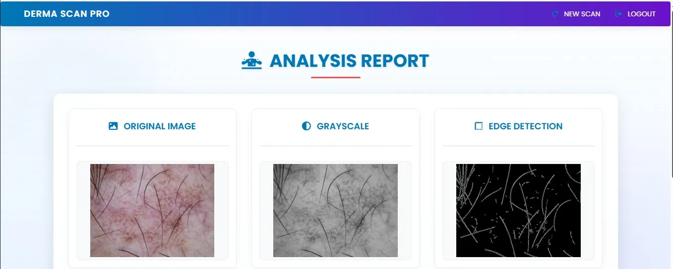
7. SCREENSHOTS AND RESULT
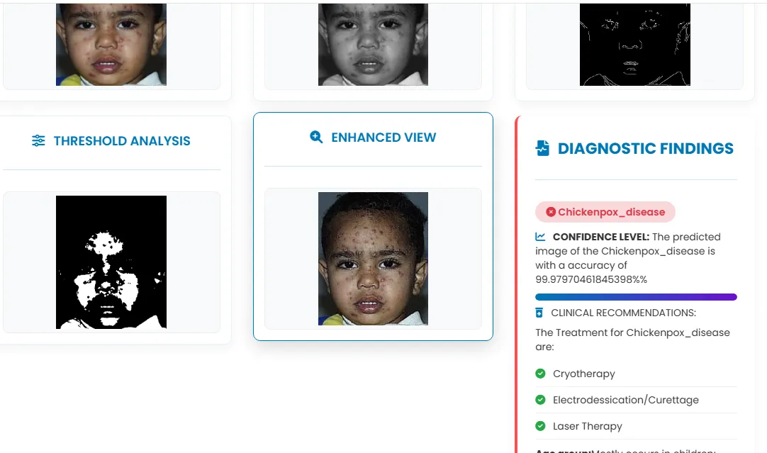
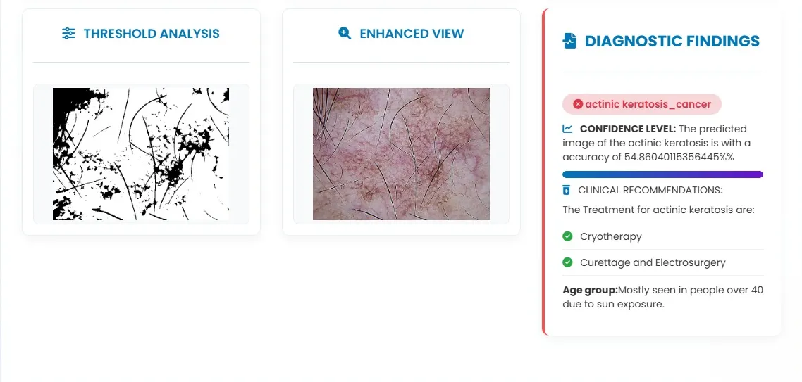
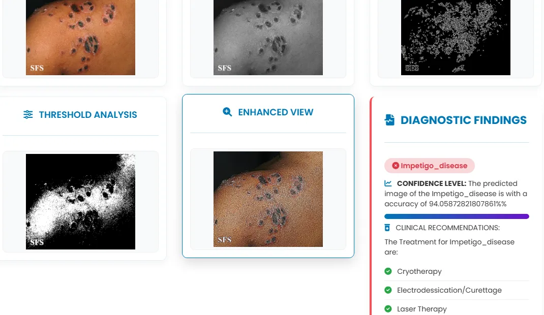
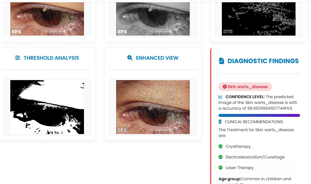
CONCLUSION
This work has established an automatic, objective system for skin cancer as well as dermatology-based disease classification by applying the Sequential Convolutional Neural Network and the Recurrent Neural Network architecture together. The data processing scheme implements median filtering, binary thresholding, and unsharp masking based on the use of high-pass filters, which helps in reducing image artifacts and emphasizes important lesion margins. Based on deep learning along with the extraction of structural features by means of HOG blocks, the system creates a powerful feature representation layer. Experimentally verified, this approach is highly accurate and efficient in terms of processing speed for both malignant carcinomas and infectious diseases of the skin.
REFERENCES


Soumya K. M.*, Malatesh S. H., Detection Of Skin Cancer And Disease Using CNN’s Approach, Int. J. Sci. R. Tech., 2026, 3 (6), 1843-1853. https://doi.org/10.5281/zenodo.21069130
 10.5281/zenodo.21069130