

We use cookies to ensure our website works properly and to personalise your experience. Cookies policy
Department of Pharmaceutics, Shivlingeshwar College of Pharmacy Almala, Ausa 413520
The present study aimed to develop a topical antifungal delivery system comprising ciclopirox olamine-loaded sodium alginate microspheres incorporated into a Carbopol 934 hydrogel. Microspheres were prepared by ionotropic gelation using calcium chloride as the cross-linking agent. Five batches (F1–F5) were assessed for particle size, entrapment efficiency, production yield, in-vitro drug release and morphology. The selected batch was incorporated into four hydrogel formulations (G1–G4), which were evaluated for appearance, pH, viscosity, spreadability, drug content, drug diffusion, antifungal activity and short-term stability. Batch F3 showed a particle size of 128 µm, entrapment efficiency of 94%, yield of 88.6% and reported cumulative release of 91.2% at the final reported time point. The gels were whitish, smooth and free from perceptible grittiness, with pH values between 5.2 and 6.3. G2 had the highest reported drug content (95.43%) and showed a 17-mm inhibition zone against Candida albicans ATCC 10231, compared with 25 mm for miconazole and 0 mm for the control. Release-kinetic analysis reported the best fit to the Higuchi model (R² = 0.993). The results suggest that the microsphere-hydrogel platform merits further investigation as a topical antifungal formulation. Conflicting release values in the source data should be reconciled before publication.
Superficial fungal infections of the skin, hair and nails are common dermatological conditions and can cause persistent symptoms, recurrence and impaired quality of life. Their clinical burden is influenced by environmental conditions, host susceptibility, delayed diagnosis and the emergence of antifungal resistance. Fungal disease remains an important global health concern, including infections caused by dermatophytes and Candida species [1–5]. Superficial mycoses are particularly common in warm, humid climates, where local environmental and behavioural factors can facilitate fungal growth [6,7]. [6–8].
Topical antifungal therapy is frequently preferred for localized cutaneous infections because it delivers the active agent directly to the affected site and can limit systemic exposure. Nevertheless, conventional creams, ointments and gels may be removed from the application site, release drug rapidly, or fail to maintain adequate drug concentrations in the relevant skin layers. The stratum corneum is a major barrier to topical drug transport, and formulation composition can substantially influence drug release, penetration and cutaneous retention [8–12]. [9–12].
Ciclopirox olamine is a hydroxypyridone antifungal used in topical preparations. Its antifungal action differs from that of azoles and allylamines and includes chelation of polyvalent metal ions, interference with metal-dependent enzymes and disruption of essential fungal cellular processes. Published clinical and formulation literature supports its use in topical antifungal products, while also demonstrating that vehicle composition affects release and skin retention [13–16]. [13–15].
Polymeric microspheres are particulate carriers that can entrap an active ingredient within or throughout a polymeric matrix and modify its release profile. Sodium alginate is a hydrophilic polysaccharide that forms ionically cross-linked networks in the presence of multivalent cations such as calcium. Ionotropic gelation is a comparatively straightforward method for producing alginate beads under mild aqueous conditions [17–22]. Microspheres can subsequently be dispersed in a hydrogel, combining particulate drug retention and release control with the ease of application, spreadability and residence characteristics of a semisolid vehicle [9,23–27].
Hydrogels are hydrated polymer networks whose rheological properties can be adjusted to provide an appropriate balance between retention and ease of spreading. Carbomer-based gels are commonly neutralized to develop viscosity, while excipients such as propylene glycol can function as humectants and may influence drug partitioning and permeation [10,11,23,24]. However, the performance of a microsphere-loaded hydrogel depends on polymer concentration, cross-linking conditions, drug loading and the final gel structure; these factors require experimental optimization rather than assumption. [23,31].
The objective of this study was to prepare ciclopirox olamine-loaded sodium alginate microspheres by ionotropic gelation, identify a suitable microsphere batch, incorporate it into a Carbopol 934 hydrogel and evaluate the physicochemical properties, in-vitro release/diffusion, antifungal activity and short-term stability of the resulting formulation. [18,24,26].
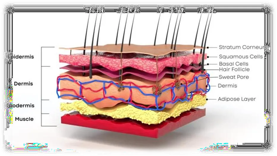
Figure 1: Cross-Sectional Representation of Human Skin Anatomy
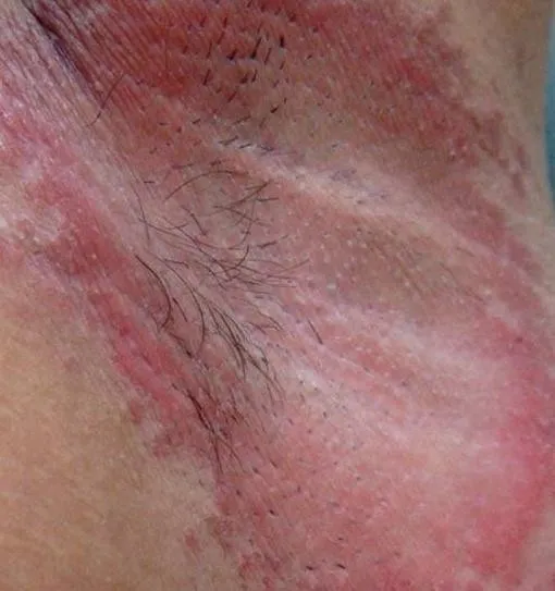
Figure 2: Cutaneous Candidiasis
MATERIALS AND METHODS
2.1 Materials
Ciclopirox olamine was obtained from Labware Chemicals. Sodium alginate and calcium chloride were obtained from Molychem, Mumbai, India. Carbopol 934 was obtained from Loba Chemie, Mumbai, India. Methyl paraben and triethanolamine were obtained from Molychem. Propylene glycol was used in the hydrogel base. All materials were used as received. The source thesis lists UV–visible spectrophotometry, FTIR, optical microscopy, a digital pH meter, Brookfield viscometer, centrifuge, USP dissolution apparatus II, Franz diffusion cell and scanning electron microscopy among the equipment used. The excipients were used as received; supplier details are reported from the source thesis.
2.2 Preformulation studies
The drug was examined for appearance and odour, and its melting point was determined by the capillary method. Solubility was examined in methanol, ethanol, distilled water and phosphate buffer (pH 7.4). Samples were sonicated, centrifuged and the supernatant analysed by UV spectrophotometry. The UV spectrum was scanned over the wavelength range of 200–400 nm, and 303 nm was selected as the analytical wavelength for drug-content estimation and microsphere-release studies [18,20].
2.3 FTIR compatibility assessment
FTIR spectra of ciclopirox olamine, selected excipients and drug–excipient mixtures were collected over 4000–400 cm⻹. The spectra were compared for retention, disappearance or displacement of characteristic bands. The source data report prominent mixture bands near 3365, 2924, 2853, 1633, 1417 and 1384 cm⻹, along with polysaccharide-associated fingerprint-region bands. These observations were interpreted as showing no major spectral evidence of incompatibility; FTIR alone does not establish chemical stability or exclude all interactions. FTIR comparison is commonly used as a preliminary screen for possible drug–excipient interactions [18,20].
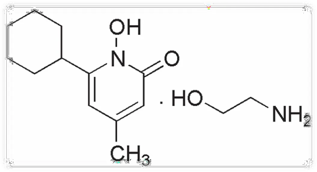
Figure 3: Ciclopirox Olamine
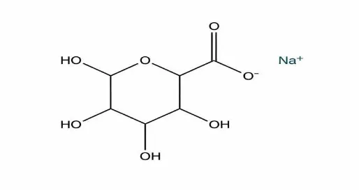
Figure 4: Sodium Alginate
2.4 Preparation of ciclopirox olamine-loaded microspheres
Microspheres were prepared by ionotropic gelation. Sodium alginate was dissolved in distilled water under continuous stirring and ciclopirox olamine was dispersed in the polymer solution. Separately, calcium chloride was dissolved in distilled water. The drug–polymer dispersion was added dropwise into the calcium chloride solution through a syringe under continuous stirring. The droplets underwent ionic cross-linking to form microspheres. The particles were allowed to harden, collected, washed and dried. Five batches were prepared by varying sodium alginate and calcium chloride while maintaining the nominal drug amount at 100 mg. This technique uses ionic cross-linking of alginate with multivalent cations to form gelled particles [24,26,28]
(F1–F5)
Table 1: Formulation Composition of Ciclopirox Olamine–Loaded Sodium Alginate Beads
| Batch | Ciclopirox olamine | Sodium alginate | Calcium chloride | Water |
|---|---|---|---|---|
| F1 | 100 mg | 0.8 g | 2.0 g | q.s. to 10 g |
| F2 | 100 mg | 0.9 g | 2.5 g | q.s. to 10 g |
| F3 | 100 mg | 1.0 g | 3.0 g | q.s. to 10 g |
| F4 | 100 mg | 1.1 g | 2.5 g | q.s. to 10 g |
| F5 | 100 mg | 1.2 g | 3.0 g | q.s. to 10 g |
2.5 Evaluation of microspheres
Particle size was estimated by optical microscopy by measuring approximately 10 particles and calculating the average diameter. Entrapment efficiency was estimated indirectly from the unentrapped drug in the supernatant after centrifugation at 6000 rpm for 30 min. The supernatant was diluted and analysed spectrophotometrically against the calibration curve. Entrapment efficiency was calculated as: EE (%) = [(total drug added − free drug detected)/total drug added] × 100. Percentage yield was calculated as: yield (%) = (practical dried microsphere mass/theoretical mass of drug plus polymer) × 100. Entrapment efficiency and production yield were calculated using the equations recorded in the source thesis; such measurements are commonly used to characterize polymeric microspheres [18,20,21]
In-vitro release was studied using USP dissolution apparatus II. Microspheres equivalent to 50 mg drug were placed in 900 mL phosphate buffer (pH 7.4) at 37 ± 0.5 °C and 50 rpm. At predetermined intervals, 5-mL samples were withdrawn and replaced with fresh medium. Samples were filtered and analysed at the stated UV wavelength. Dried microspheres were mounted on SEM stubs, coated with a thin conductive layer and examined for particle shape and surface characteristics. Release testing was used to compare the formulations under the stated experimental conditions [20,21].
2.6 Preparation of microsphere-loaded hydrogel
Carbopol 934 was dispersed in distilled water and allowed to hydrate for 30–60 min. Propylene glycol and methyl paraben were added with stirring. Triethanolamine was added dropwise to develop the gel and adjust the pH. The selected microsphere batch (F3) was incorporated slowly with gentle mixing to avoid damaging the particles. The gel was allowed to stand to release entrapped air and equilibrate. Four formulations were prepared by varying the Carbopol 934 concentration while the stated microsphere amount and other excipients were held constant. Carbomer-based hydrogel preparation typically involves polymer hydration followed by neutralization to develop the gel network [23,30,31].
Table 2: Formulation Composition of Ciclopirox Olamine (CPO)-Loaded Microsphere-Based Carbopol 934 Gel (G1–G4)
| Ingredient | G1 | G2 | G3 | G4 |
|---|---|---|---|---|
| CPO-loaded microspheres | 1 g | 1 g | 1 g | 1 g |
| Carbopol 934 | 0.15 g | 0.30 g | 0.45 g | 0.60 g |
| Propylene glycol | 1.5 g | 1.5 g | 1.5 g | 1.5 g |
| Methyl paraben | 0.006 g | 0.006 g | 0.006 g | 0.006 g |
| Triethanolamine | q.s. | q.s. | q.s. | q.s. |
| Distilled water | q.s. to 30 g | q.s. to 30 g | q.s. to 30 g | q.s. to 30 g |
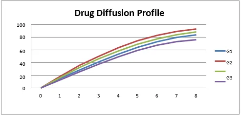
Figure 5: Drug Release Profile of Microsphere Loaded Hydrogel
2.7 Evaluation of hydrogel
The formulations were visually examined for appearance, consistency, homogeneity and grittiness. pH was measured after dispersing approximately 50 mg gel in water using a calibrated pH meter. Viscosity was measured using a Brookfield viscometer. Spreadability was assessed by the reported slide/weight method and expressed in g·cm/s. Drug content was determined after extracting a known amount of gel and analysing the diluted extract by UV spectrophotometry. In-vitro diffusion was evaluated using a diffusion-cell method; the available thesis extract does not fully specify the membrane, receptor medium, exposed area or sampling correction, and these details should be added from laboratory records. pH, viscosity, spreadability and drug content were assessed as formulation-quality attributes [9,23,28,33].
2.8 In-vitro antifungal activity
Antifungal activity was evaluated by the well-diffusion method against Candida albicans ATCC 10231. The optimized hydrogel was compared with miconazole as the reference antifungal and a control. Inhibition-zone diameter was recorded in millimetres. The source thesis does not provide the inoculum density, agar medium, well diameter, sample dose, incubation conditions or replicate number; these essential reproducibility details should be verified and included in the final manuscript. Miconazole was used as the reference antifungal agent, as stated in the source thesis; antifungal activity of ciclopirox formulations has previously been evaluated in topical-development studies [13–15].
2.9 Stability study
The optimized hydrogel was stored for 90 days under room-temperature and accelerated conditions. The thesis reports room storage and accelerated storage at 40 ± 2 °C/75 ± 5% relative humidity. Appearance, pH, viscosity and gel strength were monitored at monthly intervals. The report refers to ICH guidance; the exact applicable guideline and the stability protocol should be stated accurately in the final version. The accelerated condition reported was 40 ± 2 °C/75 ± 5% relative humidity. Stability-study design should be interpreted in the context of ICH stability guidance [32,34].
2.10 Data analysis
The source thesis presents descriptive results and release-kinetic coefficients for zero-order, first-order, Higuchi and Korsmeyer–Peppas models. No replicate-level data, standard deviations, confidence intervals or inferential statistics were provided in the material reviewed. Accordingly, results are reported descriptively and should not be interpreted as statistically significant. Model comparisons are commonly used to characterize release profiles, but the best-fitting model alone does not establish a unique release mechanism [35,36].
RESULTS AND DISCUSSION
3.1 Preformulation and FTIR compatibility
The source thesis reports that the drug and selected excipients were examined by UV spectroscopy and FTIR. Characteristic mixture bands were retained without major reported shifts or disappearance, which was interpreted as indicating no major interaction detectable by the method. This supports proceeding with the selected excipients, although a more definitive compatibility assessment would require the original spectra, peak assignments and, where appropriate, thermal or stability data.
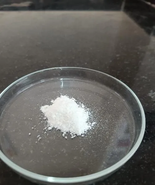
Figure 6: Ciclopirox Olamine
Table 3: Microsphere Characteristics
| Batch | Particle size (µm) | Entrapment efficiency (%) | Yield (%) | Cumulative release at final reported point (%) |
|---|---|---|---|---|
| F1 | 185 | 75.0 | 72.5 | 78.5 |
| F2 | 155 | 86.5 | 79.8 | 84.8 |
| F3 | 128 | 94.0 | 88.6 | 91.2 |
| F4 | 142 | 89.5 | 84.2 | 87.6 |
| F5 | 168 | 81.0 | 77.9 | 85.0 |
F3 was selected as the optimized microsphere batch based on the reported smallest particle size (128 µm), highest entrapment efficiency (94%), highest yield (88.6%) and highest cumulative drug release (91.2% at the final reported time point). The trend suggests that an intermediate polymer/cross-linker combination may have provided a favourable balance between droplet formation, matrix integrity and drug retention. Increased alginate concentration can raise solution viscosity and affect droplet size, while calcium-mediated cross-linking can influence matrix density and release. These explanations are plausible formulation interpretations; they were not established by a formal design-of-experiments analysis.
SEM observations in the thesis described the selected microspheres as spherical, with a relatively smooth surface and no major visible cracks. The available report does not provide particle-size distribution statistics, SEM magnification or a quantitative morphology analysis; those details should be added if the images and raw records are available.
Table 4: Physical Properties of Microsphere-Loaded Hydrogel
| Batch | Appearance/consistency | pH | Viscosity (cP) | Spreadability (g·cm/s) | Drug content (%) |
|---|---|---|---|---|---|
| G1 | Whitish, smooth; no grittiness | 5.9 | 2328 | 3.3 | 91.47 |
| G2 | Whitish, smooth; no grittiness | 5.2 | 3979 | 3.7 | 95.43 |
| G3 | Whitish, smooth; no grittiness | 6.3 | 4091 | 2.9 | 84.51 |
| G4 | Whitish, smooth; no grittiness | 5.6 | 2238 | 3.1 | 90.34 |
All four formulations were described as whitish, smooth, homogeneous and free from perceptible grittiness. The measured pH values ranged from 5.2 to 6.3. G2 had the highest reported drug content (95.43%) and spreadability (3.7 g·cm/s), while G3 had the highest viscosity (4091 cP) and lowest spreadability (2.9 g·cm/s). The general inverse relationship between viscosity and spreadability is consistent with the increase in polymer concentration, although G1 and G4 show that the relationship is not strictly monotonic across all reported values. G2 was selected as the preferred gel based on the combined reported properties.
Table 5: In-vitro Drug Diffusion and Release Kinetics
| Time (h) | G1 (%) | G2 (%) | G3 (%) | G4 (%) |
|---|---|---|---|---|
| 1 | 15.2 | 18.4 | 16.5 | 12.8 |
| 2 | 28.6 | 35.8 | 32.4 | 25.7 |
| 3 | 41.5 | 50.2 | 46.3 | 37.9 |
| 4 | 53.8 | 63.6 | 58.5 | 49.4 |
| 5 | 64.7 | 74.8 | 69.3 | 59.6 |
| 6 | 73.5 | 83.1 | 77.6 | 67.8 |
| 7 | 80.2 | 88.4 | 84.1 | 73.4 |
| 8 | 84.7 | 92.6 | 87.4 | 77.3 |
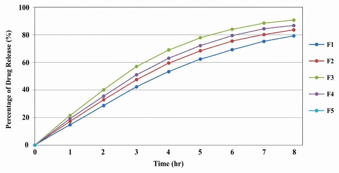
Figure 7: In-Vitro Drug Release of Microspheres
The primary hydrogel diffusion table reports that G2 reached 92.6% cumulative diffusion at 8 h, higher than G1 (84.7%), G3 (87.4%) and G4 (77.3%). The release-kinetic table also lists 92.6% at 8 h for G2.
Table 6: Release Model
| Release model | Reported R² |
|---|---|
| Zero order | 0.959 |
| First order | 0.981 |
| Higuchi | 0.993 |
| Korsmeyer–Peppas | 0.668 |
The reported regression coefficients indicate that the Higuchi model provided the best fit (R² = 0.993), followed by first-order (0.981) and zero-order (0.959) models. This pattern is consistent with a diffusion-associated release profile in the tested conditions. A high R² alone does not establish the underlying mechanism; model comparison should use the same validated dataset, and the Korsmeyer–Peppas exponent and fitting range should be reported if mechanistic interpretation is intended.
Table 7: In-vitro Antifungal Activity
| Sample | Inhibition-zone diameter (mm) |
|---|---|
| Control | 0 |
| Miconazole reference | 25 |
| Optimized formulation G2 | 17 |
G2 produced a reported inhibition zone of 17 mm against C. albicans ATCC 10231, while miconazole produced 25 mm and the control produced no inhibition. This indicates measurable antifungal activity in the agar-diffusion assay, but the zone was smaller than that of the reference. The result should be considered preliminary because only single values are presented and key assay conditions and replicate variability are not available. Agar diffusion is influenced by drug release and diffusion through agar as well as intrinsic antifungal potency; it cannot by itself demonstrate superior clinical efficacy or improved skin penetration.
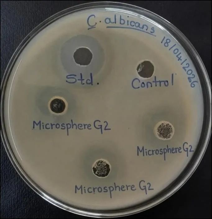
Figure 8: In-vitro Antifungal Studies
3.6 Short-term stability
During the reported 90-day study, no visible change in appearance was recorded under either storage condition. At room storage, pH changed from 5.2 at month 1 to 5.9 at month 3 and viscosity from 3975 to 4298 cP. Under accelerated storage, pH changed from 5.3 to 5.8 and viscosity from 4010 to 4395 cP. Gel strength was reported to change from ++ to +++ by month 3. These findings suggest that the formulation retained its gross appearance while some rheological and pH drift occurred. Because drug content, degradation products and release performance were not tabulated at each stability interval, the data support only preliminary physical stability, not a complete shelf-life claim.
CONCLUSION
Ciclopirox olamine-loaded sodium alginate microspheres were prepared by ionotropic gelation and incorporated into a Carbopol 934 hydrogel. Based on the reported particle size, entrapment efficiency, yield and release results, F3 was selected as the optimized microsphere batch. Of the hydrogel formulations, G2 showed the highest reported drug content and favourable spreadability, with a pH within the reported range of 5.2–6.3. The formulation showed an inhibition zone against C. albicans ATCC 10231 and a reported best fit to the Higuchi release model. These findings indicate potential for further development as a topical antifungal delivery system. Nevertheless, the conflicting in-vitro diffusion values, incomplete assay-method details and absence of replicate statistics must be resolved before journal submission. Further work should include repeat experiments with appropriate controls, validated analytical methods, skin permeation/retention assessment, irritation testing and a stability-indicating assay.
REFERENCES


Vitthal Pawar, Dr. Ajay Fugate, Dr. Vishweshwar Dharashive, Pramod Naraynkar, Vinod Hajare, Yashwant Lohar, Development and Evaluation of Ciclopirox Olamine-Loaded Microsphere Hydrogel for Topical Antifungal Delivery, Int. J. Sci. R. Tech., 2026, 3 (11), 178-185. https://doi.org/10.5281/zenodo.23941842
 10.5281/zenodo.23941842