

We use cookies to ensure our website works properly and to personalise your experience. Cookies policy
Department of Pharmacy, TVES Hon'ble Loksevak Madhukarrao Chaudhari College Of Pharmacy, Faizpur (MH.), India
The present study focuses on the formulation and evaluation of Diclofenac Sodium transdermal patches using the solvent casting method. The prepared patches were characterized for physical appearance, weight uniformity, thickness, folding endurance, surface pH, moisture content, drug content uniformity, in-vitro drug release, and stability. The results demonstrated that all formulations exhibited a smooth, flexible, and uniform film with good mechanical strength and minimal variation in thickness and weight. The folding endurance values indicated sufficient durability, while the surface pH (6.9–7.1) confirmed skin compatibility. Drug content analysis showed uniform distribution (95–98%), ensuring consistent drug delivery. The in-vitro dissolution studies revealed a biphasic release pattern, with an initial burst phase followed by sustained drug release. The optimized formulation (F2) exhibited 93.75% cumulative drug release over 10 hours, highlighting its potential for prolonged therapeutic action. Stability studies over three months confirmed no significant changes in physical properties, pH, or thickness, demonstrating the robustness of the formulation. These findings suggest that Diclofenac Sodium transdermal patches can serve as an effective alternative for controlled drug delivery, enhancing patient compliance and therapeutic efficacy.
​Transdermal drug delivery systems (TDDS) have emerged as an effective alternative to conventional drug delivery methods, offering advantages such as improved patient compliance, controlled drug release, and reduced systemic side effects. These systems facilitate the direct absorption of drugs through the skin into the systemic circulation, bypassing the gastrointestinal tract and first-pass metabolism. TDDS has been widely explored for the delivery of nonsteroidal anti-inflammatory drugs (NSAIDs), including Diclofenac Sodium, due to their potential to provide sustained therapeutic effects while minimizing gastrointestinal irritation.
Diclofenac Sodium, a potent NSAID, is commonly prescribed for the management of pain and inflammation associated with conditions such as osteoarthritis, rheumatoid arthritis, and musculoskeletal disorders. However, oral administration of Diclofenac Sodium is often associated with adverse effects such as gastric ulceration, hepatic toxicity, and poor bioavailability due to extensive first-pass metabolism. To overcome these limitations, transdermal patches offer a promising approach by providing a controlled and sustained release of Diclofenac Sodium, enhancing therapeutic efficacy and reducing the frequency of administration.
The present study focuses on the formulation and evaluation of Diclofenac Sodium transdermal patches using the solvent casting method. The objective is to develop a stable, effective, and patient-friendly transdermal patch capable of delivering Diclofenac Sodium in a controlled manner over an extended period. The patches were prepared using different polymeric compositions to optimize mechanical strength, drug release kinetics, and skin permeability. Various evaluation parameters, including physical characterization, drug content uniformity, in-vitro drug release, and stability studies, were conducted to assess the suitability of the formulated patches for transdermal drug delivery.
This research aims to establish a formulation that ensures consistent drug release, maintains stability under storage conditions, and provides a viable alternative to oral administration of Diclofenac Sodium. The findings from this study are expected to contribute to the advancement of transdermal drug delivery systems and enhance the therapeutic application of Diclofenac Sodium for pain management.
MATERIALS AND METHODS
Materials
The drug used in the study included Diclofenac Sodium, procured from Cipla Pharmaceuticals, India and various polymers, excipients and solvents obtained from reputable suppliers.
Methods
FORMULATION TABLE
|
Ingredient |
F1 |
F2 |
F3 |
|
Diclofenac Sodium (mg) |
10 |
10 |
10 |
|
Ethyl Cellulose (mg) |
200 |
300 |
400 |
|
PEG-400 (ml) |
1.2 |
1.2 |
1.2 |
|
Chloroform: Methanol |
1:4 |
1:4 |
1:4 |
|
Eucalyptus oil (ml) |
0.3 |
0.3 |
0.3 |
|
Menthol (ml) |
0.5 |
0.2 |
0.5 |
Table 1: Ingredients used
Solvent Casting Method
Ethyl Cellulose was used for the formulation of Transdermal Patch. Polyethylene glycol (PEG 400) was used as a plasticizer. Menthol and eucalyptus oil is used as penetration enhancer. The polymer was dissolved in chloroform: methanol (1:4) solvent. The drug was dispersed uniformly in the viscous solution with continuous stirring. The resulting mass was poured into leveled mercury surface in a Petri dish covered with inverted funnel. The Petri dish was left undisturbed at room temperature for one day. The patch was obtained intact by slowly lifting from the Petri dish and transdermal patches were cut into radius of 2cm2 [28].
EVALUATION OF TRANSDERMAL PATCHES
Physical appearance
All the transdermal patches are visually inspected for colour, clarity, flexibility and smoothness.
Weight uniformity
Four patches from each batch are accurately weighed using a digital balance. The average weight and the standard deviation values are calculated from the individual weights.
Thickness of the films
The thicknesses of the drug loaded polymeric films are measured using a digital vernier caliper. The measurements are made at five different points, four at the corners and one at the centre of the patch. The average and standard deviation of five readings were calculated for each formulation.
Folding endurance
To measure the film's folding endurance, a small 2 x 2 cm² strip was folded frequently at the same point until it broke. Alternatively, the film was manually folded at an angle of 1800 until it broke, and the number of breaks was recorded. The three readings' average and standard deviation for all films were calculated.
Surface pH
Patches were kept in glass tubes containing 10 ml phosphate buffer (pH -7.4) and the pH of the surface measured after 1, 2, 3, 4, 5, 6, 7 and 8 hours by placing the tip of the glass microelectrode of a digital pH meter close to the surface of the patch and allowing it to equilibrate for 1 min prior to recording [29].
Percentage moisture content
The prepared films are weighed individually and kept in a desiccator containing silica gel at room temperature for 24 hours. The films were again weighed and the percentage moisture content is calculated using the formula:
Percentage moisture content = [(Initial weight – Final weight)/Final weight] x 100
Drug content uniformity
The consistent distribution of drug content can be determined using any standard test procedure established in a standard pharmacopoeia for that particular API. The API contents in each patch is evaluated to ensure content uniformity. The limit for content homogeneity is 85-115%. Each batch's 2x2 cm2 films were sliced and mixed with 50 ml of phosphate buffer with a pH of 6.8 (1000 g/ml). The solution was filtered using Whatman filter paper and diluted by pipetting 1 ml into a 100 ml volumetric flask with phosphate buffer pH 6.8 (10 g/ml). The resultant solution was examined at 323 nm with a Shimadzu spectrophotometer, and the drug concentration was calculated by comparing it to the standard drug absorbance of Ketorolac mixture [30,31].
In-vitro dissolution test
In-vitro drug diffusion studies was carried out using the 20ml Franz diffusion cell. The membrane was stabilized before mounting to remove the soluble components. The membrane was mounted between the donor and receptor compartments. The receptor compartment was filled with 20 ml of isotonic phosphate buffer of pH 7.4 which was maintained at 37± 0.2oC and hydrodynamics were maintained using magnetic stirrer. One patch of dimension 2 cm × 2 cm was previously moistened with a few drops of pH 7.4 phosphate buffer and placed in donor compartment. 1 ml samples from receptor compartment were withdrawn at suitable time interval of 1, 2, 3, 4, 6, 8 and 10 hours which was then replaced with 1 ml of pH 7.4 phosphate buffer. The percentage of drug permeated was determined by measuring the absorbance in UV Visible spectrophotometer at λ max of 323 nm [32].
Stability Studies:
Patches were conducted for a duration of three months. The formulations were kept in lid tight container at the following conditions as per ICH guidelines: 30ËšC ± 2Ëš C / 75 ± 5 % RH. The samples were taken Initially 0 month and 3rd month and evaluated (Appearance, pH and Thickness) [32,33].
RESULTS AND DISCUSSION
Evaluation of Transdermal Patches
Physical Appearance
The formulated patches were observed as white, amorphous films with a smooth surface, confirming uniform polymer distribution. The patches exhibited transparency, indicating proper formulation without air entrapment or crystallization. Additionally, the patches were non-sticky, ensuring ease of application and removal. These characteristics confirm that the prepared transdermal patches possess the necessary aesthetic and handling properties, making them suitable for transdermal drug delivery.
Weight Uniformity
The weight uniformity of transdermal patches was evaluated to ensure consistent formulation and drug distribution across different batches. The recorded weights for batches F1 to F3 ranged from 84 ± 2.36mg to 90 ± 3.41mg, with minimal deviations. The results indicate that all batches maintained uniform weight within an acceptable range, suggesting reproducibility in the solvent casting method. Slight variations in weight could be attributed to differences in polymer composition, solvent evaporation, or film thickness. However, the low standard deviation values confirm that the patches were prepared with high precision, ensuring batch-to-batch consistency. These findings are essential for maintaining formulation uniformity, which directly impacts drug release and therapeutic efficacy.
Thickness of the Films
The thickness values ranged from 0.24 ± 0.001 mm to 0.26 ± 0.002 mm, with minimal standard deviation, indicating precise formulation. The slight variations observed may be due to differences in polymer concentration, solvent evaporation rate, or casting technique. However, the low standard deviation values confirm that the patches exhibited uniform thickness across different formulations, ensuring reproducibility and consistent drug diffusion. Maintaining a uniform thickness is essential for achieving controlled drug release and optimal therapeutic efficacy.
Folding Endurance
Folding endurance was evaluated to determine the mechanical strength and flexibility of the transdermal patches. The results for different batches are as shown in table 2. The values indicate that all batches exhibited sufficient mechanical resistance, ensuring durability and flexibility. The highest folding endurance was observed in batch F2 (190±7), suggesting a stronger and more elastic film, likely due to optimized polymer composition and plasticizer content. The lowest value was recorded for batch F1 (175±6), which, while slightly lower, still meets the required mechanical strength for handling and application. Overall, all batches demonstrated satisfactory mechanical properties, confirming their suitability for transdermal application without the risk of brittleness or breakage during use.
Surface pH
The surface pH of the patches was determined at different time intervals (1 to 8 hours) using a digital pH meter. The recorded pH values remained between 6.9 and 7.1, which is close to skin pH, indicating that the patches are non-irritating and suitable for transdermal application.
Percentage Moisture Content
The percentage moisture content of the transdermal patches was evaluated to assess their stability and susceptibility to moisture absorption. The results for different batches were as shown in table 2. The moisture content ranged from 2.8 to 3.5%, indicating minimal variation among batches. Batch F3 (3.5%) exhibited the highest moisture content, which may be attributed to differences in polymer composition or environmental factors during formulation. Conversely, batch F1 (2.8%) had the lowest moisture content, suggesting better resistance to moisture absorption. Lower moisture content is desirable as it enhances the stability of the patch and prevents microbial contamination. The overall results confirm that the formulated patches maintain an acceptable moisture content, ensuring product stability and integrity.
Drug Content Uniformity
The drug content uniformity of the transdermal patches was evaluated to ensure consistent distribution of Diclofenac Sodium across different batches. The results obtained were shown in table 2. The drug content ranged from 95 to 98%, indicating minimal variation and confirming uniform drug distribution within the patches. Batch F2 (98%) exhibited the highest drug content, while batches F1 (95%) and F3 (96%) had slightly lower but acceptable values. The uniformity in drug content across all batches ensures reproducibility in formulation and reliable drug delivery. These results confirm that the solvent casting method effectively incorporated Diclofenac Sodium into the polymeric matrix, meeting the standard requirements for content uniformity in transdermal drug delivery systems.
|
Batch |
Weight (mg) |
Folding Endurance |
Surface pH |
Thickness (mm) |
% Moisture content |
% Drug Content |
|
F1 |
84 ± 2.36 |
180±5 |
7.0 |
0.25 ± 0.001 |
3.2 |
95 |
|
F2 |
90 ± 3.41 |
190±7 |
6.9 |
0.26 ± 0.002 |
2.8 |
98 |
|
F3 |
87 ± 3.1 |
175±6 |
7.1 |
0.24 ± 0.001 |
3.5 |
96 |
Table 2: Evaluations of Transdermal Patches
In-vitro Dissolution Test
The in-vitro drug diffusion study was performed using a Franz diffusion cell to evaluate the release of Diclofenac Sodium from the transdermal patches. The receptor compartment was filled with 20 mL of isotonic phosphate buffer (pH 7.4) and maintained at 37 ± 0.2°C. A magnetic stirrer was used to ensure proper mixing. The transdermal patch (2 × 2 cm) was moistened with a few drops of phosphate buffer and placed in the donor compartment. Samples of 1 mL were withdrawn at predetermined intervals (1, 2, 3, 4, 6, 8, and 10 hours) and replaced with an equal volume of fresh phosphate buffer to maintain sink conditions. The drug content in the samples was analyzed using a UV-Visible spectrophotometer at 323 nm. The cumulative percentage of drug release was calculated and plotted against time to assess the release kinetics.
The results indicated a biphasic drug release pattern, with an initial burst release followed by sustained diffusion. The cumulative drug release for the optimized formulation (F2) reached 93.75% at 10 hours, demonstrating a controlled and prolonged release profile suitable for transdermal delivery.
|
Time (hr) |
% Cumulative Drug Release |
||
|
|
F1 |
F2 |
F3 |
|
1 |
14.1 |
17.46 |
25.76 |
|
2 |
16.3 |
19.30 |
28.12 |
|
3 |
26.64 |
27.87 |
37.50 |
|
4 |
27.46 |
39.60 |
39.32 |
|
5 |
43.7 |
43.90 |
42.09 |
|
6 |
49.2 |
49.91 |
49.78 |
|
7 |
68.02 |
57.45 |
58.03 |
|
8 |
78.43 |
78.39 |
79.45 |
|
9 |
90.05 |
89.76 |
86.75 |
|
10 |
92.36 |
93.75 |
91.02 |
Table 3: % Cumulative Drug Release of Transdermal Patches of All Batches
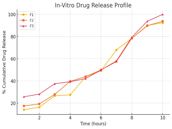
Figure 1: % Cumulative Drug Release of Transdermal Patches of All Batches
Stability Studies
The results confirm that the patches remained stable over three months under ICH-specified storage conditions, demonstrating their suitability for extended use.
|
Batch |
Time (Months) |
Appearance |
pH |
Thickness (mm) |
|
F2 |
0 |
Smooth, No cracks |
6.9 |
0.26 ± 0.002 |
|
3 |
Smooth, No cracks |
6.8 |
0.26 ± 0.001 |
Table 4: Stability Studies
CONCLUSION
The present study successfully formulated and evaluated Diclofenac Sodium transdermal patches using the solvent casting method. The formulated patches were subjected to extensive physicochemical characterization, including physical appearance, weight uniformity, thickness, folding endurance, surface pH, moisture content, drug content uniformity, in-vitro drug release, and stability studies, to assess their suitability for transdermal drug delivery.
All prepared patches exhibited a smooth, flexible, and homogeneous surface, with no visible cracks or imperfections, ensuring uniform polymer distribution. The weight uniformity analysis showed minimal variations across batches, confirming the consistency of the formulation process. The thickness values were found to be within an acceptable range, ensuring uniformity in film formation, which is essential for controlled drug release. Folding endurance testing demonstrated that the patches had excellent mechanical strength and flexibility, indicating their ability to withstand handling and application without breaking.
The surface pH of the patches remained within the range of 6.9–7.1, which is close to the physiological pH of human skin. This suggests that the patches are non-irritating and suitable for transdermal administration. The percentage moisture content of the patches was maintained at a low level (2.8–3.5%), which is beneficial for enhancing formulation stability and preventing microbial growth. The drug content uniformity results revealed that the Diclofenac Sodium was distributed homogeneously within the polymeric matrix, with drug content ranging between 95–98%, ensuring dose accuracy and reproducibility.
The in-vitro drug release study conducted using a Franz diffusion cell demonstrated a biphasic release pattern, with an initial burst release followed by sustained drug diffusion. Among the different formulations, batch F2 exhibited the most desirable release profile, achieving 93.75% cumulative drug release over 10 hours. This indicates that the formulation successfully prolonged the release of Diclofenac Sodium, which is crucial for maintaining therapeutic drug levels in systemic circulation over an extended period.
Stability studies conducted over three months under ICH-specified storage conditions confirmed that the transdermal patches retained their physical integrity, pH, and thickness, with no significant degradation. The results indicate that the formulation is stable over time, making it suitable for long-term use.
Overall, the results demonstrate that the formulated Diclofenac Sodium transdermal patches exhibit favourable physicochemical properties, controlled drug release, and stability, making them a promising alternative for sustained drug delivery. These patches offer advantages such as improved patient compliance, reduced dosing frequency, and minimized gastrointestinal side effects compared to oral administration. Future in-vivo pharmacokinetic and clinical studies are necessary to further validate the therapeutic efficacy and safety of these patches for pain management and inflammatory conditions.
REFERENCES

Pavan Kasar*, Aayush Nemade, Tamanyu Patil, Yash Bhavsar, Mansi Patil, Ankita Thakare, Formulation And Evaluation Of Diclofenac Sodium Transdermal Patches For Controlled And Sustained Drug Delivery, Int. J. Sci. R. Tech., 2026, 3 (7), 219-227. https://doi.org/10.5281/zenodo.21273949
 10.5281/zenodo.21273949