

We use cookies to ensure our website works properly and to personalise your experience. Cookies policy
Rashtrasant Janardhan Swami College of Pharmacy Kokamthan, Kopargaon.
Niosomes are mixtures of cholesterol and nonionic Surfactant. It develops sustain release Formulation of Diclofenac Niosomes in order to provide Better therapeutic effect. Diclofenac Niosomes was prepared By different techniques. These are : Ether injection method , Ethanol injection method, Sonication method, Thin film hydration and Reverse phase evaporation Technique. The main Aim of this study is to formulate Niosomes as carriers for delivery of diclofenac sodium. Niosomes are composed of non ionic surfactants and Cholesterol. Niosomes were prepared by ether injection method By using drug, non ionic surfactant (Span 80).The formulations were optimized From the above methods with respect to vesicle shape, Particle size, entrapment efficiency, drug content and drug release. Formulation containing span 80 prepared by ether injection method was found to be best with drug content of 62 %,: entrapment efficiency of 83.10%, mean vesicular diameter of 2000nm, zeta potential of 29 mv. Overall, the results Demonstrate that Diclofenac-loaded niosomes possess suitable physicochemical Characteristics and can serve as a promising vesicular carrier for controlled and effective Analgesic and Anti-anti-Inflammatory drug delivery.
Diclofenac sodium (DS), known as potent NSAID with pronounced analgesic Properties, has been so far utilized in the prolonged treatments of Osteoarthritis Rheumatoid arthritis. The drug is Considerably metabolized In the liver and has to be administered frequently Since its biological half-life is only 1 to 2 hours Gastrointestinal Consequences such as ulcer, bleeding, or perforation of the Intestinal walls Have thus been reported with the long term use of DS . Because of its Shorter Biological half-life as well as adverse effects, it is considered as a perfect Candidate for controlled transdermal drug delivery (1)
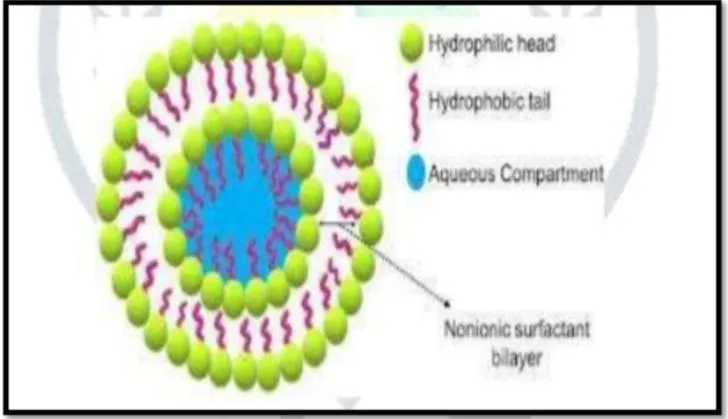
Niosomal drug delivery was opted for site specific action of Diclofenac in the Treatment Of rheumatoid arthritis. Rheumatoid arthritis is a systemic, Chronic, long-term auto Immune disorder that leads to inflammation of the Synovial, joints, and surrounding Tissues gradually leading to joint Destruction. The pattern of Joints affected is usually Symmetrical which Effects the small joints of the hand and Feet. It can also affect other Organs Including heart, lungs and Eyes. The drugs which Are employed for the Treatment of rheumatoid arthritis are non-steroidal anti-Inflammatory drugs (NSAIDS), corticosteroids, Disease modify anti-rheumatic drug(DMARD’S) Etc. Non- Steroidal anti-Inflammatory drugs (NSAIDs) are most commonly Used to reduce Inflammation and Pain. NSAIDs acts by cyclooxygenase Enzyme inhibition resulting In anti-inflammatory Action, the main factor Limiting the oral use of NSAIDs is the Development of Gastrointestinal (GI)_ Adverse events, ranging from dyspepsia to Serious life-threatening Events. Several studies have shown the effectiveness of topical NSAIDs in treating Acute and chronic soft tissue conditions. Conventional oral Administration causes gastrointestinal Side effects and low bioavailability due to first- Pass Metabolism .Transdermal delivery using niosomes improves drug Penetration, Provides sustained release, and reduces side Effects. Incorporating diclofenac-loaded Niosomes into topical gel Enhances patient Compliance and therapeutic efficacy .Diclofenac sodium is a nonsteroidal anti-inflammatory Drug with potent Analgesic. Moderate anti-inflammatory Action. Diclofenac sodium comes Under Class-I1 of the (BCS) Biopharmaceutical Classification System. It has Poor Solubility and absorption From the oral route of administration It also Causes gastric bleeding as a side effect, We May prepare topical hydrogel Niosomal preparation of Diclofenac sodium.(2)
Niosomes can increase the Penetration Of drug through the stratum carenum hence Increase local Analgesic action of the drug. Niosomal preparation of Diclofenac sodium Overcomes its side effect of gastric Bleeding and also increases its physical Stability. Niosomes are microscopic vesicles of non-ionic surfactant that are produced by Modified ether injection method non-ionic surfactant, with cholesterol. They resemble Liposomes. Both liposomes and niosomes function as active transporters of lipophilic And amphiphilic medications. The phospholipids that make up the liposomal bilayer And the non-ionic surfactant that forms the niosomal bilayer distinguish the two systems According to the preparation technique and the inverse structure in the event of a non- Aqueous solvent, niosomes are created by the self-assembly of non-ionic surfactants in Aqueous media as spherical, unilamellar, bilayer, multilamellar system, and polyhedral Structures, Hydrophilic ends of the surfactant are exposed outwardly in the noisome, Whereas hydrophobic ends face one another, forming a bilayer of the surfactant, Both Hydrophilic and hydrophobic drugs can be contained in this. known as vesicular delivery_ systems, these nanoparticles function as niosomes to increase a drug’s Therapeutic effectiveness .These carriers (niosomes) are biologically inert in Nature, Devoid Of any antigenic, pyrogenic or allergic reactions and their components can be Utilized as the component of Biological membrane .Drugs incorporated in liposomes, Niosomes were not activated under Physiological Conditions and did not cause Unfavorable side effects as well. Compare to liposomes, Niosomes have the advantage that the components Are Extremely cheap compared to Phospholipids, and both the lipids and Non-ionic Surfactants are similarly stable.(3)
Advantages :-
1.The vesicle suspension being water-based vehicle offers high patient Compliance When compared to other Dosage forms.
2. Drug molecules of wide range of solubilities can be accommodated in the Niosomes Provided by the Infrastructure consisting of hydrophilic, lipophilic And amphiphilic Moieties.
3.Vesicle characteristics can be controlled by altering the composition of Vesicle, size Lamellarity, surface Charge, tapped volume and concentration.
4. They can release the drug in sustained/controlled manner.
5.Storage and handling of surfactants Special conditions like low Temperature and inert Atmosphere.
6- They can act as a depot formulation, thus allowing the drug release in a Controlled manner.
7.They possess stable structure even in emulsion form.
8.They enhance the oral bioavailability of poorly soluble drugs
9.Surfactants are biodegradable, biocompatible, non-toxic and Non-Immunogenic.
10.They are economical for large scale production.
11.Niosomes are used for parenteral, oral, and topical routes.
12. No special conditions are needed for handling and storing surfactants
13. Controlled and targeted drug delivery
14.Stable and osmotically active
15.Increased dermal penetration and oral bioavailability.
General characteristics of niosomes :-
• Biodegradable
• Biocompatible
• Non-toxic
• Non-immunogenic
• Non-carcinogenic
• High resistance to hydrolytic degrade.
Method of Preparation :- (4)
1.Ether Injection
2. Film Method/Hand Shaking Method
3. Sonication
4.Heating Method
5. Multiple Membrane Extrusions Method
6. Reverse Phase Evaporation
7. Bubble Method
1. Ether Injection Method -
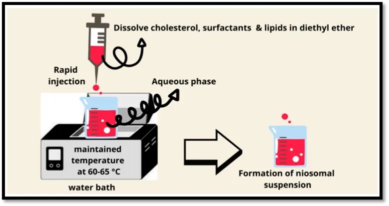
Fig 2. Ether Injection Method (5)
2. Film Method/ Hand Shaking Method –
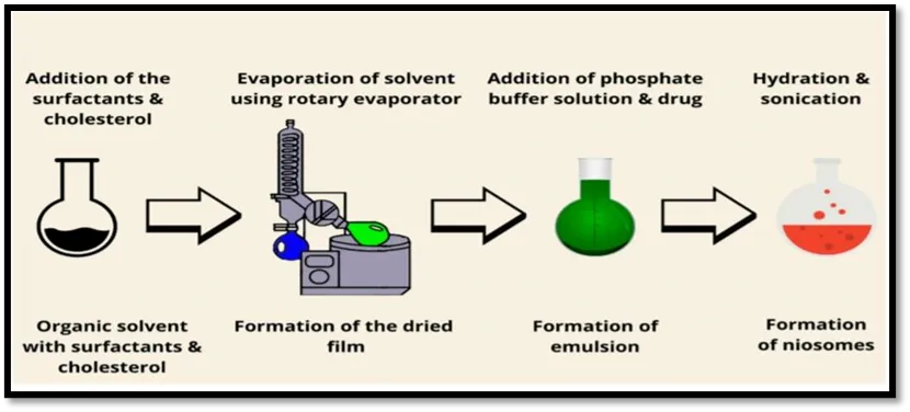
Fig 3. Hand shaking method (5)
3. Sonication –
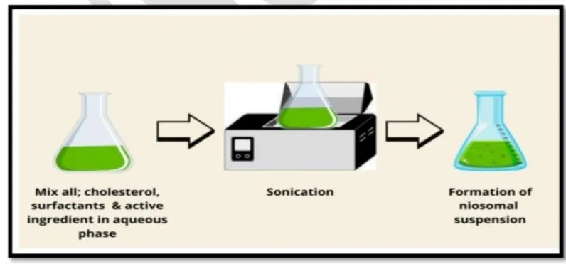
Fig 4. Sonication Method (5)
4. Bubble Method -
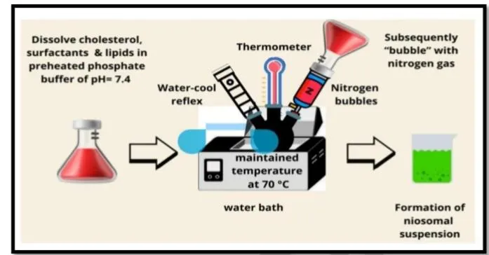
Fig 5. Bubble Method (5)
5. Heating Method –
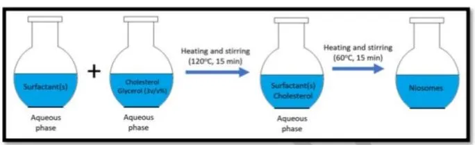
Fig 6. Heating Method (6)
6. Multiple Membrane Extrusions Method –
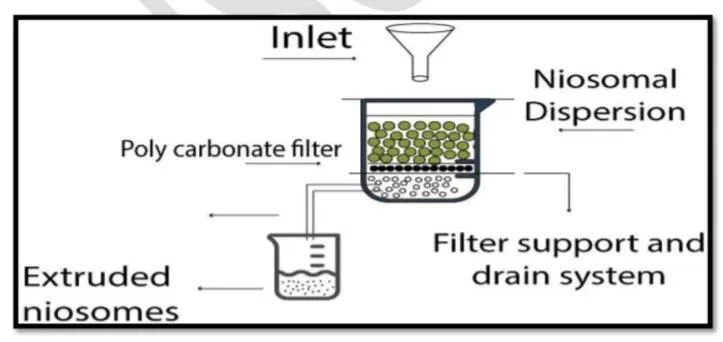
Fig 7. Multiple Membrane Extrusions Method (7)
7. Reverse Phase Evaporation –
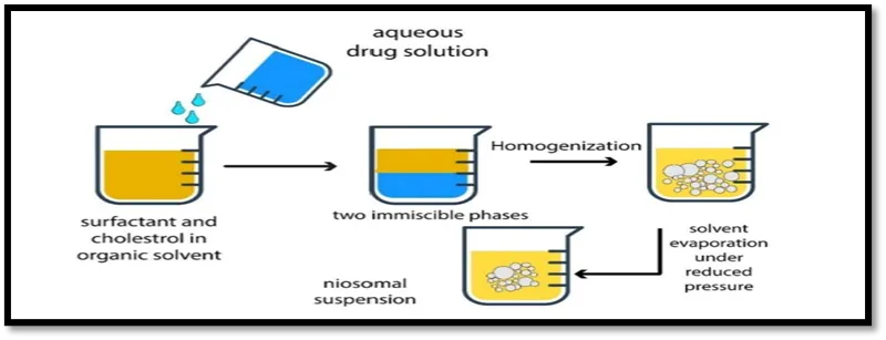
Fig 8. reverse phase evaporation (7)
Applications –
1.Controlled and sustained release of drugs for long-lasting action
2. Transdermal delivery to enhance drug penetration through the skin.
3.Ocular delivery to increase eye drug retention and reduce dosing
4.Pulmonary delivery for treating lung diseases via inhalation.
5.Oral delivery to protect drugs from degradation and improve absorption
6.Parenteral delivery (V/IM/SC) for long-circulating drug action. (8)
Material and method –Material
Method – Ether Injection Method
Principle –
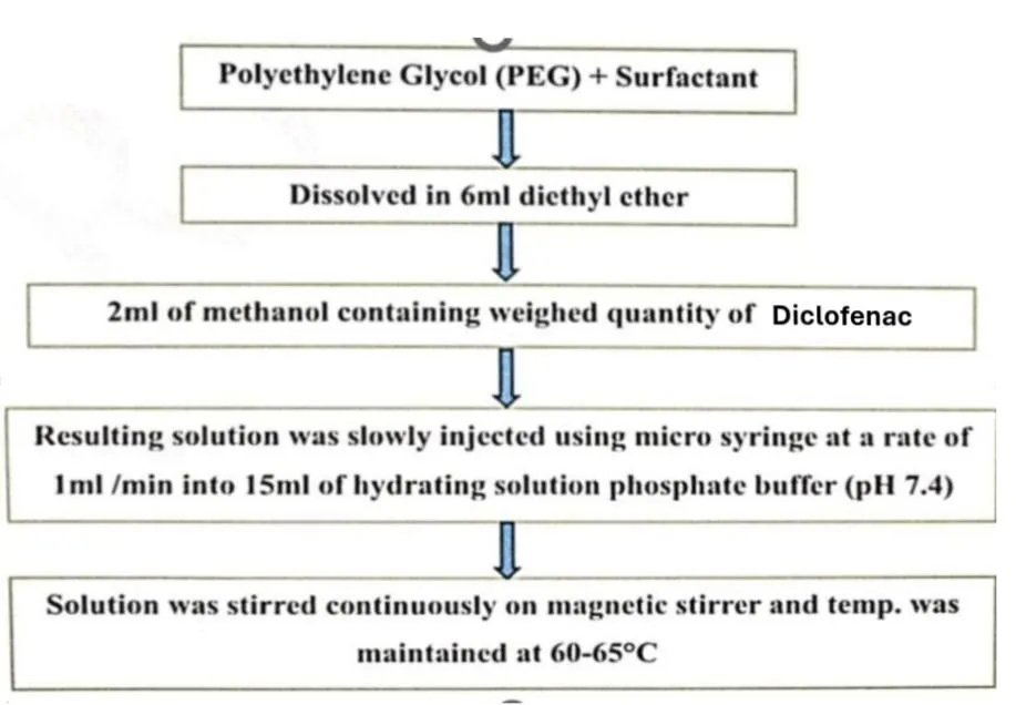
The ether injection method is a Technique used to prepare niosomes, The method Involves injecting a solution mixture of ether, surfactant, PEG, and Drug into a hot Aqueous solution, resulting in the formation of niosomes . (9)
Advantages -
Disadvantages –
Experimental Work -
Formula –
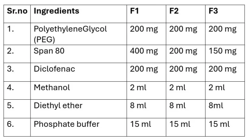
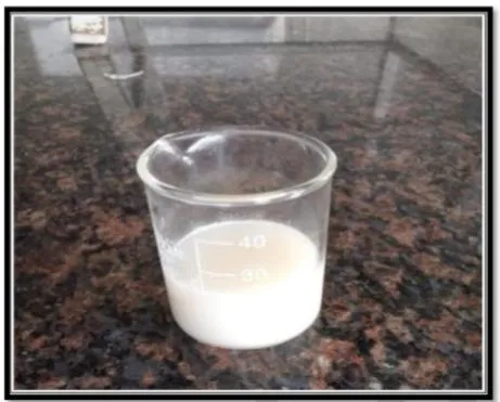
Fig 9. Niosomal suspension
Procedure –
Optimization
To optimize the ether injection method, several parameters can
Be adjusted, including:
1.Surfactant concentration.
2. Injection rate
3.Temperature of the aqueous solution
4.Stirring speed By adjusting these parameters, it is possible to optimize the cther injection Method for Specific applications and improve the quality and characteristics of the resulting Niosomes .
Evaluation Test:-
The particle shape analysed by using optical microscope.
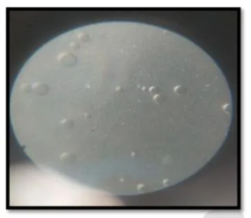
Fig 10. Particle shape
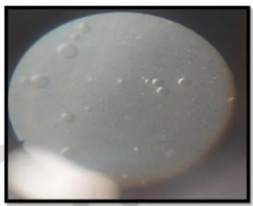
Fig 11. Particle shape
2. Drug contents -
Niosomal suspension equivalent to 10mg taken in a volumetric flask of 100ml and Volume was make up by phosphate buffer pH 7.4, after that 1ml of this mixture was Diluted to 10ml by phosphate buffer 7.4 and the % drug content was calculated or Observed at using UV spectrophotometer.(10)
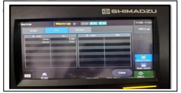
Fig. 12 Drug content
3. pH – The pH of the formulations was determined by using digital pH Meter. The Measurement of pH of the formulation was done in triplicate and average values are Calculated.
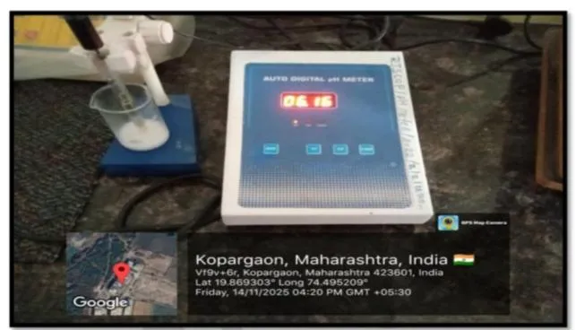
Fig 13. pH of niosomal suspension
4. Entrapment efficacy –
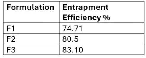
Entrapment efficiency- is defined as the ratio of drug molecules encapsulated into The niosomes nanoparticles to the total used drug, and can be determined by the Following equation
EE (Amount of trapped drug/Total amount of initially added-drug) x100% after Preparing niosomal dispersion unentrapped drug is separated by dialysis, gel filtration And centrifugation .
Centrifugation-
1. Prepare the noisome suspension: 10 mg equivalent drug From formulation. Of Prepared niosomal suspension by ether injection method were dissolved in1 0ml of 7.4 pH phosphate buffer. Ensure the niosomal suspension is homogeneous and well- dispersed . (10)
2. Centrifugation: Centrifuge the noisome suspension at a high speed for a sufficient Time (eg.30 min ) to separate the niosomes from the free drug.
3. Separate the supernatant: Carefully collect the supernatant, which contains the free Drug.
4. Determine the amount of free drug: Measure the absorbance of the supernatant using A UV-Vis spectrophotometer.
5. Calculate the entrapment efficiency: Using the formula: Entrapment Efficiency (%) (Total amount of drug added-Amount of free drug)/Total amount of drug added x 100
5. Partical size and zeta potential- Particle size shows the average diameter of the niosomal vesicle. Zeta potential Represents the surface charge of the vesicles, which helps to predict their stability. These Parameters were measured using a Zetasizer instrument.
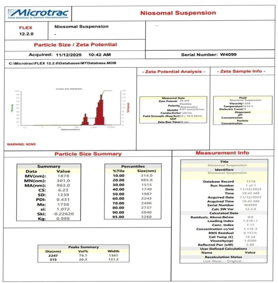
Fig 14. Particle size and zeta potential
RESULT AND DISCUSSION-
The niosomes prepared using fluconazole drug was studied under microscope to Observe the formation of niosomal vesicles. The niosomal vesicles were found to be Uniform in size and shape. The shape of the niosomes was observed spherical.
The drug content was Studied for the formulation prepared by ether injection method .The drug content was found to be (62.5%), which may have optimum surfactant Polyethylene Glycol (PEG) ratio to provide highest drug content of ibuprofen in Niosomal vesicles.
The standard range of the pH of the niosomal solution was 5.5-7.5 and the pH Of the Niosomal solution was found to be 6.16 which is ideal pH.
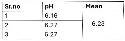
The prepared Diclofenac-loaded niosomes showed an average particle size Of 2 um (2000 nm) The zeta potential was found to be 31 mV, confirming good Stability of the Niosomal suspension.
CONCLUSION
Incorporation in the niosomes to target the Niosomes to the specific site In Promising drug delivery model, Niosomes are considered to be better Candidate for Drug delivery as compared to liposomes due to various factors Like cost, stability etc. These advantages over the liposomes make it a better Targeting, agent, Ophthalmic, Topical, parenteral and various other routes are Used for targeting, the drug to the site Of action for better efficacy, Transdermal delivery appears to be an attractive route Of administration to Maintain the drug blood levels of ibuprofen for and extended Period of time. Formulation of niosomal suspension showed entrapment efficiency (83.10%), pl1 (6.17). particle size (2000 um), the shape of the niosomes was Observed spherical, and the drug content (62.5%). Hence it was considered To be Good niosomal formulation. In conclusion, the cther injection method Is a widely used and effective method for Preparing niosomes. The method Offers several advantages, including simplicity and High entrapment Efficiency. However, the method also has some limitations, and the Choice of Surfactant, and drug is critical to the success of the method.
REFERENCES

Aparna Sawant*, Sakshi Ghorpade, Shruti Gosavi, Murtadak Bhagyashri, Thorat Suvarna S., Formulation And Evaluation Of Diclofenac Loaded Niosomes By Ether Injection Method, Int. J. Sci. R. Tech., 2026, 3 (5), 945-954. https://doi.org/10.5281/zenodo.15179749
 10.5281/zenodo.20409997