

We use cookies to ensure our website works properly and to personalise your experience. Cookies policy
1Department of Pharmacology, JES's SND College of Pharmacy, Babhulgaon (Yeola), India
2Department of Pharmaceutical Quality Assurance, JES's SND College of Pharmacy, Babhulgaon (Yeola), India.
3Department of Pharmaceutics, JES's SND College of Pharmacy, Babhulgaon (Yeola), India
Gut dysbiosis is increasingly associated with neurodegenerative diseases through the gut–brain axis. Altered gut microbiota may influence neuroinflammation, intestinal and blood–brain barrier integrity, oxidative stress, protein aggregation, and neurotransmitter signaling through microbial metabolites. This review summarizes the current evidence linking gut dysbiosis with Alzheimer’s disease, Parkinson’s disease, multiple sclerosis, amyotrophic lateral sclerosis, and Huntington’s disease. It also discusses experimental models, microbiome-targeted therapies, multi-omics approaches, and artificial intelligence in microbiome research. Emerging strategies include dietary interventions, probiotics, prebiotics, postbiotics, fecal microbiota transplantation, phage therapy, precision microbiome therapy, and microbiome engineering. Despite promising findings, variability between individuals, lack of standardized methods, limited clinical evidence, and uncertainty regarding causality remain major challenges. Future research should emphasize standardized, longitudinal, and personalized approaches to establish the therapeutic potential of microbiome modulation in neurodegenerative diseases.
1.1. Definition of gut dysbiosis
Dysbiosis Specifically, GD can be defined as an increased number of pathogenic microbiota or pathobionts (microbes producing pro-inflammatory cytokine) and decreased number of beneficial microbes or symbiont (microbes producing anti-inflammatory cytokines). The pathogenic representation of GD-induced NPDs [1]. Decreased expression of tight junction proteins (including claudia-5 and occluding) between the intestinal epithelial cells leading to increased permeability, resulting in leaky gut (also known as leaky gut syndrome) [2]. Altered neuroimmune status is marked by alterations in microglial maturation, neurogenesis, myelination, neurotrophin expression, neurotransmitters, and their respective receptors [3]. The indirect effects of intestinal microbes include modified levels of neurotransmitter precursors in the gut lumen along with modulation in the synthesis of neurotransmitters like GABA, 5-HT, dopamine (DA), and noradrenaline (NA) [4].
1.2. Definition and significance of gut dysbiosis
1.3. Overview of neurodegenerative disease
Neurodegenerative diseases (ND) are a heterogeneous group of progressive disorders characterized by the gradual loss of structure and function of neurons, ultimately leading to neuronal death. These disorders primarily affect the central and peripheral nervous systems and are among the leading causes of disability and mortality worldwide. The increasing prevalence of neurodegenerative diseases is closely associated with population ageing, making them a significant global public health concern.
The most common neurodegenerative diseases include Alzheimer's disease, Parkinson's disease, Amyotrophic lateral sclerosis, Huntington's disease, and Multiple sclerosis. Although these disorders differ in their clinical manifestations, they share several common pathological mechanisms, including abnormal protein aggregation, oxidative stress, mitochondrial dysfunction, neuroinflammation, impaired protein clearance, excitotoxicity, and neuronal apoptosis.
1.4 Introduction of gut-brain axis
The term gut microbiota brain axis is defined as a bidirectional communication between the brain and gut bacterial community through multiple systems forming a network. It has a significant role in maintaining the homeostasis of the CNS and gastrointestinal system [12]. The interaction pathways in this network include both indirect and direct signaling via the neuronal pathway, immune system, and chemical transmitter [13]. As different biological systems are engaged in these networks, their complexity needs to be explored.
1.5 Why gut microbiota has emerged as a therapeutic target
Gut microbiota alterations in the small bowel and colon have been evidenced extensively in patients with FGIDs Several studies showed that the gut microbiota profile of IBS patients significantly differs from that of healthy subjects. A recent meta-analysis showed that IBS patients presented higher levels of pathogenic bacteria of the genera Enterobacter and lower commensal bacteria of the general Bifidobacterium and Lactobacillus compared with the healthy controls [14]. A microbiome signature from patients with IBS was found to be decrease in microbial diversity, and the severity of the symptoms was associated with Clostridial species and/or methanogenic microbes. Another study demonstrated the overrepresentation of Methanobravibacter smithii in fecal samples from patients with functional constipation, and constipation-predominant IBS (IBS-C) [15]. One study showed an increased ratio of Firmicutes to Bacteroidetes, increased Streptococcus and Ruminococcus, and decreased Lactobacillus and Bifidobacterium species in patients with IBS [16].
1.6. Objectives of the review
1.To understand the concept of gut dysbiosis and its causes.
2.To examine the role of the gut microbiota in maintaining neurological health.
3.To investigate the relationship between gut dysbiosis and neurodegenerative diseases such as Alzheimer's disease, Parkinson's disease, and Multiple sclerosis.
4.To explore the mechanisms of the gut brain axis, including inflammation, immune modulation, microbial metabolites, and neurotransmitter signaling.
5.To evaluate current and emerging therapeutic strategies, such as probiotics, prebiotics, dietary interventions, and fecal microbiota transplantation, for preventing or managing neurodegeneration.
6.To identify gaps in current research and suggest future directions for understanding the gut microbiome's role in neurological disorders.
1.7. Scope of the review
1.Covers the composition and functions of the normal gut microbiota and the factors leading to gut dysbiosis.
2.Focuses on the gut brain axis as a communication network between the gastrointestinal tract and the central nervous system.
3. Reviews evidence linking gut dysbiosis with major neurodegenerative diseases.
3.Discusses biological mechanisms such as neuroinflammation, oxidative stress, intestinal barrier dysfunction, microbial metabolites, and immune responses.
4.Includes recent advances in microbiome-based diagnostics and therapeutic approaches.
5.Highlights the potential of personalized microbiome-targeted interventions in improving neurological health while acknowledging current limitations and ongoing research
2.The healthy gut microbiota and the gut brain axis
2.1 Composition of the healthy gut microbiota
2.1.1. Dominant bacterial phyla
The healthy adult human gut microbiota is dominated primarily by the phyla Firmicutes (Bacillota) and Bacteroidetes (Bacteroidota), which together generally account for ~80–90% of the bacterial community, although their relative abundance varies substantially between individuals and populations [17]. Other consistently detected phyla include Actinobacteria (Actinomycetota) and Proteobacteria (Pseudomonadota), usually at considerably lower abundances. Verrucomicrobia (Verrucomicrobiota) and Fusobacteria are also present but generally constitute smaller fractions of the healthy gut microbiota. Recent metaproteomic data, for example, found an average of approximately 60% Bacillota, 35% Bacteroidota, and ~2% each Actinomycetota and Pseudomonadota in the subjects studied [18].(Table 1).
Table 1: Examples of Bacterial Phylum and Their Role in Gut
| Bacterial phylum | Current name | Examples | Main role in gut | Reference |
|---|---|---|---|---|
| Bacillota | Firmicutes | Faecalibacterium, roseburia, lactobacillus | Fiber fermentation, butyrate/SCFA production | [19] |
| Bacteroidata | Bacteroidetes | prevotella, bacteroides | Breakdown of complex carbohydrates | [20] |
| Actinomycetota | Actinobacteria | bifidebacterium | Carbohydrates fermentation; gut barrier support | [21] |
| Pseudomonadota | Proteobacteria | escherichla, klebsiella | Minor component; facultative anaerobic metabolism | [22] |
2.1.2. Age related changes in microbiota
As human life expectancy increases, aging has become a public health issue. Numerous studies demonstrate that gut microbiota plays key roles in aging process. Compared with young adults, the gut microbiota of elderly lacks diversity and is less stable combined with increased abundance of Bacteroidetes, Proteobacteria,particularly Gammaproteobacteria, and decreased abundance of Firmicutes and bifidobacteria [23]. The aging-related dysbiosis of gut microbiota not only causes intestinal disorders but also extra-intestinal diseases, such as reduction of innate immunity, sarcopenia, and cognitive decline. Midlife is a transition between young adulthood and elderly, when many physiological and psychological changes occur, leading to high morbidity rate, such as obesity and type 2 diabetes (T2D). Further more, overweight/obesity and the vascular risk factors in midlife are demonstrated [24].
2.2 Components of the gut-brain axis
The gut brain axis (GBA) represents the bidirectional communication between gut microbiota and the brain. This intricate communication involves the coordinated interaction of the nervous system, immune system, and endocrine system (Figure 1). It plays a pivotal role in regulating both physical and mental health while maintaining homeostasis within the GI tract, CNS, and microbial systems. This regulation is achieved through the direct or indirect influence of chemical transmitters, including microbial hormones and metabolites [25]. Although not fully delineated, the following are some of the main aspects of the GBA and potential therapeutic effects on NDDs.
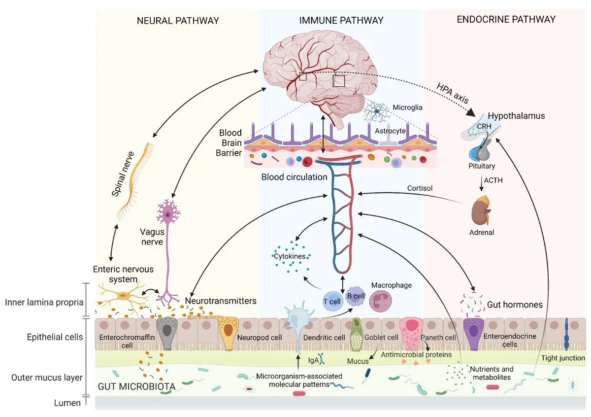
Figure 1: Gut -Brain Axis
within the GI
direct or indirect
2.2.1. Neural pathways (vagus nerve)
The vagus nerve is a major PNS pathway, comprising mainly afferent fibers that transmit visceral signals from the GI, cardiovascular, and respiratory systems to the CNS, while efferent fibers provide feedback to peripheral organs. Vagal pathways regulate appetite, stress, inflammation, and cognition. Vagal afferents detect mechanical, chemical, and hormonal signals through specialized terminals and connections with enteroendocrine cells (neuropods) [26,27].
2.2.2. Endocrine pathway
Enteroendocrine cells (EECs), although comprising only about 1% of intestinal epithelial cells, form a major endocrine signaling system linking the gut and brain. Different EECs release hormones such as GIP, CCK, and GLP-1. They sense nutrients, microbiota, and microbial metabolites through transporters and GPCRs, thereby influencing energy and glucose homeostasis and food intake [28,29].
2.2.3. Immune pathway
The gut immune system maintains intestinal homeostasis by recognizing harmful microbial signals and coordinating innate and adaptive immune responses. Enterocytes and immune cells express pattern-recognition receptors (PRRs), including TLRs, which detect microbial-associated molecular patterns such as LPS and peptidoglycan. Their activation triggers immune and inflammatory responses that can influence gut–brain communication [30,31].
2.2.4. Enteric nervous system
The ENS contains approximately 200–600 million neurons organized mainly into the myenteric and submucosal plexuses. It regulates intestinal motility, secretion, absorption, and immune functions and communicates with the CNS through vagal and sympathetic pathways. Due to its structural and functional complexity, the ENS is often termed the “second brain” and plays an important role in gut–brain communication [32,33].
2.2.5. Microbial metabolites
The gut microbiota is a diverse community of bacteria, fungi, archaea, parasites, and viruses that contributes substantially to host metabolism. Its collective genetic repertoire, the microbiome, contains millions of genes and greatly expands the metabolic capacity of the human host. Microbial metabolites therefore represent important mediators of gut–brain communication and host physiological functions [34,35].
2.3. Physiological Functions
2.3.1. Maintenance of intestinal barrier
The intestinal barrier acts as a selective interface that allows nutrient absorption and immune surveillance while limiting the entry of pathogens and harmful molecules. It consists mainly of the mucus layer, intestinal epithelial cells, and the underlying lamina propria containing innate and adaptive immune components [36,37].
2.3.2. Neurotransmitter synthesis
Neurons and glial cells regulate neurotransmitter synthesis through specific enzymes, and disruptions in these processes are associated with neurological and psychiatric disorders. Gut microbiota can also produce neurotransmitters, their precursors, or metabolites that influence neurotransmitter synthesis and potentially affect brain function [38,39].
2.3.3. Immune regulation
Balanced production of pro- and anti-inflammatory cytokines is essential for effective immune defense and intestinal homeostasis. Dysregulated cytokine production can alter the gut microbial environment, while excessive systemic cytokines may influence brain function by affecting the blood–brain barrier and neural signaling [40,41].
2.3.4. Energy metabolism
Glucose is the brain’s primary energy source under normal conditions, whereas ketone bodies produced by the liver during prolonged fasting can serve as an alternative fuel. Gut-derived metabolites and signals contribute to brain energy metabolism and neuronal function, while gut–brain pathways also participate in the regulation of insulin, glucagon, hepatic glucose production, and peripheral glucose utilization [42,43].
3.Gut dysbiosis: causes and consequences
Disruption of the gut microbiota ecosystem has many consequences, which can be divided into disruption of the gut barrier and imbalance of the host immune and metabolic systems. The integrity of the intestinal wall could be compromised by acetaldehyde produced by the microbiota from exogenous or endogenous ethanol , direct mucolytic activity, and other mechanisms. The host immune system can be modulated by microbiota-derived molecules via inflammasome signalling or Toll-like receptor (TLR) and NOD-like receptor (NLR) signalling [44]. Dysbiosis can be caused by host-specific factors such as genetic background, health status (infections, inflammation), and lifestyle habits or—more importantly—environmental factors such as diet (high sugar, low fibre), xenobiotics (antibiotics, drugs, food additives), and hygiene [45].
3.1 Factors contributing to gut dysbiosis
Changes in gut motility, immune function, stomach acidity, diet, and medication use can reduce microbial diversity and alter beneficial bacteria. Diets high in ultra-processed foods, refined sugars, saturated fats, and low in fiber can reduce microbial diversity and beneficial short-chain-fatty-acid–producing bacteria. A diverse, fiber-rich diet generally supports a healthier microbiome. Antibiotics can eliminate susceptible beneficial bacteria along with pathogens, potentially reducing diversity and allowing resistant or opportunistic organisms to expand [46]. Exposure to pollutants, pesticides, heavy metals, and other environmental chemicals may alter microbial composition and microbial metabolism. Persistent psychological stress can influence the gut through the gut–brain axis, affecting intestinal motility, permeability, immune signaling, and microbial composition. Physical inactivity, poor sleep, smoking, excessive alcohol consumption, and irregular eating patterns can negatively influence the gut microbiome. Host genetics can influence immune responses, intestinal barrier function, metabolism, and the types of microbes that can colonize the gut [47].
3.2 Biomarkers of dysbiosis
Dysbiosis refers to an imbalance in the composition, diversity, or functional activity of the gut microbiota. Biomarkers of dysbiosis help identify alterations in microbial communities and their associated metabolic functions. These biomarkers can provide valuable information about changes in gut microbial composition and host–microbiota interactions. Their assessment may support the early detection, monitoring, and understanding of dysbiosis-associated diseases (Table 2).
Table 2: Biomarkers of Dysbiosis
| Biomarker | What is measured | Role in dysbiosis | Reference |
|---|---|---|---|
| Microbial diversity indices | α-diversity: shannon, simpson β-diversity: Bray-curtis |
Reduced richness/evenness is often associated with dysbiosis, although direction depend on disease | [48] |
| Short - chain fatty acids (SCFAs) | Acetate, propionate, butyrate; sometimes valerate/isobutyrate | Reduced butyrate and altered total SCFAs concentrations may indicate impaired microbial fermentation and intestinal metabolic function | [49] |
| Lipopolysaccharides (LPS) | Plasma/serum LPS or LPS related markers | Increased circulating LPS can indicate increased exposure to bacterial endotoxin and impaired gut barrier function; measurement is technically challenging | [50] |
| Fecal metabolites | Untargeted or targeted metabolomics: bile acids, indoles, phenols, lactate, succinate, amino acid metabolism | Reflects functional consequences of microbial community changes and can reveal alterations not capturing by taxonomic profiling | [51] |
4. Molecular mechanism linking gut dysbiosis and neurodegeneration
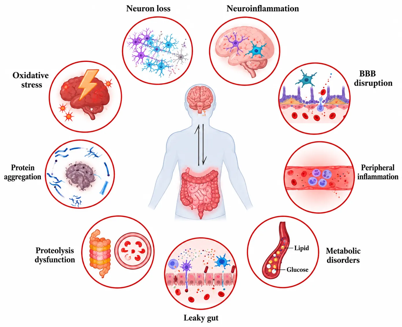
Figure 2: Various Mechanisms Linking Gut Dysbiosis and Neurodegeneration
4.1 Increased intestinal permeability (leaky gut)
Zonulin regulates intestinal tight junctions, which include proteins such as occludin, claudins, and ZO-1. Inflammation, oxidative stress, infection, and dietary or environmental factors can disrupt these junctions and increase intestinal permeability. This allows microbial products to enter the circulation and activate inflammatory pathways such as TLR/NF-κB, potentially contributing to systemic inflammation [52–54]. Increased permeability may also facilitate the translocation of bacterial endotoxins and other microbial components into the bloodstream. Persistent exposure to these molecules can promote systemic inflammation and indirectly affect BBB integrity and neuronal homeostasis.
4.2 Blood–brain barrier dysfunction
The blood–brain barrier (BBB) maintains CNS homeostasis by regulating the passage of nutrients and harmful substances between the blood and brain. It is composed of endothelial cells, tight junctions, pericytes, the basement membrane, and astrocytes. Alterations in these components can increase BBB permeability and are associated with neurodegenerative disease progression. Pericytes and astrocytes are particularly important for maintaining endothelial integrity, vascular function, and BBB permeability [55,56]. Systemic inflammatory mediators and microbial products may contribute to endothelial dysfunction and disruption of BBB tight junctions. Increased BBB permeability can consequently expose neural tissue to circulating inflammatory molecules, potentially enhancing neuroinflammatory responses.
4.3 Neuroinflammation
Microglia are the principal immune cells of the CNS and play important roles in synaptic maintenance and inflammatory responses. Microbial products can activate microglial pattern-recognition receptors, triggering NF-κB/MAPK signaling and the NLRP3 inflammasome. Activated microglia release inflammatory mediators such as TNF-α, IL-1β, IL-6, ROS, and chemokines, which can promote oxidative stress, mitochondrial dysfunction, synaptic impairment, and neuronal injury. Astrocyte activation can further amplify these inflammatory processes, creating a persistent neuroinflammatory cycle [57,58]. Chronic activation of glial cells may impair neuronal communication and reduce the ability of the CNS to resolve inflammation. Prolonged neuroinflammation can therefore contribute to progressive synaptic dysfunction and neuronal loss characteristic of neurodegenerative disorders.
4.4 Oxidative stress and mitochondrial dysfunction
Oxidative stress occurs when the production of reactive oxygen species exceeds the capacity of antioxidant systems. Mitochondria are major sources of ROS, particularly at electron transport chain complexes I and III. Excessive ROS can damage proteins, lipids, mitochondrial DNA, and cellular membranes, impair oxidative phosphorylation, and promote apoptosis. Oxidative stress and mitochondrial dysfunction can therefore reinforce each other and contribute to neuronal damage and neurodegeneration [59,60]. Neurons are particularly vulnerable because of their high energy requirements and dependence on mitochondrial ATP production. Gut-derived inflammatory signals and microbial metabolites may further disturb mitochondrial function, increasing ROS production and reducing cellular energy availability.
4.5 Protein misfolding and aggregation
Accumulation of misfolded proteins is a characteristic feature of many neurodegenerative disorders, including amyloid-β and tau in AD and α-synuclein in PD. Certain gut bacteria produce amyloid-like proteins that may influence host protein aggregation or activate innate immune responses. Experimental studies also suggest that gut microbial composition and microbial metabolites can modulate α-synuclein aggregation and microglial activation, although the precise mechanisms remain under investigation [61,62]. Microbial alterations may also affect the cellular mechanisms responsible for protein folding and clearance. Impaired autophagy and proteasomal degradation can further promote the accumulation of abnormal proteins and increase neuronal vulnerability.
4.6 Neurotransmitter dysregulation
Gut microbiota can influence neurotransmitter synthesis, metabolism, and signaling. Certain bacterial species can produce or modify neurotransmitters and their precursors, including GABA, dopamine, norepinephrine, and serotonin. Microbiota depletion studies have demonstrated changes in neurotransmitter levels and receptor expression in the gut, blood, and brain. These findings suggest that microbial alterations may influence neuronal signaling, cognition, mood, and gastrointestinal functions [63,64]. Changes in microbial composition may therefore alter the availability of neurotransmitter precursors and their metabolic pathways. Such alterations can affect communication between the enteric and central nervous systems and may contribute to changes in cognitive and behavioral functions.
4.7 Proteolysis dysfunction
Gut dysbiosis may disturb microbial and host protease activity, promoting abnormal protein degradation and the generation of inflammatory peptides. Excessive proteolytic activity can damage intestinal barrier proteins and increase gut permeability. Subsequent systemic inflammation and oxidative stress may affect the BBB and activate glial cells. Chronic inflammation can also impair the ubiquitin–proteasome and autophagy–lysosomal systems, reducing the clearance of misfolded proteins such as amyloid-β, tau, and α-synuclein and potentially promoting their accumulation [65,66]. Disruption of normal proteolytic balance may therefore connect intestinal barrier dysfunction with impaired neuronal protein homeostasis. Reduced clearance of damaged proteins can increase cellular stress and contribute to progressive synaptic and neuronal dysfunction.
4.8 Peripheral inflammation
Gut dysbiosis can promote chronic, low-grade systemic inflammation by reducing beneficial microbial metabolites and increasing intestinal permeability. Microbial products such as LPS can enter the circulation and activate immune cells through receptors such as TLR4, stimulating NF-κB/MAPK signaling and production of pro-inflammatory cytokines including TNF-α, IL-1β, and IL-6. Persistent peripheral inflammation may contribute to oxidative stress, endothelial dysfunction, BBB impairment, and neuroinflammation [67]. Circulating inflammatory mediators can communicate with the CNS through neural, humoral, and vascular pathways. Repeated or prolonged inflammatory signaling may consequently maintain glial activation and increase susceptibility to neuronal damage.
4.9 Microbial metabolites
Gut microbiota produce or modify several biologically active metabolites, including SCFAs, tryptophan metabolites, secondary bile acids, and TMAO. SCFAs such as acetate, propionate, and butyrate support intestinal barrier integrity and regulate immune and metabolic functions. Tryptophan-derived indoles can activate AhR and influence intestinal and immune homeostasis. Secondary bile acids act through receptors such as FXR and TGR5 to regulate metabolism and inflammation, while dietary choline and carnitine can be converted by bacteria to TMA and subsequently to TMAO in the liver. Collectively, these metabolites provide important molecular pathways through which gut dysbiosis can influence systemic and neurological functions [68,69]. Alterations in the production or balance of these metabolites may therefore modify inflammatory, metabolic, and neuronal signaling. Their effects on the gut barrier, immune system, vascular function, and CNS may collectively contribute to mechanisms involved in neurodegeneration.
5. Gut Dysbiosis in Major Neurodegenerative Diseases
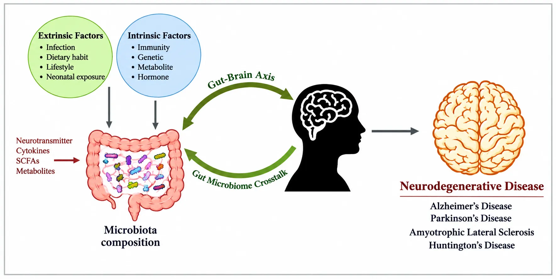
Figure 3: The Microbiota Composition Is Altered by External and Internal Factors
5.1. Alzheimer’s disease
AD is the most common form of dementia and is characterized by amyloid-β (Aβ) plaques, tau tangles, and progressive cognitive decline. Studies report gut microbiome alterations in AD, including reduced microbial diversity, decreased beneficial SCFA-producing bacteria, and increased potentially pro-inflammatory taxa such as Escherichia/Shigella [70,71].
Gut dysbiosis may contribute to AD through increased intestinal permeability, systemic inflammation, altered microbial metabolites, and impaired microglial function. Reduced SCFAs, particularly butyrate, may affect microglial maturation and amyloid clearance, potentially promoting Aβ accumulation [72].
5.2. Parkinson’s disease
PD is characterized by dopaminergic neuronal loss and α-synuclein aggregation. Gastrointestinal dysfunction, particularly constipation, is common and may occur years before motor symptoms. PD patients show alterations in gut microbiota, including reductions in some SCFA-producing bacteria and changes in potentially pro-inflammatory taxa [73,74].
Gut dysbiosis may influence intestinal inflammation, barrier integrity, and α-synuclein pathology. In addition, certain gut bacteria, such as Enterococcus faecalis, can metabolize levodopa in the intestine and potentially reduce its availability for absorption [75,76].
5.3. Multiple sclerosis
MS is an immune-mediated disease involving CNS inflammation, demyelination, and neurodegeneration. Experimental studies have shown that specific gut bacteria can promote Th17-cell responses and induce MS-like disease in animal models [77]. Human studies have also reported alterations in SCFA-producing and potentially pro-inflammatory bacterial groups [78].
Changes in gut microbiota have been associated with increased pro-inflammatory Th17 responses in MS patients. These findings suggest that dysbiosis may influence MS through effects on intestinal barrier function and systemic immune regulation [79].
5.4. Amyotrophic lateral sclerosis
ALS is a progressive neurodegenerative disease characterized by motor neuron loss and paralysis. Gut microbiome studies have identified dysbiosis in ALS, although findings vary according to diet, disease stage, and other factors. Some studies report reduced beneficial SCFA-producing bacteria and increased potentially inflammatory taxa [80,81].
Animal studies provide further evidence for microbiome involvement. In SOD1^G93A^ mice, microbiome alterations occur before major symptoms, and colonization with Akkermansia muciniphila was reported to improve disease-related outcomes [82]. These findings suggest a potential role for microbial and metabolic pathways in ALS progression.
5.5. Huntington’s disease
Huntington’s disease (HD) is an autosomal dominant neurodegenerative disorder characterized by progressive motor, cognitive, and psychiatric abnormalities. Dysphagia in advanced disease can contribute to weight loss and aspiration complications [83,84].
Recent studies suggest that HD may be associated with alterations in gut microbiome composition and diversity. Animal studies have identified differences in bacterial abundance, with some changes varying between male and female HD models [85,86]. However, the functional significance of these microbial alterations and their contribution to HD pathology require further investigation.
Table 3: Gut Microbiota Alterations and Proposed Gut–Brain Mechanisms in Major Neurodegenerative Diseases
| Disease | Gut microbiota changes | Metabolic changes | Gut–brain mechanism | Major consequences | Reference |
|---|---|---|---|---|---|
| Alzheimer’s disease | ↓ diversity; altered Firmicutes/Bacteroidetes | Altered SCFAs, bile acids, LPS | Gut leak → inflammation → BBB/microglia | Aβ accumulation, neuroinflammation | [87] |
| Parkinson’s disease | Altered Prevotella, Akkermansia, Enterobacteriaceae | Altered SCFAs and bile acids | Gut inflammation, vagal signaling, α-synuclein | α-Synuclein aggregation, neuronal loss | [88] |
| Multiple sclerosis | ↓ beneficial; ↑ pro-inflammatory taxa | Altered SCFAs and tryptophan metabolites | Gut leak → Th17/Treg imbalance | Inflammation, demyelination | [89] |
| ALS | Altered diversity and SCFA producers | Altered SCFAs and inflammatory metabolites | Barrier dysfunction and immune activation | Oxidative stress, motor-neuron loss | [90] |
| Huntington’s disease | Altered composition and diversity | Altered SCFAs and microbial metabolites | Gut permeability and immune signaling | Neuroinflammation, neuronal dysfunction | [91] |
6. Experimental Models for Studying Gut–Brain Interactions
6.1 Germ-free animal models
Germ-free rodents, particularly mice, are widely used to study the role of gut microbiota in brain function and disease. Other models, including zebrafish, C. elegans, pigs, and non-human primates, provide complementary approaches. These models help investigate microbiota effects on behavior, stress, cognition, and neuroinflammation [92,93].
6.2 Fecal microbiota transplantation models
FMT involves transferring fecal microbiota from a donor into a recipient to modify the gut microbial community. In animal studies, FMT has been used to investigate whether microbiota from healthy or diseased individuals can influence neurodegenerative phenotypes, including amyloid pathology and cognition. However, clinical evidence in neurological disorders remains limited and safety requires careful consideration [94–96].
6.3 Antibiotic-induced dysbiosis models
Antibiotics such as ampicillin, vancomycin, neomycin, and metronidazole are used to disrupt gut microbiota in experimental animals. These models help examine the effects of dysbiosis on microbial metabolites, intestinal permeability, immune signaling, neuroinflammation, and behavior [97].
6.4 Humanized microbiome models
Humanized microbiome models are commonly generated by colonizing germ-free mice with microbiota from human donors. They allow researchers to investigate how microbiota from healthy or diseased individuals affect host physiology, metabolism, immunity, and neurological outcomes under controlled conditions [98,99].
6.5 Organoid models
Intestinal and brain organoid systems provide controlled platforms for studying interactions among epithelial cells, immune cells, microbes, and microbial metabolites. Advanced 3D and microfluidic models can reproduce aspects of the gut environment and help investigate the effects of SCFAs and immune signaling on neuronal, astrocytic, and microglial functions [100,101].
7. Therapeutic Strategies Targeting Gut Dysbiosis
Growing evidence linking gut dysbiosis with neurodegenerative mechanisms has led to several therapeutic approaches targeting the microbiota–gut–brain axis (MGBA), including dietary modification, probiotics, prebiotics, synbiotics, postbiotics, phage therapy, precision microbiome interventions, and nanotechnology-based delivery systems (Fig. 4).
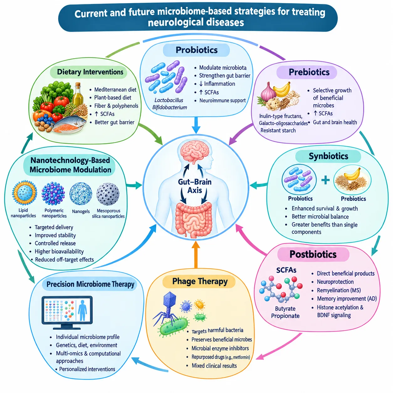
Figure 4: Therapeutic Strategies Targeting Microbiota–Gut–Brain Axis (MGBA)
7.1 Dietary Interventions
Diet is a major determinant of gut microbiome composition. Plant-rich diets, particularly Mediterranean and plant-based patterns, provide fiber and polyphenols that promote beneficial microbes, increase short-chain fatty acid (SCFA) production, and support intestinal barrier integrity. Higher adherence to Mediterranean-type diets has been associated with reduced risk of Alzheimer’s disease (AD) and Parkinson’s disease (PD) and slower cognitive decline [102,103].
7.2 Probiotics
Probiotics, including Lactobacillus and Bifidobacterium strains, can modulate gut microbial composition, strengthen the intestinal barrier, and reduce inflammation. Their metabolites, particularly SCFAs, may influence neuroimmune signaling, blood–brain barrier integrity, neurogenesis, and CNS inflammation [104,105].
7.3 Prebiotics
Prebiotics are non-digestible substrates that selectively promote beneficial gut microorganisms. Compounds such as inulin-type fructans, galacto-oligosaccharides, and resistant starch can enhance beneficial microbial growth and SCFA production, potentially supporting intestinal and brain health [106,107].
7.4 Synbiotics
Synbiotics combine probiotics with complementary prebiotics to enhance microbial survival, growth, and metabolic activity. By increasing beneficial microbial populations and improving gut microbial balance, synbiotics may provide greater benefits than probiotics or prebiotics alone [108,109].
7.5 Postbiotics
Postbiotics provide beneficial microbial products directly, offering more controlled dosing than live microorganisms. SCFAs such as butyrate and propionate have shown neuroprotective effects in experimental models, including improved remyelination in multiple sclerosis and enhanced memory in AD, potentially through histone acetylation and BDNF signaling [110,111].
7.6 Phage Therapy
Phage-based approaches aim to selectively target harmful bacteria or microbial pathways while preserving beneficial microbes. Other microbiome-modulating strategies include microbial enzyme inhibitors and repurposed drugs such as metformin, which may alter SCFAs, bile acids, and GLP-1 signaling. However, clinical trials have produced mixed results, with some synbiotic and antibiotic interventions showing no significant improvement in neurological outcomes [112,113].
7.7 Precision Microbiome Therapy
Individual differences in microbiome composition, genetics, diet, and environmental factors can influence responses to microbiome-targeted therapies. Precision approaches therefore aim to tailor interventions according to individual or disease-specific microbiome profiles using computational and multi-omics methods [114,115].
7.8 Nanotechnology-Based Microbiome Modulation
Nanoparticles can enable targeted delivery of probiotics, prebiotics, antimicrobial agents, bacteriophages, and bioactive compounds to specific gastrointestinal regions. Lipid nanoparticles, polymeric nanoparticles, nanogels, and mesoporous silica nanoparticles can improve stability, controlled release, bioavailability, and microbial targeting while potentially reducing off-target effects [116–118].
8. Multi-omics and Artificial Intelligence in Gut Microbiome Research
Multi-omics approaches integrate information from microbial genes, transcripts, proteins, and metabolites to provide a comprehensive understanding of gut microbiome composition and function. Artificial intelligence (AI) and machine learning further enable the analysis of complex microbiome datasets and support biomarker discovery and personalized medicine.
8.1 Metagenomics
Metagenomics characterizes the genetic composition of microbial communities and their associations with health and disease. Shotgun metagenomics sequences the overall genetic content of a sample, whereas targeted-amplicon sequencing focuses on specific microbial groups, providing complementary approaches for microbiome profiling and disease investigation [119].
8.2 Metabolomics
Metabolomics identifies and quantifies metabolites produced by gut microorganisms and the host. These metabolites reflect microbial metabolic activity and microbiota–host interactions, providing important insights into gut function and disease-associated metabolic changes [120,121].
8.3 Transcriptomics
Transcriptomics examines RNA transcripts to characterize gene expression and regulatory activity under specific physiological or pathological conditions. RNA sequencing enables the detection of coding and non-coding transcripts, differential gene expression, and alternative splicing, thereby helping elucidate molecular mechanisms of disease [122,123].
8.4 Proteomics
Metaproteomics identifies proteins produced by microbial communities and provides functional information about active metabolic and regulatory pathways. When integrated with metabolomics, it links microbial proteins to their metabolic products, offering a more comprehensive understanding of microbiome function [124].
8.5 Machine Learning and Artificial Intelligence
Machine learning enables computational analysis of complex microbiome datasets without explicit rule-based programming. Supervised and unsupervised algorithms can identify microbial patterns, biomarkers, co-abundance groups, and community structures such as enterotypes, supporting disease classification and prediction [125,126].
8.6 Personalized Microbiome-Based Medicine
Personalized microbiome medicine combines individual microbial profiles with clinical, genetic, dietary, and immune data to understand disease risk and treatment responses. Techniques such as 16S rRNA sequencing, shotgun metagenomics, and metabolomics can guide individualized dietary and microbiome-based interventions, including probiotics, prebiotics, synbiotics, postbiotics, and fecal microbiota-based therapies. However, interindividual variability, methodological differences, and limited clinical validation remain important challenges [127–129].
9. Current Challenges
9.1 Inter-individual Microbiome Variability
Gut microbiome composition varies considerably among individuals due to diet, age, geography, genetics, medications, lifestyle, and host physiology. This variability makes it difficult to define a universal “healthy” microbiome and limits the generalizability of findings across populations.
9.2 Lack of Standardized Methodologies
Differences in sample collection, storage, DNA extraction, sequencing technologies, laboratory procedures, bioinformatic pipelines, and clinical metadata can lead to inconsistent results. Standardized protocols and clinical endpoints are therefore needed to improve reproducibility and comparability.
9.3 Establishing Causality Versus Association
Although microbiome studies frequently identify disease-associated microbial changes, these associations do not establish causality. Microbial alterations may result from disease, medications, diet, or inflammation. Longitudinal studies, controlled interventions, and mechanistic validation are required to establish causal relationships.
9.4 Limited Long-Term Clinical Data
Many microbiome studies are cross-sectional or short-term, limiting understanding of microbiome stability, disease progression, and treatment responses. Longitudinal studies are needed but remain challenging because of high costs, participant retention, repeated sampling, and the need for comprehensive clinical data.
9.5 Ethical and Regulatory Considerations
Microbiome research involves biological and potentially sensitive health data, raising concerns regarding informed consent, privacy, data ownership, and secondary data use. In addition, regulatory frameworks for microbiome-based diagnostics and therapeutics are still evolving, creating challenges for safety assessment, validation, and clinical implementation.
10. Future Perspectives
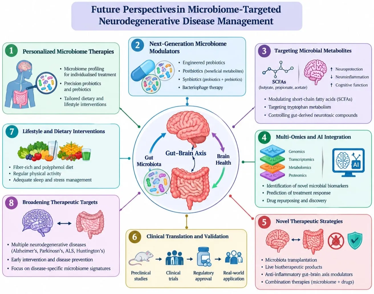
Figure 5: Future Perspectives in Microbiome-Targeted Neurodegenrative Disease Management
Future microbiome research in neurodegenerative diseases will focus on personalized and precision-based therapies. Microbiome-derived biomarkers may support early diagnosis, disease monitoring, and prediction of treatment responses. Microbiome engineering, CRISPR-based editing, next-generation probiotics, and personalized nutrition may enable targeted modulation of microbial communities. Integration of multi-omics, artificial intelligence, and machine learning could improve understanding of microbiome–brain interactions and guide individualized treatments. Combining microbiome-based interventions with conventional therapies may further enhance therapeutic strategies. However, clinical validation, standardization, long-term safety, and ethical considerations remain essential.
CONCLUSION
Gut dysbiosis is increasingly recognized as an important factor potentially contributing to neurodegenerative diseases through the gut–brain axis. Microbial metabolites, immune and inflammatory pathways, intestinal barrier dysfunction, and neuronal signaling may collectively influence brain health. Alterations in gut microbial composition have been associated with diseases such as Alzheimer’s disease and Parkinson’s disease; however, current evidence remains insufficient to establish dysbiosis as a direct cause of neurodegeneration.
Microbiome-targeted approaches, including dietary modification, probiotics, prebiotics, synbiotics, postbiotics, and fecal microbiota transplantation, show promising therapeutic potential, but clinical findings remain heterogeneous. Individual differences in age, diet, genetics, medications, lifestyle, and microbiome composition further complicate treatment development. Future research should focus on well-designed longitudinal and multicenter clinical trials, standardized microbiome methodologies, identification of disease-specific microbial biomarkers, and clarification of underlying mechanisms.
Integration of multi-omics, artificial intelligence, and personalized microbiome-based therapies may help develop more precise interventions. Overall, the gut microbiome represents a promising therapeutic avenue for neurodegenerative diseases, but robust mechanistic evidence, clinical validation, and long-term safety data are essential before routine clinical application.
REFERENCES


Diksha Devkate, Pooja B. Rasal, Vikas S. Shinde, Amol U. Gayke, Gut Dysbiosis and Neurodegeneration: Current Evidence and Future Directions, Int. J. Sci. R. Tech., 2026, 3 (11), 848-869. https://doi.org/10.5281/zenodo.24300201
 10.5281/zenodo.24300201