

We use cookies to ensure our website works properly and to personalise your experience. Cookies policy
1Department of Pharmacology, JES's SND College of Pharmacy, Babhulgaon (Yeola), India
2Department of Pharmaceutical Quality Assurance, JES's SND College of Pharmacy, Babhulgaon (Yeola), India.
3Department of Pharmaceutics, JES's SND College of Pharmacy, Babhulgaon (Yeola), India
The gut–liver axis is a complex bidirectional communication network that plays an essential role in maintaining hepatic and intestinal homeostasis. The gut microbiota, intestinal barrier, portal circulation, bile acids, microbial metabolites, and immune signaling collectively regulate hepatic metabolism, inflammation, and immune responses. Alterations in gut microbial composition and function, commonly referred to as dysbiosis, can impair intestinal barrier integrity, increase microbial translocation and endotoxemia, alter bile acid metabolism, and promote oxidative stress and inflammatory signaling, thereby contributing to the development and progression of various liver diseases. This review discusses the structural and functional components of the gut–liver axis and summarizes the mechanisms linking gut dysbiosis with liver injury, including intestinal permeability, lipopolysaccharide-mediated inflammation, oxidative stress, and immune dysregulation. The role of gut microbiota in drug-induced liver injury, alcohol-associated liver disease, metabolic dysfunction-associated steatotic liver disease, steatohepatitis, viral hepatitis, fibrosis, cirrhosis, and hepatocellular carcinoma is also highlighted. Particular emphasis is placed on microbiome-derived molecules, including short-chain fatty acids, bile acids, indole derivatives, tryptophan metabolites, bacteriocins, and extracellular vesicles, as potential hepatoprotective mediators. Furthermore, emerging microbiome-based strategies such as probiotics, prebiotics, synbiotics, postbiotics, fecal microbiota transplantation, precision nutrition, dietary polyphenols, herbal medicines, and nanotechnology-based targeted delivery systems are discussed. Despite promising evidence, challenges related to microbiome variability, disease-specific dysbiosis, safety, standardization, and limited clinical validation remain. Future advances in personalized microbiome therapy, engineered probiotics, microbiome-derived therapeutics, artificial intelligence, and gut–liver organoid models may facilitate the development of precise and effective hepatoprotective interventions.
Liver disease is a severe global health problem with high morbidity and mortality. Alcohol, drugs, toxicants, viruses, and obesity can cause liver disorders, including fatty liver, hepatitis, fibrosis, cirrhosis, and liver cancers [1,2]. The liver is in close association with the intestine, both anatomically and functionally, through the hepatic portal venous system. It has been reported that 10–100 trillion microorganisms from 300–500 different species estimated worldwide reside in the adult human gut [3,4]. The gut microbiota has been recently found to beinvolvedin liver disease pathogenesis [5,6]. Epidemiological studies have indicated that liver diseases constitute an important part of global morbidity and mortality and have become a huge economic burden and an urgent public health crisis [7]. However, due to the lack of understanding of their pathogenesis, late diagnosis and rapid progression, the clinical therapeutic strategies for liver diseases are still insufficient, which directly leads to unsatisfactory treatment effects [8]. Therefore, the clinical treatment and management of liver diseases remain a considerable challenge, and there is an urgent need to deeply explore the mechanism and develop promising therapeutic drugs and strategies on this basis. Many dietary natural products possess the abilities to protect against liver diseases [9]. They have shown the potential in improving the integrity of the gut mucosa, modulating the composition of the microbiota, reducing the toxic metabolites and translocated bacteria in the liver [10]. Figure 1 indicates modulation of gut microbiota and its role in the gut-liver axis.
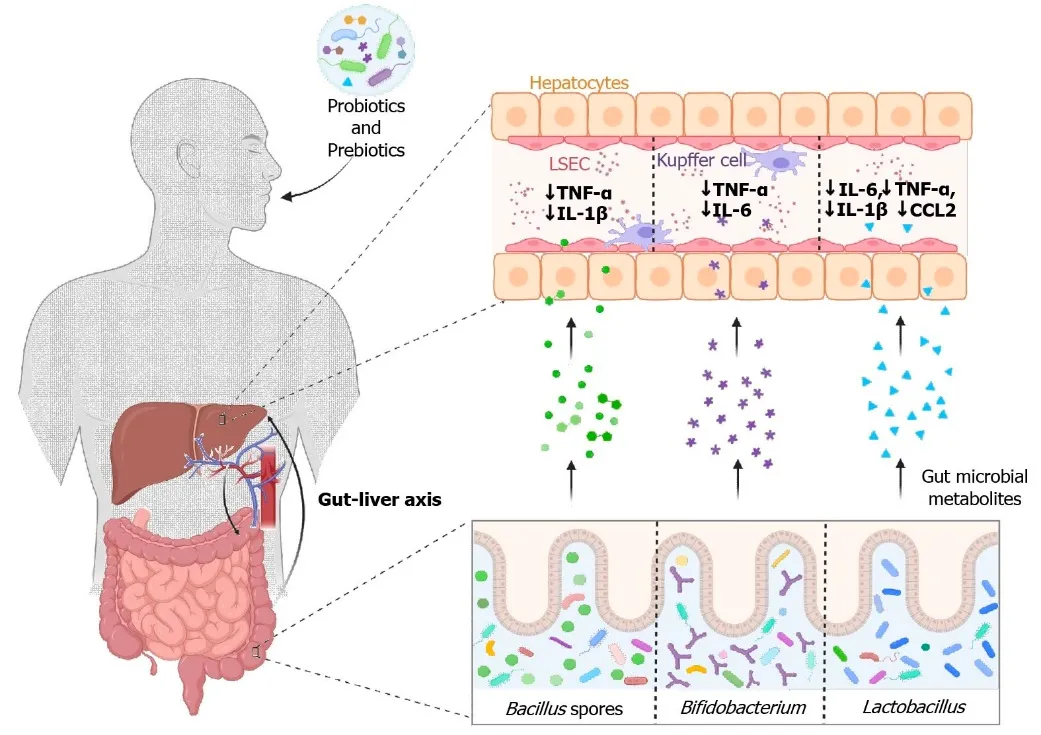
Figure 1: Modulation of gut microbiota and its role in the gut-liver axis during coronavirus disease 2019. Probiotics and prebiotics could be used as potential therapeutics to lower coronavirus disease 2019 symptom severity by producing various bioactive metabolites, which are absorbed into the liver mainly via the hepatic portal vein, for regulation of hepatic function by reducing inflammatory cytokines. CCL2: Chemokine (C-C motif) ligand 2; IL-1β: Interleukin-1 beta; IL-6: Interleukin 6; LSEC: Liver sinusoidal endothelial cells; TNF-α: Tumor necrosis factor-α. https://www.wjgnet.com/1007-9327/full/v29/i11/1708.
1.1. Global Burden of Liver Diseases
Liver disease accounts for over two million deaths annually (cirrhosis, viral hepatitis, and liver cancer) and accounts for 4% of all deaths worldwide (1 out of every 25 deaths); 1 out of 3 liver-related deaths occur among females [11]. Currently liver disease is the eleventh-leading cause of death, but liver deaths may be underestimated [12]. Cirrhosis is currently the tenth-leading cause of death in Africa (thirteenth-leading cause in 2015); ninth-leading cause in Southeast Asia and Europe; and the fifth-leading cause of death in the
Eastern Mediterranean. Cirrhosis also has a major impact on global health due to the high number of disability-adjusted life-years (DALYs) that it generates, representing the 15th leading cause of DALYs globally. The highest impact of liver disease is on the young, where it represents the 12th leading cause of DALYs in those aged 25 to 49 years [13]. The potential years of life lost may be higher, especially in Europe [14]. Cirrhosis is also an expensive disease. In the US in 2016, liver-related expenditure was $32.5 billion (95% CI $27.0-$40.4 billion) with two-thirds of these costs attributable to inpatient or emergency department care. Over 20 years, healthcare spending has increased by 4% per year primarily driven by hospital-based services.[15] We are currently at a pivotal juncture in terms of awareness, recognition and management of liver diseases at the global level. We propose collectively focusing on three goals that may blunt the global burden of liver disease. These include HCV elimination, increasing awareness of ALD and NASH and education/early screening for cirrhosis. The WHO has called on countries to eliminate viral hepatitis as a public health threat by 2030, with elimination defined as a 90% reduction in incidence, treatment of 80% of eligible people and a 65% reduction in mortality.296,297 This goal may be particularly challenging in developing countries. All major liver societies have endorsed and supported a comprehensive HCV elimination plan [16].
1.2. Need for Novel Hepatoprotective Strategies Targeting the Gut Microbiome
Conventional hepatoprotective therapies mainly focus on reducing oxidative stress, inflammation, lipid accumulation, or hepatocyte injury. However, increasing evidence shows that gut microbiome dysbiosis and disruption of thegut–liver axis play important roles in the development and progression of liverdiseases such as metabolic dysfunction-associated steatotic liver disease (MASLD), alcoholic liver disease, drug-induced liver injury, and cirrhosis. Therefore, approaches that specifically target the gut microbiome may provide a promising alternative or complementary strategy.[17]
1.2.1. Major reasons for developing novel microbiome-based hepatoprotective strategies include:
Limitations of conventional hepatoprotective agents – Existing therapies may provide incomplete protection and often do not directly address the underlying gut–liver axis dysfunction.[18]
Gut microbiome dysbiosis – Alterations in microbial diversity and composition can increase intestinal permeability and promote the transfer of bacterial products to the liver [19].
Reduction of microbial-derived toxins – Dysbiotic microbiota can increase endotoxins such as lipopolysaccharide (LPS), which activate hepatic inflammatory pathways. Modulating microbiomes may reduce this inflammatory burden.
Restoration of intestinal barrier integrity – Novel probiotics, prebiotics, postbiotics, and microbial metabolites may strengthen the intestinal barrier and reduce bacterial translocation [20].
Modulation of short-chain fatty acids (SCFAs) – Beneficial microbial metabolites such as butyrate, acetate, and propionate can influence intestinal integrity, inflammation, lipid metabolism, and hepatic function [21].
Bile acid–microbiome regulation – Gut bacteria modify bile acids, which regulate hepatic metabolism and inflammation through pathways such as FXR–FGF19 and TGR5. Targeting these pathways represents a promising hepatoprotective approach [22].
Personalized therapy – Individual differences in gut microbiota may influence disease progression and therapeutic response, creating opportunities for microbiome-based personalized hepatoprotection [23].
Novel therapeutic platforms – Next-generation probiotics, engineered bacteria, fecal microbiota transplantation, postbiotics, bacteriophages, microbial metabolites, and precision prebiotics are emerging approaches that could selectively modify disease-associated microbial communities [24].
Multi-targeted action – Microbiome-directed therapies may simultaneously influence oxidative stress, inflammation, intestinal permeability, lipid metabolism, bile-acid signaling, and immune responses [25].
1.2. Concept of the Gut-Liver Axis
Liver disease is responsible for more than 2 million deaths annually and accounts for approximately 4% of global deaths [26]. It is currently the 11th leading cause of death worldwide, although its burden may be underestimated [27]. The major causes of liver-related mortality include cirrhosis and hepatocellular carcinoma (HCC), commonly associated with viral hepatitis [28, 29], alcohol-associated liver disease (ALD) [30], and metabolic dysfunction-associated fatty liver disease (MAFLD). MAFLD is the most common chronic liver disease worldwide, affecting more than one-third of adults, and is also a rapidly increasing cause of primary liver cancer [31, 32].
The gut-liver axis refers to the bidirectional communication between the intestine, its microbiota, and the liver through the intestinal barrier and portal circulation [33, 34]. The liver influences intestinal health through bile and immune factors, while the gut affects liver function through microbial metabolites and translocated microbial products [34]. Alcohol, high-fat diets, pathogens, and other environmental factors can disrupt the intestinal barrier, promoting bacterial and toxin translocation into the portal circulation [35]. Bacterial toxins such as cytolysin from Enterococcus faecalis and candidalysin from Candida albicans can further damage intestinal epithelial cells [36].
Translocated pathogen-associated molecular patterns (PAMPs), particularly lipopolysaccharide (LPS), activate Toll-like receptor 4 (TLR4) on Kupffer cells and hepatic stellate cells. This activates MyD88- and TRIF-dependent signaling pathways, leading to NF-κB and AP-1 activation, production of pro-inflammatory cytokines such as TNF-α, IL-6, and IL-1β, and subsequent hepatic inflammation and fibrogenesis [37].
Gut microbial metabolism also influences liver disease progression. Bacteria such as Desulfovibrionaceae and Enterobacteriaceae convert dietary choline into trimethylamine (TMA), which is converted in the liver to trimethylamine N-oxide (TMAO); elevated TMAO is associated with altered bile acid metabolism and inflammation in MASLD [41]. Bacteria such as Clostridium scindens convert primary bile acids into secondary bile acids, including deoxycholic acid (DCA), which at high concentrations can cause hepatotoxicity, DNA damage, and promote HCC [38, 39]. In addition, Klebsiella pneumoniae and Escherichia coli can produce endogenous ethanol through fermentation, potentially contributing to oxidative stress and MASLD pathogenesis. Thus, disruption of the gut-liver axis plays an important role in intestinal barrier dysfunction, microbial translocation, inflammation, metabolic disturbances, and progression of liver disease. Figure 2 illustrates the role of the gut–liver axis in regulating gut microbiome composition and function, intestinal barrier integrity, microbial metabolite production, and their subsequent effects on liver health.
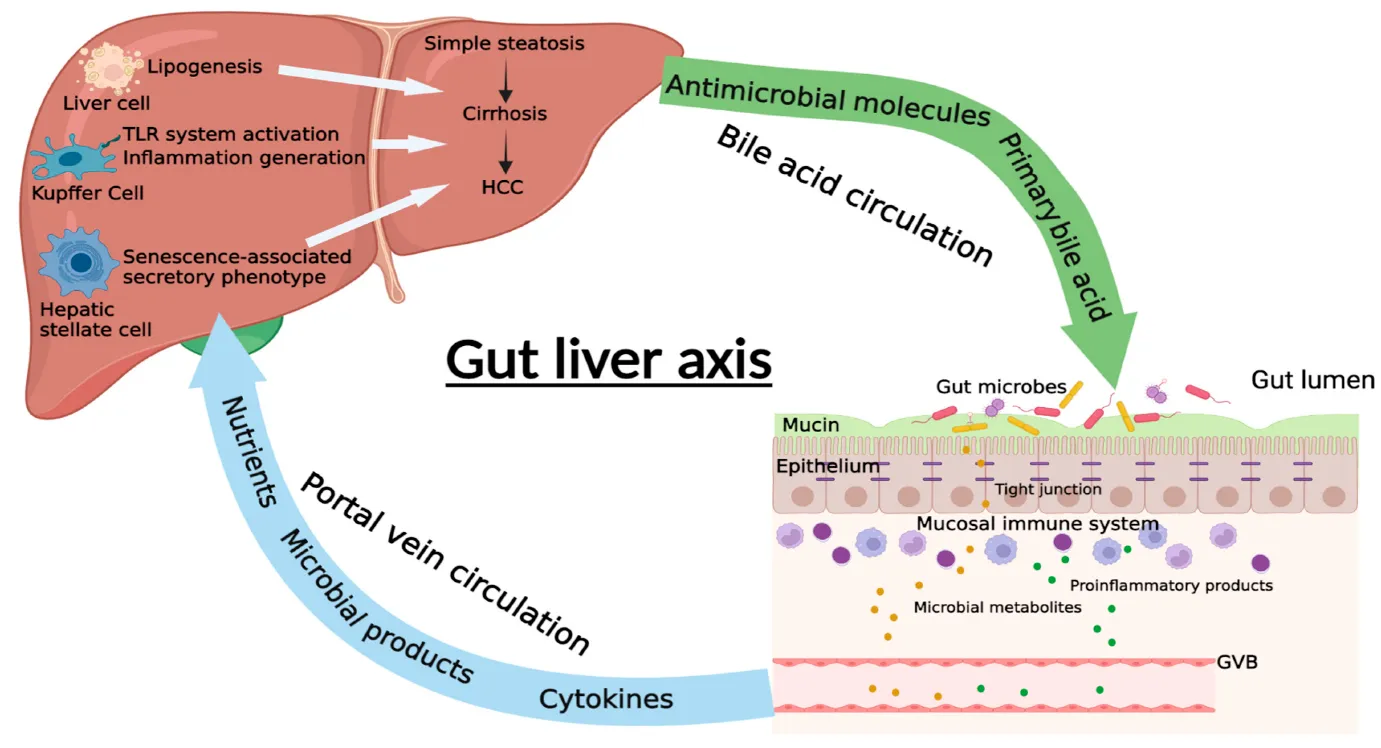
Figure 2: The Role of Gut–Liver Axis in Gut Microbiome (Https://Www.Mdpi.Com/2227-9059/10/3/524)
1.4. Importance of Intestinal Microbiota in Liver Health
The human gastrointestinal tract contains over 100 trillion microorganisms, including bacteria, fungi, viruses, and archaea [40]. Their composition varies throughout the gastrointestinal tract according to factors such as pH and oxygen availability, with microbial density highest in the colon [41]. Bacteroidetes and Firmicutes are the predominant bacterial phyla, accounting for approximately 90% of the gut microbiota [42,43]. The microbiota is dynamic and influenced by diet, physical activity, medications, and circadian rhythms. It performs essential functions, including nutrient extraction, drug and xenobiotic metabolism, immune regulation, and production of bioactive metabolites [44,45]. In particular, microbial fermentation of dietary fiber produces short-chain fatty acids (SCFAs), including acetate, propionate, and butyrate, which support intestinal epithelial health and contribute to immune regulation [46].
1.5. Objectives and scope of the review:
Objective:
1.Explain the gut–liver axis and its bidirectional communication between the intestinal microbiota and liver.
2.Describe the role of gut microbiota dysbiosis in the development and progression of liver diseases such as MASLD/NAFLD, alcoholic liver disease, viral hepatitis, and cirrhosis.
3.Examine microbiome-derived metabolites, including short-chain fatty acids, bile acids, indole derivatives, and microbial endotoxins, and their influence on hepatic inflammation, oxidative stress, and metabolism.
4.Evaluate microbiome-based hepatoprotective strategies, including probiotics, prebiotics, synbiotics, postbiotics, dietary interventions, fecal microbiota transplantation, and microbiota-targeted drugs.
5.Assess the mechanisms of action through which these interventions may restore intestinal barrier integrity, reduce inflammation, regulate immune responses, and protect hepatocytes.
6.Critically analyze recent preclinical and clinical evidence regarding the efficacy, safety, and limitations of microbiome-targeted hepatoprotective approaches.
7.Identify current research gaps and challenges and discuss future prospects for developing personalized microbiome-based therapies for liver disease.
Scope:
1.Gut–liver axis: intestinal barrier, portal circulation, bile acids, immune signaling, and microbial metabolites.
2.Gut microbiome and liver disease: contribution of dysbiosis to MASLD, alcoholic liver disease, cirrhosis, hepatic inflammation, fibrosis, and hepatocellular carcinoma.
3.Microbial mechanisms of hepatotoxicity: endotoxemia, altered bile-acid metabolism, intestinal permeability, oxidative stress, and inflammatory signaling.
4.Microbiome-based hepatoprotective interventions: probiotics, prebiotics, synbiotics, postbiotics, dietary modification, FMT, bacteriophages, microbial metabolites, and targeted microbial modulation.
5.Novel therapeutic approaches: next-generation probiotics, engineered microorganisms, precision microbiome therapeutics, and combination therapies.
6.Clinical translation: evidence from animal models and human clinical trials, safety considerations, regulatory challenges, and limitations.
7.Future perspectives: integration of metagenomics, metabolomics, artificial intelligence, and personalized medicine to develop patient-specific microbiome-based hepatoprotective strategies.
2. ANATOMY AND PHYSIOLOGY OF THE GUT-LIVER AXIS.
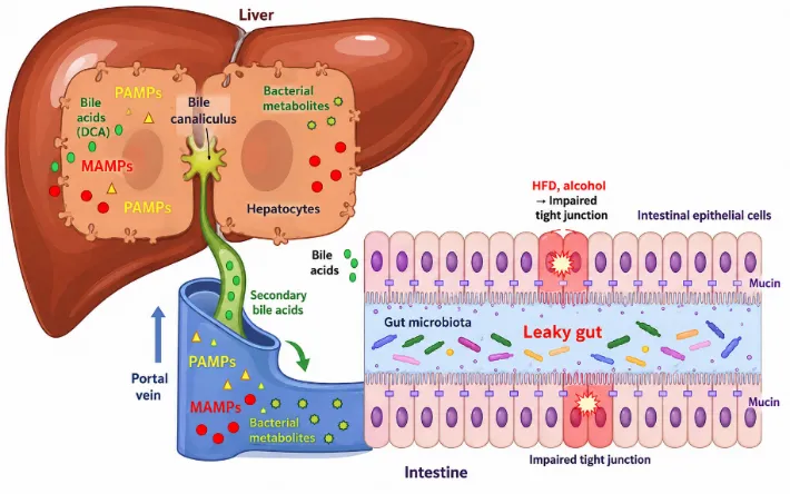
Figure 3: Gut–Liver Axis: Interplay of Gut Microbiota, Bile Acids and Intestinal Barrier Dysfunction
The gut and liver are anatomically and physiologically connected through the portal vein, which transports nutrients, microbial metabolites, and microbial components from the intestine to the liver. Bile and liver-derived substances return to the intestine through the bile duct, forming enterohepatic circulation [47]. Disruption of intestinal tight junctions increases gut permeability, allowing microbial products, metabolites, and bacteria to reach the liver. The liver also functions as an immune organ containing Kupffer cells, NK/NKT cells, T and B lymphocytes, hepatic stellate cells (HSCs), and liver sinusoidal endothelial cells (LSECs), which respond to gut-derived factors and regulate inflammation. Gut dysbiosis, bacterial overgrowth, and impaired intestinal barrier function can therefore contribute to liver inflammation, fibrosis, and cancer development [47,48]. Gut microbiota, bile acids, and intestinal barrier dysfunction contribute to bidirectional gut–liver communication (Figure 3).
2.1. Structural Components
The gut–liver axis consists of interconnected anatomical and functional components that facilitate continuous communication between the intestine and liver. Major structural components include the intestinal barrier, gut microbiota, portal circulation, liver, and biliary system. Together, these components regulate the transport of nutrients and microbial metabolites, bile acid circulation, immune responses, and maintenance of intestinal and hepatic homeostasis. Disruption of any component can disturb gut–liver communication and contribute to metabolic dysfunction and inflammation [49].Table 1 explains structural components of Gut-Liver Axis.
Table 1: Structural Components of Gut–Liver Axis
| Component | Structural Overview | Key Features | Primary Functions |
|---|---|---|---|
| Intestinal barrier | Epithelial cells, mucus layer, tight junctions, immune cells and microbiota | Claudins, occludin, mucus, GALT | Nutrient absorption; prevents pathogen/toxin entry; regulates immunity |
| Portal vein | Major venous vessel connecting intestine to liver | Formed by mesenteric and splenic veins; carries nutrient-rich blood | Transports nutrients, toxins and microbial products to the liver |
| Liver | Lobular organ with hepatocytes and portal triads | Dual blood supply; hepatic lobules | Metabolism, detoxification, protein and bile synthesis |
| Bile ducts | Network of ducts connecting liver to intestine | Lined by cholangiocytes; forms intra- and extrahepatic ducts | Transports bile; eliminates bilirubin and cholesterol; supports bile-acid circulation |
2.2. COMMUNICATION BETWEEN GUT AND LIVER
The gut–liver axis is a bidirectional communication system that maintains metabolic, immune, and physiological homeostasis between the intestine and liver. The gut delivers nutrients, microbial metabolites, microbial products, and immune signals to the liver mainly through the portal circulation, allowing the liver to process and detoxify these substances. In the reverse direction, the liver communicates with the intestine through bile acids, antibodies, and immune-regulatory factors, which influence intestinal microbiota, barrier integrity, and intestinal metabolism [50]. Disruption of this communication can lead to intestinal barrier dysfunction, microbial dysbiosis, inflammation, and altered bile acid metabolism, thereby contributing to liver and metabolic disorders. Table 2. Shows microbial metabolites Involved in Gut–Liver Communication.
The gut–liver axis involves coordinated communication through portal circulation, bile acid signalling, immune interactions, and microbial metabolites. The portal circulation transports nutrients, microbial products, and metabolites from the intestine to the liver, where they undergo metabolism, detoxification, and immune surveillance. Meanwhile, bile acids synthesized in the liver enter the intestine and are modified by gut microbiota into secondary bile acids, which activate receptors such as FXR and TGR5 to regulate metabolism, inflammation, and bile acid homeostasis. Gut microbiota also produce metabolites such as short-chain fatty acids (SCFAs), indole derivatives, and other bioactive compounds, which influence intestinal barrier integrity, immune responses, and hepatic metabolism. In addition, gut-derived microbial molecules interact with hepatic immune cells, including Kupffer cells and macrophages, while liver-derived bile, antibodies, and immune mediators influence the intestinal microbiota and mucosal immune system. Together, these pathways maintain gut and liver homeostasis, whereas their disruption can promote dysbiosis, barrier dysfunction, inflammation, and liver disease. Figure 4 illustrates Integrated Pathways of the Gut-Liver Axis.
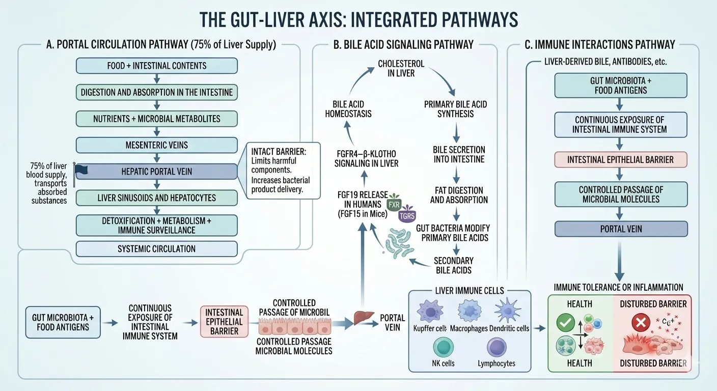
Figure 4: Integrated Pathways of the Gut-Liver Axis
3. HUMAN GUT MICROBIOME
The gut microbiome is a complex and diverse microbial community residing mainly in the gastrointestinal tract. It consists predominantly of bacteria, along with archaea, fungi, viruses, and protozoa. Bacteria represent the most abundant and functionally diverse group, with major phyla including Firmicutes, Bacteroidota, Actinobacteriota, and Proteobacteria. These microorganisms contribute to dietary digestion, production of short-chain fatty acids (SCFAs), vitamin synthesis, maintenance of intestinal barrier integrity, and regulation of host immunity. The composition of the gut microbiome varies among individuals and is influenced by diet, age, lifestyle, medications, and environmental factors (Table 2).
The composition and activity of gut microbiota are influenced by several host, dietary, environmental, and lifestyle factors. Diet is one of the major determinants; high-fiber diets generally support beneficial microbial fermentation and SCFA production, whereas diets rich in saturated fat and processed foods may alter microbial diversity. Age, from infancy to old age, is associated with changes in microbial composition. Antibiotics and other medications can significantly modify microbial populations and may reduce microbial diversity. Other important factors include genetics, geographical location, physical activity, stress, sleep, infections, health status, and alcohol or tobacco exposure. These factors can promote either maintenance of a balanced microbiota (eubiosis) or disruption of microbial composition and function (dysbiosis), potentially affecting intestinal and systemic health (Figure 5).
Table 2: Composition of Gut Microbiome
| Microbial Group | Major Members | Main Characteristics | Functions |
|---|---|---|---|
| Bacteria | Firmicutes, Bacteroidetes, Actinobacteria, Proteobacteria | Most abundant and diverse group | Digestion, SCFA production, vitamin synthesis and immune regulation |
| Archaea | Methanobrevibacter smithii | Methane-producing microorganisms | Hydrogen utilization and contribution to fermentation |
| Fungi | Candida, Saccharomyces | Eukaryotic microorganisms | Modulate microbial diversity, metabolism and immune responses |
| Viruses | Bacteriophages, eukaryotic viruses | Viral component of the gut ecosystem | Regulate bacterial populations and microbial community structure |
| Protozoa | Blastocystis and other intestinal protozoa | Less abundant microbial group | Interact with gut bacteria and influence host immune responses |
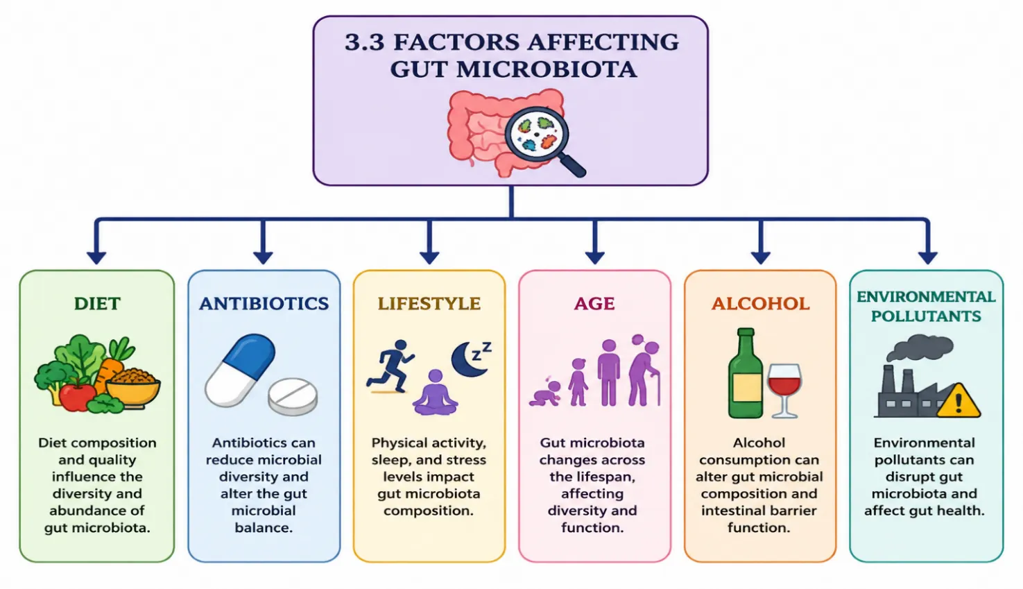
Figure 5: Factors Affecting Gut Microbiota [51]
4. MECHANISMS LINKING GUT DYBIOSIS AND LIVER INJURY
Gut dysbiosis can disrupt the gut–liver axis by impairing intestinal barrier integrity and altering microbial metabolites. Increased microbial translocation and endotoxin exposure activate hepatic inflammatory and immune pathways, while oxidative stress further promotes hepatocellular damage. Collectively, these mechanisms contribute to inflammation, metabolic dysfunction, and progressive liver injury [52].
4.1 Increased Intestinal Permeability (“Leaky Gut”)
Gut dysbiosis can impair the integrity of the intestinal epithelial barrier, resulting in increased intestinal permeability, commonly referred to as “leaky gut.” Under normal conditions, intestinal epithelial cells are connected by tight junction proteins such as occludin, claudins, and zonula occludens (ZO) proteins, which regulate the movement of substances between epithelial cells [53-57]. Dysbiosis can alter the abundance and activity of beneficial microorganisms and reduce microbial metabolites such as short-chain fatty acids (SCFAs) that support epithelial integrity. These changes can weaken tight junctions and increase paracellular permeability. Consequently, bacterial components, endotoxins, microbial metabolites, and other luminal products can cross the intestinal barrier and enter the portal circulation. Because portal blood directly delivers intestinal contents to the liver, increased permeability exposes hepatic cells to higher concentrations of microbial products. This persistent exposure can activate hepatic immune responses, promote inflammation, and contribute to hepatocellular injury [58,59].
4.2 Endotoxemia
Endotoxemia refers to the increased presence of bacterial endotoxins, particularly lipopolysaccharide (LPS), in the circulation. LPS is a major component of the outer membrane of Gram-negative bacteria. During gut dysbiosis, an increased abundance of LPS-producing bacteria, together with impaired intestinal barrier function, can facilitate the transfer of LPS from the intestinal lumen into the portal bloodstream. In the liver, LPS is recognized primarily by Toll-like receptor 4 (TLR4) in association with accessory proteins such as CD14 and MD-2. Activation of TLR4 initiates intracellular signaling pathways that stimulate inflammatory responses in Kupffer cells, hepatic macrophages, and other liver cells. This process promotes the release of inflammatory mediators and can enhance oxidative stress, hepatocyte dysfunction, and tissue injury. Persistent low-grade endotoxemia has therefore been implicated in the progression of metabolic and inflammatory liver disorders [60, 61].
4.3 Inflammatory Pathways
Gut dysbiosis promotes hepatic inflammation through activation of multiple innate immune and intracellular signaling pathways. Microbial products such as LPS, bacterial DNA, peptidoglycans, and other pathogen-associated molecular patterns (PAMPs) can activate pattern-recognition receptors on hepatic immune cells. One of the major downstream pathways is the nuclear factor-kappa B (NF-κB) signaling pathway. Activation of NF-κB promotes transcription of several pro-inflammatory genes and increases the production of cytokines including tumor necrosis factor-alpha (TNF-α), interleukin-1 beta (IL-1β), and interleukin-6 (IL-6). These mediators promote recruitment and activation of inflammatory cells and can interfere with normal hepatocyte function. Persistent inflammatory signaling may also stimulate stellate-cell activation and extracellular matrix deposition, thereby contributing to the development of hepatic fibrosis. Thus, continuous gut-derived inflammatory stimulation can transform transient hepatic immune activation into chronic liver inflammation and progressive tissue damage [62, 63].
4.4 Oxidative Stress
Gut dysbiosis can contribute to oxidative stress by increasing the production of reactive oxygen species (ROS) and weakening antioxidant defenses in the liver. Increased exposure to microbial products, inflammatory cytokines, and altered microbial metabolites can stimulate ROS generation in hepatocytes and immune cells. Excessive ROS can attack cellular lipids, proteins, DNA, and mitochondrial components. Lipid peroxidation is particularly important because oxidation of membrane lipids can impair cellular membranes and generate secondary reactive molecules that further promote inflammation. Mitochondrial dysfunction may additionally increase ROS production and reduce cellular energy metabolism. Normally, antioxidant systems such as glutathione, superoxide dismutase, catalase, and glutathione peroxidases help maintain redox balance. When ROS generation exceeds these protective mechanisms, oxidative stress develops. This oxidative imbalance can amplify inflammatory signaling, promote hepatocyte injury, and contribute to the progression of steatosis, inflammation, and fibrosis [64-66].
4.5 Immune Dysregulation
Gut microbiota have an important role in regulating both intestinal and hepatic immune responses; therefore, dysbiosis can result in immune dysregulation within the gut–liver axis. Increased translocation of microbial products through a compromised intestinal barrier can continuously stimulate Kupffer cells and hepatic macrophages, which act as important sensors of gut-derived microbial signals. Activated immune cells can release inflammatory cytokines and chemokines that promote further recruitment of immune cells to the liver. Dysbiosis can also influence T-cell differentiation and activity, including changes in regulatory and pro-inflammatory T-cell responses. Alterations in microbial metabolites, particularly SCFAs and bile acid derivatives, may further modify immune-cell function. Persistent immune activation can maintain chronic hepatic inflammation and promote hepatocyte damage and fibrogenic responses. Therefore, disruption of normal microbiota–immune interactions represents an important mechanism through which gut dysbiosis can contribute to the initiation and progression of liver injury [67, 68].
5.Role of Gut Microbiota in liver diseases
The gut microbiota plays an important role in maintaining gut–liver axis homeostasis and regulating hepatic metabolism, immunity, and inflammation [69-72]. Alterations in microbial composition and function can promote intestinal barrier dysfunction, endotoxemia, abnormal bile acid metabolism, and production of harmful microbial metabolites, thereby contributing to liver injury and disease progression. Gut dysbiosis has been associated with various liver diseases, including drug-induced liver injury, alcohol-associated liver disease, MASLD, MASH, viral hepatitis, liver fibrosis, cirrhosis, and hepatocellular carcinoma [73] (Table 3).
Table 3: Role of Gut Microbiota and Key Microbiota-Related Mechanisms in Different Liver Diseases
| Liver Disease | Role of Gut Microbiota | Reference |
|---|---|---|
| Drug-Induced Liver Injury | Alters drug metabolism and hepatic inflammatory responses. | [74] |
| Alcohol-Associated Liver Disease | Promotes endotoxemia and hepatic inflammation. | [75] |
| Non-Alcoholic Fatty Liver Disease | Contributes to steatosis, inflammation and metabolic dysfunction. | [76, 77] |
| Metabolic Dysfunction-Associated Steatotic Liver Disease | Influences metabolic signaling and hepatic fat accumulation. | [78] |
| Metabolic Dysfunction-Associated Steatohepatitis | Enhances inflammation, oxidative stress and disease progression. | [78] |
| Viral Hepatitis | Modulates hepatic immune responses and inflammation. | [79] |
| Liver Fibrosis | Promotes inflammatory and fibrogenic responses. | [80] |
| Liver Cirrhosis | Increases bacterial translocation and systemic inflammation. | [81] |
| Hepatocellular Carcinoma | Influences inflammation, immunity and carcinogenic signaling. | [82] |
6. Microbiome-Derived Molecules as Hepatoprotective Mediators
Gut microorganisms produce a wide range of bioactive molecules that participate in gut–liver communication and may exert hepatoprotective effects. These include short-chain fatty acids, secondary bile acids, indole derivatives, tryptophan metabolites, bacteriocins, and extracellular vesicles. These molecules can regulate intestinal barrier integrity, inflammation, oxidative stress, immune responses, and hepatic metabolism, thereby influencing liver health (Table 4).
Table 4: Examples of Microbiome-Derived Molecules as Hepatoprotective Mediators
| Microbiome-Derived Molecule | Major Examples | Potential Hepatoprotective Effects | References |
|---|---|---|---|
| Short-Chain Fatty Acids (SCFAs) | Butyrate, acetate, propionate | Maintain intestinal barrier integrity, regulate inflammation and hepatic metabolism | [83] |
| Secondary Bile Acids | Deoxycholic acid (DCA), lithocholic acid (LCA) | Modulate FXR/TGR5 signaling, bile acid homeostasis and immune responses | [84] |
| Indole Derivatives | Indole, indole-3-acetaldehyde | Support intestinal barrier function and regulate inflammatory signaling | [85] |
| Tryptophan Metabolites | Indole-3-propionic acid, indole-3-acetic acid | Activate AhR-related pathways and influence oxidative stress and inflammation | [85] |
| Bacteriocins | Microbially produced antimicrobial peptides | Suppress pathogenic bacteria and help maintain microbial balance | [86] |
| Extracellular Vesicles | Bacterial membrane vesicles | Transport microbial proteins, lipids and nucleic acids that modulate host immune and metabolic pathways | [87] |
7. Microbiome-Based Hepatoprotective Strategies
Microbiome-based hepatoprotective strategies aim to restore gut microbial balance and improve communication along the gut–liver axis. Probiotics can support beneficial microorganisms, strengthen intestinal barrier integrity, and reduce inflammation and endotoxin translocation. Prebiotics promote the growth of beneficial bacteria and enhance short-chain fatty acid production, while synbiotics combine probiotics and prebiotics for complementary effects. Postbiotics provide beneficial microbial metabolites or components without requiring viable microorganisms. Fecal microbiota transplantation (FMT) may help restore microbial diversity and correct dysbiosis in selected liver disorders. Precision nutrition uses individual dietary and microbiome characteristics to develop personalized interventions for metabolic and hepatic health. Dietary polyphenols from fruits, vegetables, tea, and other plant sources can modulate gut microbial composition, improve microbial metabolite production, and exert antioxidant and anti-inflammatory effects, thereby supporting liver protection (Figure 6).
Herbal medicines are promising microbiome-targeted approaches because their bioactive constituents can simultaneously influence gut microbiota and hepatic pathways. Phytochemicals such as flavonoids, phenolic compounds, terpenoids, and polysaccharides may promote beneficial bacteria, reduce dysbiosis, strengthen intestinal barrier function, and regulate bile acid metabolism. They may also decrease endotoxin-associated inflammation, oxidative stress, hepatic steatosis, and fibrotic responses. Thus, herbal medicines may exert hepatoprotective effects through combined microbiota modulation, antioxidant, anti-inflammatory, and metabolic regulatory mechanisms. However, further well-designed clinical studies are needed to establish their efficacy, safety, optimal doses, and microbiome-specific effects (Figure 7).
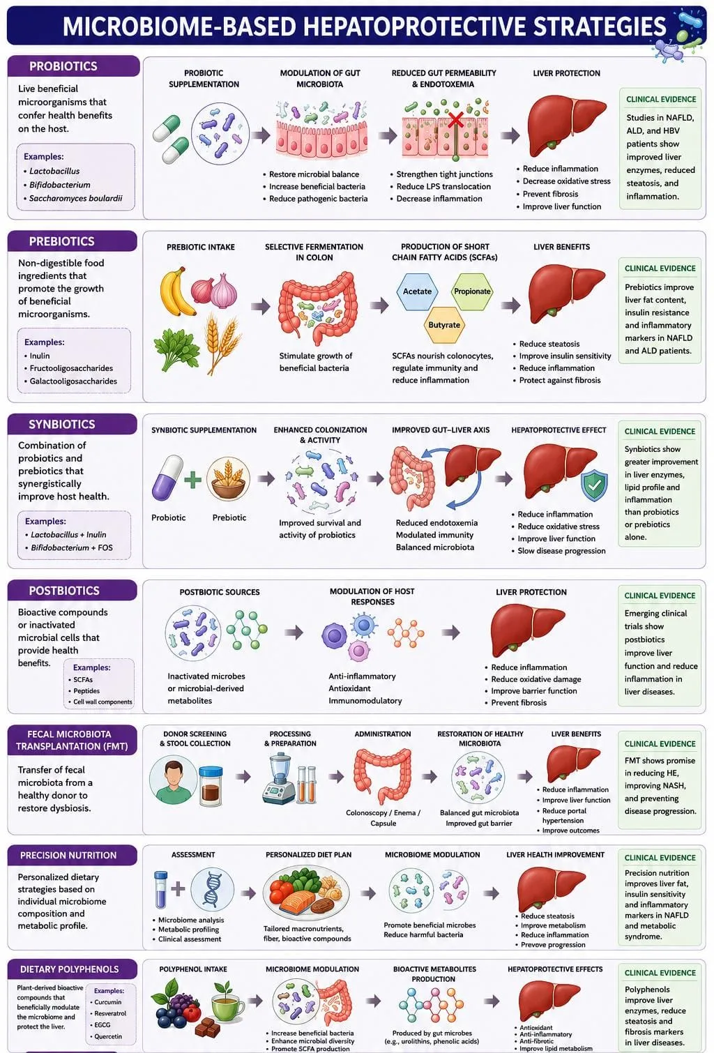
Figure 6: Microbiome-based Hepatoprotective Strategies
Herbal medicines are promising microbiome-targeted approaches because their bioactive constituents can simultaneously influence gut microbiota and hepatic pathways. Phytochemicals such as flavonoids, phenolic compounds, terpenoids, and polysaccharides may promote beneficial bacteria, reduce dysbiosis, strengthen intestinal barrier function, and regulate bile acid metabolism. They may also decrease endotoxin-associated inflammation, oxidative stress, hepatic steatosis, and fibrotic responses. Thus, herbal medicines may exert hepatoprotective effects through combined microbiota modulation, antioxidant, anti-inflammatory, and metabolic regulatory mechanisms. However, further well-designed clinical studies are needed to establish their efficacy, safety, optimal doses, and microbiome-specific effects (Figure 7).
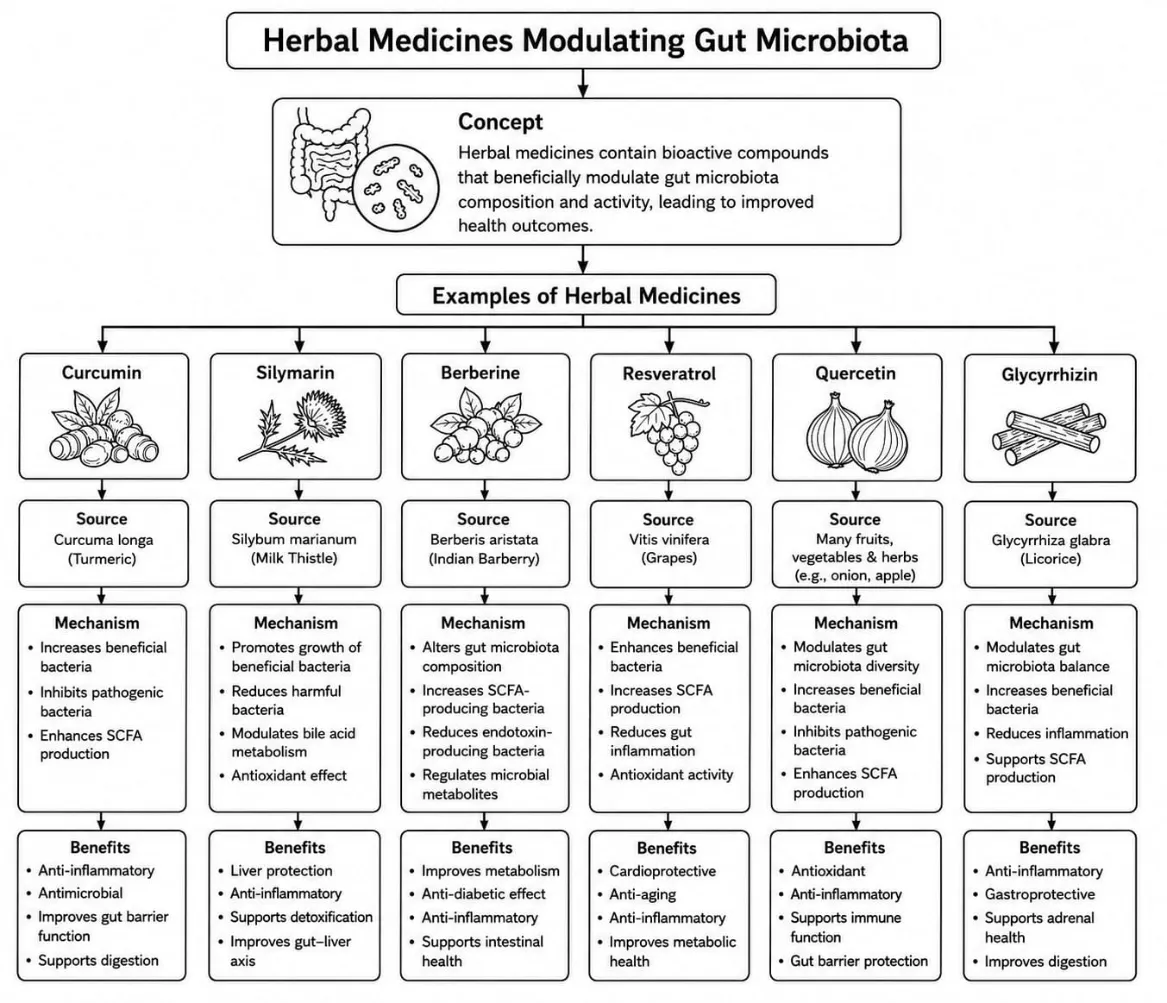
Figure 7: Herbal Medicines Modulating Gut Microbiota
8. Nanotechnology and Targeted Microbiome Therapy
8.1. Nano-probiotics: Nano-probiotics involve the use of nanotechnology to improve the stability, survival, and targeted delivery of probiotics in the gastrointestinal tract. Encapsulation within nanosystems can protect probiotics from unfavorable gastric conditions and enhance their ability to reach the intestine, thereby supporting restoration of microbial balance and gut barrier function [87].
8.2. Polymeric nanoparticles: Polymeric nanoparticles can encapsulate probiotics, microbial metabolites, or therapeutic agents and protect them from premature degradation. Their controlled-release properties allow targeted delivery within specific regions of the gastrointestinal tract and may improve the therapeutic effects of microbiome-modulating agents.
8.3. Liposomes: Liposomes are lipid-based vesicular systems capable of carrying both hydrophilic and lipophilic compounds. They can improve the stability and bioavailability of microbiome-targeting agents and facilitate their delivery to intestinal tissues, potentially enhancing modulation of gut–liver signaling [88].
8.4. Colon-targeted drug delivery: Colon-targeted delivery systems are designed to release therapeutic agents specifically in the colon, where microbial populations are highly diverse and abundant. Such systems may use pH-, enzyme-, or microbiota-responsive mechanisms to deliver probiotics, metabolites, or other therapeutic compounds at the desired site [89].
8.5. Smart drug delivery systems: Smart delivery systems respond to specific physiological or pathological conditions such as pH, enzymes, microbial metabolites, or inflammation to control drug release. These systems provide opportunities for precise microbiome modulation and may improve therapeutic efficacy while reducing unwanted systemic effects [90].
9. Current Challenges
9.1. Complexity and Inter-individual Variability: The gut microbiome contains a highly diverse microbial community that varies among individuals due to diet, age, genetics, lifestyle, medications, and environmental factors. This variability can lead to differences in response to microbiome-based hepatoprotective therapies.
9.2. Etiology-Specific Dysbiosis: Microbial alterations differ considerably among liver diseases such as MASLD, alcoholic liver disease, cirrhosis, and hepatocellular carcinoma. Therefore, a single microbiome-targeted intervention may not be equally effective across different liver conditions.
9.3. Unclear Mechanisms and Causality: Although microbial metabolites such as SCFAs, bile acids, and LPS are linked with liver health, their precise molecular mechanisms and interactions with hepatic pathways remain incompletely understood. It is also difficult to establish whether dysbiosis is a cause or consequence of liver injury.
9.4. Standardization and Safety Concerns: Variations in probiotics, herbal preparations, FMT, and other microbiome-modulating therapies can result in inconsistent outcomes. Excessive or inappropriate manipulation of the microbiome may also disturb microbial balance and produce undesirable effects.
9.5. Limited Clinical Evidence and Need for Personalized Therapy: Many microbiome-based approaches have shown promising results in experimental studies, but large-scale, long-term human clinical trials remain limited. Individual microbiome profiles may therefore need to be considered to develop safe and effective personalized hepatoprotective therapies [91].
10. Future Perspectives
Future hepatoprotective research is expected to focus on personalized microbiome therapy and precision hepatology, where individual microbial profiles, metabolic characteristics, and disease status guide treatment selection. Engineered probiotics may be developed to deliver specific therapeutic metabolites, while microbiome-derived drugs could provide novel bioactive molecules for regulating inflammation and hepatic metabolism. AI-assisted microbiome therapeutics may help identify disease-associated microbial patterns and predict individual treatment responses. In addition, organoid and organ-on-chip models can provide advanced platforms for studying gut–liver interactions, screening microbiome-targeted therapies, and accelerating the development of safe and effective personalized interventions.
CONCLUSION
The gut–liver axis plays a central role in maintaining hepatic homeostasis through interactions involving gut microbiota, microbial metabolites, bile acids, intestinal barrier function, and immune signaling. Dysbiosis can contribute to inflammation, endotoxemia, oxidative stress, and metabolic disturbances associated with liver injury. Therefore, modulation of the gut microbiome using probiotics, prebiotics, postbiotics, dietary approaches, herbal medicines, and advanced targeted delivery systems represents a promising hepatoprotective strategy. However, variations in microbiome composition, limited clinical evidence, safety concerns, and incomplete understanding of underlying mechanisms remain important challenges. Future research integrating precision medicine, artificial intelligence, engineered probiotics, microbiome-derived therapeutics, and advanced gut–liver models may facilitate the development of safer and more effective microbiome-based interventions for liver diseases.
REFERENCES

Sani Gaikwad, Pooja B. Rasal, Vikas S. Shinde, Amol U. Gayke, Gut–Liver Axis and Microbiome-Based Hepatoprotective Strategies: Emerging Therapeutic Approaches for Liver Diseases, Int. J. Sci. R. Tech., 2026, 3 (11), 38-55. https://doi.org/10.5281/zenodo.23909005
 10.5281/zenodo.23909005