

We use cookies to ensure our website works properly and to personalise your experience. Cookies policy
Sree Abirami College Of Pharmacy, Machegoundanpalayam, Seerapalayam Village, Echanari (Po), Coimbatore 641-021
Solanum nigrum L. (Manathakkali), a member of the family Solanaceae, is a small herbaceous plant that has long occupied a place in folk and traditional medicine on account of its reported antioxidant, anti-inflammatory and mucosal-protective properties. In the present investigation, a churnam (fine herbal powder) was prepared from the leaves of S. nigrum and subjected to a systematic pharmacognostical and physicochemical standardization protocol, followed by preliminary screening for anti-inflammatory and antioxidant activity. The dried leaf powder was characterized through organoleptic and powder-microscopic examination, and its physicochemical constants — loss on drying, total ash, acid-insoluble ash, water-soluble ash, extractive values in chloroform, methanol, acetone and water, foaming index and swelling index — were determined and compared against acceptable pharmacopoeial ranges. Qualitative phytochemical screening of the ethanolic leaf extract confirmed the presence of carbohydrates, proteins, alkaloids, glycosides, flavonoids, tannins and saponins. The bovine serum albumin (BSA) protein-denaturation-inhibition assay was employed to gauge anti-inflammatory potential, while the DPPH free-radical-scavenging assay was used to estimate antioxidant capacity, both assays being run in parallel with aspirin and ascorbic acid, respectively, as reference standards. A clear, concentration-dependent response was recorded for the test churnam in both assays: 74.70% inhibition of protein denaturation and 65.57% radical-scavenging activity were achieved at the highest concentration tested (50 µl), corresponding to roughly 85% and 80% of the respective standard drug responses. These findings lend preliminary scientific credibility to the ethnomedicinal use of S. nigrum in inflammatory and ulcer-related gastric conditions and support its candidacy as a low-cost, plant-derived adjunct or alternative to conventional anti-ulcer therapy. The standardization parameters generated in this work may additionally serve as quality-control benchmarks for future formulation and regulatory work involving this drug. Confirmation of an actual gastroprotective effect, however, will require follow-up in vivo investigation using established ulcer models such as pylorus ligation or ethanol-induced gastric injury.
1.1 Herbal Medicine in Traditional Healthcare
Plant-derived remedies remain central to healthcare in regions where indigenous systems such as Ayurveda, Siddha, Unani and Traditional Chinese Medicine have been practiced for millennia. Their therapeutic value stems from secondary metabolites — alkaloids, flavonoids, tannins, glycosides, saponins and phenolic acids — that act individually or synergistically on biological targets. However, the clinical reliability of any herbal product depends on rigorous identification and standardization, since batch-to-batch variability and adulteration remain the principal obstacles to wider acceptance of phototherapeutics.
1.2 Churnam as a Traditional Dosage Form
Churnam (choornam), a fine, uniformly sieved powder prepared from dried plant material, is among the simplest and most versatile dosage forms described in Siddha and Ayurvedic practice. Its preparation involves cleaning, shade-drying, size reduction, sieving to a uniform mesh, and storage under conditions that protect the finished powder from moisture, light and microbial contamination. An ideal churnam is finely and evenly powdered, free from foreign matter and excess moisture, stable on storage, free-flowing, and retains the natural colour, odour and active-constituent content of the source drug.
1.3 Peptic Ulcer Disease
A peptic ulcer is a breach in the mucosal lining of the stomach or duodenum that penetrates into the deeper submucosal tissue, arising when aggressive factors — gastric acid, pepsin, Helicobacter pylori and NSAID use — overwhelm the mucosa's protective mucus-bicarbonate barrier, prostaglandin-mediated cytoprotection and blood flow. Presentation typically includes burning epigastric pain related to meal timing, along with bloating, nausea and, in severe cases, gastrointestinal bleeding or perforation. H. pylori infection and chronic NSAID use are the two dominant causes, with alcohol, smoking, stress and genetic susceptibility as contributory factors. Gastric ulcers occur mainly along the lesser curvature, while duodenal ulcers — the more common type — reflect elevated acid secretion; stress-related and mucocutaneous ulcers (e.g., diabetic foot or pressure ulcers) arise through distinct mechanisms.
Conventional management centres on acid suppression with proton pump inhibitors, H2-antagonists or mucoprotective agents, together with H. pylori eradication using triple antibiotic therapy where indicated. Lifestyle modification (avoiding NSAIDs, alcohol and smoking) is an important adjunct, and prevention relies on judicious NSAID use, H. pylori screening in high-risk groups, and stress and dietary management.
1.4 Rationale for Churnam-Based Anti-Ulcer Therapy
Herbal churnams occupy an important niche among traditional anti-ulcer remedies owing to their simple preparation and the combined action of their constituent phytochemicals. Anti-ulcer formulations are typically derived from plants with mucosal-protective, antioxidant, anti-inflammatory and acid-neutralizing properties that reinforce the mucosal barrier, blunt acid-pepsin aggression and hasten epithelial repair. Solanum nigrum, with its documented content of steroidal alkaloids, flavonoids and saponins and long history of use in gastric and hepatic disorders, provides a plausible scientific basis for inclusion in anti-ulcer churnam formulations, provided its quality can be consistently standardized.
1.5 Inflammation: Definition, Classification and Relevance
Inflammation is the body's protective response to injury, infection or harmful stimuli, mediated through changes in blood vessels, immune cells and chemical mediators. Acute inflammation is a rapid, short-lived response marked by vasodilation and leukocyte recruitment, whereas chronic inflammation persists over weeks or longer and can drive progressive tissue damage and fibrosis. Its cardinal signs — redness, heat, swelling, pain and loss of function — arise from increased blood flow, fluid extravasation and mediator-driven nerve stimulation, and may be accompanied by systemic features such as fever and fatigue. Common triggers include infection, physical or chemical injury, allergic and autoimmune reactions, and chronic metabolic disturbance.
1.6 Anti-inflammatory Agents
Anti-inflammatory agents reduce inflammation and its accompanying pain, swelling and redness. Conventional options include NSAIDs and corticosteroids, while natural alternatives such as turmeric, ginger and Boswellia species have been widely investigated. Herbal agents combining antioxidant and anti-inflammatory activity, such as Solanum nigrum, are of particular interest because oxidative stress and inflammatory signalling often act together to sustain tissue injury, including at the gastric mucosal level — the mechanistic basis explored in the present study.
2. REVIEW OF LITERATURE
Earlier investigations addressing the pharmacognostical characterization, phytochemical composition and pharmacological potential of Solanum nigrum and related species are summarized below, providing the scientific backdrop for the present standardization work.
Yadav et al. (2018) highlighted the widespread problem of adulteration in Ayurvedic raw drugs and demonstrated how micromorphological, anatomical, physicochemical and HPTLC analysis together verify botanical identity. Jagtap et al. (2016) established macroscopic and microscopic reference standards for S. nigrum leaves, identifying single-layered palisade parenchyma, anisocytic stomata and warty/glandular trichomes as diagnostic features, with phytochemical screening confirming alkaloids, glycosides, tannins and saponins, and HPTLC resolving eleven and nine spots at 254 nm and 366 nm respectively.
Gupta (2024) extended anatomical characterization to the leaf, stem and root, describing dorsiventral mesophyll with anisocytic stomata in the leaf, unicellular trichomes and scattered vascular bundles in the stem, and typical dicotyledonous root anatomy — findings proposed as authentication benchmarks. Goel et al. (2022) compared the physicochemical profile of S. nigrum berries with Tribulus terrestris fruit, recording moisture, ash and density parameters and using TLC/UV data to distinguish the two drugs. Liu et al. (2022) and Chen et al. (2022) each compiled comprehensive reviews of the botanical background, phytochemistry, pharmacology and toxicology of S. nigrum.
Jani et al. (2012) confirmed that S. nigrum (Kakamachi) met the ash-value and extractive limits of the Ayurvedic Pharmacopoeia of India and was free from harmful heavy-metal levels, with qualitative tests confirming saponins, tannins and alkaloids. Hameed et al. (2017) catalogued diverse ethnomedicinal uses of different plant parts — leaves for skin and respiratory conditions, fruit juice for diarrhoea, and root for ophthalmic and bone disorders — and reported antimicrobial activity of methanolic extracts against both gram-positive and gram-negative bacteria. Shori et al. (2012) identified glycoalkaloids, glycoproteins and phenolic acids (gallic acid, catechin, caffeic acid, rutin, among others) as principal constituents, associating the plant with antibacterial, anti-inflammatory, anticancer and antioxidant activity, while cautioning against the toxicity of unripe berries.
Collectively, these studies establish that reliable pharmacognostical and physicochemical benchmarks are essential for authenticating S. nigrum, and that its alkaloid, flavonoid, saponin and phenolic content provides a credible mechanistic basis for its traditional use in inflammatory and gastric disorders — a basis directly tested in the present work through in vitro anti-inflammatory and antioxidant assays.
3. PLANT PROFILE
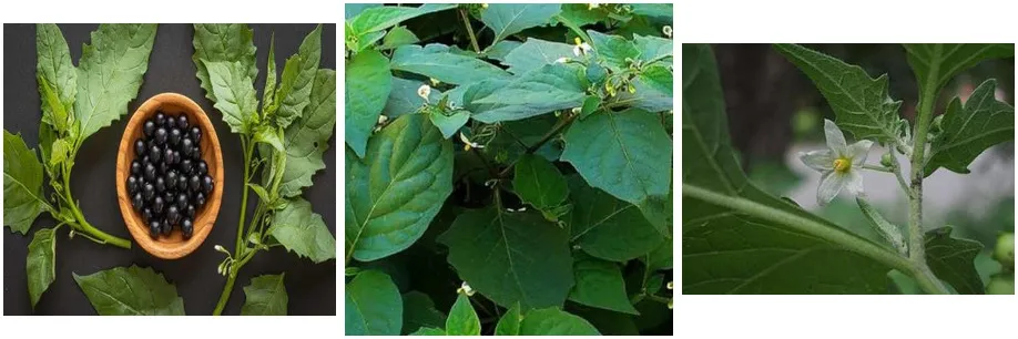
Fig. 1: Solanum nigrum L. — flower, foliage and ripe berries
3.1 Botanical Identity
Solanum nigrum L., known widely as Black Nightshade and, in Tamil, as Manathakkali (மணதà¯à®¤à®•à¯à®•à®¾à®³à®¿), is a small herbaceous plant of the family Solanaceae, distributed across tropical and temperate regions and long used in traditional medicine. Its therapeutic reputation is underpinned by steroidal glycoalkaloids, flavonoids, phenolic compounds, tannins and saponins.
3.2 Taxonomic Classification
|
Rank |
Classification |
|
Kingdom |
Plantae |
|
Phylum |
Tracheophyta (Magnoliophyta) |
|
Class |
Magnoliopsida |
|
Order |
Solanales |
|
Family |
Solanaceae |
|
Genus |
Solanum |
|
Species |
Solanum nigrum |
Table 1: Taxonomical classification of Solanum nigrum L.
S. nigrum is an annual herb of the nightshade family, believed native to Eurasia but now distributed worldwide as a common weed and medicinal plant, with three recognized subspecies differing mainly in pubescence and growth habit. The leaves are ovate, the flowers small and white with a star-like corolla, and the berries turn black on ripening; unripe (green) berries are toxic and must not be consumed.
3.3 Botanical Description
S. nigrum is an erect annual herb reaching 0.25–1 m in height, with a well-developed, often lignified taproot. The stem is green or purplish and finely pubescent. Leaves are ovate, 2.5–10 cm long and 1.5–5.5 cm wide, with a shortly pointed apex, a cuneate base narrowing into a 1–2 cm petiole, and an irregularly toothed margin bearing sparse soft hairs. Small white, star-shaped flowers are borne in extra-axillary cymes of three to ten; the mature berry is globose, about 8 mm in diameter, turning glossy black at ripeness and containing numerous small, laterally compressed seeds.
3.4 Vernacular Names
|
Language |
Common Name |
|
Hindi |
Mokoi, Makoi |
|
Tamil |
Manathakkali |
|
Malayalam |
Mulaku-thakkali |
|
Telugu |
Kasaka |
|
Kannada |
Kaaki, Ganike |
|
Marathi |
Laghukavali |
|
Manipuri |
Leipungkhangga |
Table 2: Vernacular names of Solanum nigrum across Indian languages
This diversity of regional names reflects the plant's long-standing familiarity across the Indian subcontinent as both a weed and a recognized medicinal resource. While ripe fruits are generally edible, unripe berries contain higher concentrations of steroidal glycoalkaloids and should be avoided.
3.5 Traditional Therapeutic Applications by Plant Part
|
Plant Part |
Traditional Applications |
|
Leaves |
Management of skin conditions (scabies, ringworm, vitiligo), poultices for local swelling, expressed juice for cough and asthma, ear drops for pain relief, and gargles for oral ulceration. |
|
Fruits |
Used as a diuretic, antispasmodic and mild emetic in diarrhoea and febrile illness; also employed as a tonic and laxative in indigenous practice. |
|
Roots |
Juice preparations used in asthma and whooping cough. |
|
Whole plant |
Applied broadly for hepatic disorders, fever and general detoxification, and valued in Ayurveda and Traditional Chinese Medicine for its anti-inflammatory action. |
Table 3: Traditionally reported uses of different parts of Solanum nigrum
Leaf material is most commonly processed into pastes or expressed juices for topical use, while internal administration — where practised — is typically limited to small volumes (10–20 ml). Given the documented toxicity of certain plant parts, particularly unripe fruit, supervision by a qualified practitioner is advisable.
4. AIM AND OBJECTIVES
4.1 Aim
Peptic ulcer disease is a widespread gastrointestinal disorder rooted in an imbalance between mucosal-aggressive and mucosal-protective mechanisms. While conventional pharmacotherapy is generally effective, it is often accompanied by long-term adverse effects and recurrence on discontinuation. Solanum nigrum, valued in Siddha and folk medicine for its antioxidant, anti-inflammatory and hepatoprotective properties, offers a scientifically credible, plant-based option for gastric mucosal protection — provided its quality can be consistently standardized. The present study therefore aimed to carry out the pharmacognostical and physicochemical standardization of Solanum nigrum leaf churnam and to evaluate its preliminary anti-inflammatory and antioxidant activity as a scientific basis for its proposed anti-ulcer application.
4.2 Objectives
5. MATERIALS AND METHODS
5.1 Collection of Plant Material
Healthy, disease-free fresh leaves of Solanum nigrum were collected during July 2026 from Coimbatore, Tamil Nadu.
5.2 Authentication of the Plant
The collected material was identified as Solanum nigrum Hook. f. & Thomson and formally authenticated by Dr. S. S. Hameed, Scientist 'F' and Head of Office (I/C), Botanical Survey of India, Southern Regional Centre, whose authentication certificate is reproduced in Fig. 6.
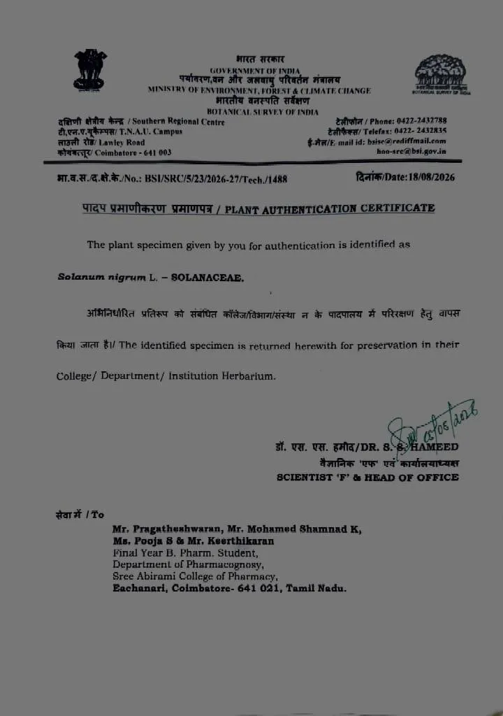
Fig. 2: Plant authentication certificate issued by the Botanical Survey of India, Southern Regional Centre
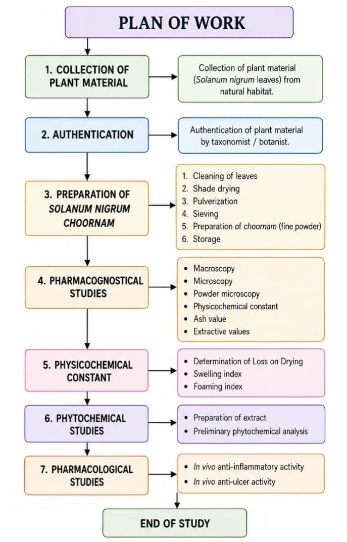
Fig. 3: Overall plan of work followed in the present study
5.3 Preparation of Solanum nigrum Churnam
The churnam was prepared from properly authenticated, dried plant material through a sequence of collection, cleaning, shade-drying, size reduction, sieving and storage. Fresh, healthy leaves were sourced from a pollution-free location, with damaged, diseased or discoloured material excluded, and washed to remove soil, dust and other extraneous matter. Cleaned leaves were spread thinly on trays and dried under shade at ambient temperature — direct sunlight was avoided to prevent degradation of heat- and light-sensitive phytoconstituents — until fully brittle. The dried leaves were then coarsely broken and powdered in a clean, dry mechanical grinder, avoiding excessive frictional heating, and passed through a suitable sieve to achieve a uniform, free-flowing powder. The finished churnam was accurately weighed, labelled with batch details, and stored in a well-sealed, airtight container in a cool, dry location protected from moisture, light and contamination.
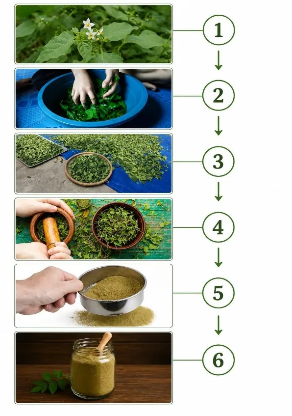
Fig. 4: Sequential steps in the preparation of Solanum nigrum leaf churnam — (1) plant collection, (2) cleaning, (3) shade drying, (4) size reduction, (5) sieving, (6) storage of the finished churnam
Precautions Observed During Preparation
5.5 Pharmacognostical Studies
5.5.1 Macroscopic (Organoleptic) Evaluation
Organoleptic examination offers the simplest and most rapid means of establishing the identity, purity and general quality of a crude drug. Colour, odour, taste and other physical attributes such as size, shape, surface texture, fracture and thickness of the powdered material were recorded.
5.5.2 Powder Microscopy
Powder microscopy was carried out to reveal the diagnostic anatomical features characteristic of Solanum nigrum leaf powder, an approach widely used in pharmacognosy for authentication purposes. The powder, olive-green to dark green in colour, was mounted in glycerin and examined under a bright field trinocular microscope.
The combination of cluster calcium-oxalate crystals with paracytic stomata was found to reliably distinguish S. nigrum powder from likely adulterants, while the presence of lignified elements indicated the inclusion of stem fragments in whole-plant preparations.
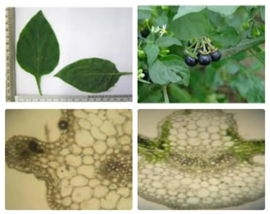
Fig. 5: Powder-microscopic characteristics of Solanum nigrum leaf — leaf lamina, fruiting twig, and transverse-section detail showing vascular and parenchymatous elements
5.6 Determination of Physicochemical Constants
Shade-dried, powdered Solanum nigrum leaf material was used throughout for the determination of physicochemical constants, following procedures consistent with World Health Organization guidelines for the quality control of herbal materials.
5.6.1 Loss on Drying (Moisture Content)
Approximately 3 g of the powdered drug was weighed into a tared dish and dried in a hot-air oven at 105 °C for 3–5 hours, cooled in a desiccator, and re-weighed; the process was repeated until a constant weight was obtained. Percentage moisture was calculated as the proportional loss in weight relative to the initial sample weight. For a herbal powder of this type, moisture content in the range of 7–10% is generally considered optimal for discouraging microbial growth; values exceeding 12% raise the risk of degradation of heat-labile constituents such as solanine-type alkaloids, while values below 6% can adversely affect subsequent extractive-value determinations. Maintaining low moisture is important for preventing microbial and fungal contamination, extending shelf life and preserving overall powder quality.
5.6.2 Ash Values
Ash-value determination provides a measure of the inorganic content of the crude drug and is a key indicator of purity and possible adulteration.
(a) Total Ash Value
Two to three grams of powder were weighed into a tared silica dish and ignited gradually to 450–600 °C in a muffle furnace until a carbon-free, white-to-grey ash was obtained; the residue was cooled in a desiccator and weighed, the process being repeated to constant weight. The percentage total ash was calculated as 100 × (W3 − W1) / (W2 − W1), where W1, W2 and W3 denote the weights of the empty dish, the dish with sample, and the dish with ash, respectively. Total ash value serves to establish quality standards for herbal drugs, detect adulteration or contamination and support pharmacognostical standardization.
(b) Acid-Insoluble Ash
The total ash obtained above was boiled with approximately 25 ml of dilute hydrochloric acid (2–10%) for five minutes, and the insoluble residue collected on ashless filter paper, washed with hot water until neutral, dried and re-ignited at 500–550 °C to constant weight. The percentage acid-insoluble ash was calculated relative to the original sample weight. This parameter is particularly useful for detecting contamination with sand, soil or other earthy material, since it primarily reflects silica content; an elevated value may indicate adulteration with inorganic matter.
(c) Water-Soluble Ash
Total ash was boiled with 25 ml of hot water for 5–10 minutes, and the insoluble residue filtered off, washed, and ignited at approximately 450 °C for 15 minutes to constant weight. Water-soluble ash was calculated as the difference between total ash and water-insoluble ash, expressed as a percentage of sample weight, and provides an index of the water-soluble mineral content — principally carbonates and oxalates — useful for comparing different batches or samples of the same drug.
5.6.3 Extractive Values
Extractive value reflects the proportion of a crude drug that is soluble in a given solvent and thereby indicates the quantity of chemically extractable constituents present, since different solvents preferentially dissolve different classes of phytoconstituent. In each case, 3 g of air-dried powder (40–60 mesh) was macerated with 100 ml of the respective solvent for 24 hours — with frequent agitation during the first six hours followed by 18 hours undisturbed — filtered rapidly through Whatman No. 41 paper, and 25 ml of the filtrate evaporated to dryness in a tared dish on a water bath, dried at 105 °C to constant weight, cooled in a desiccator and weighed. The percentage extractive value, relative to air-dried material, was calculated as (weight of residue / weight of sample) × (1000/25) × 100. This procedure was repeated using chloroform, methanol, acetone and water in turn, the solvent polarity guiding the class of constituents preferentially extracted, as summarized in Table 4a.
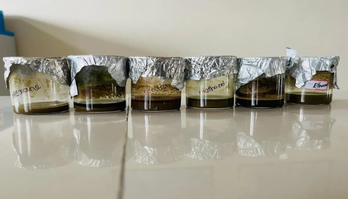
Fig. 6: Set-up used for determination of solvent extractive values (hexane, chloroform, methanol, ethanol)
|
Solvent Used |
Relative Polarity |
Constituent Class Preferentially Extracted |
|
Chloroform |
Non-polar |
Fats, waxes, resins, volatile oils |
|
Acetone |
Moderately polar |
Tannins, flavonoids, some glycosides |
|
Methanol |
Moderately polar |
Glycosides, flavonoids, resins |
|
Water |
Highly polar |
Sugars, polysaccharides, organic acids, gums |
Table 4a: Solvents used for extractive-value determination and their target constituent classes
5.6.4 Foaming Index
Steroidal saponins, well documented in Solanum nigrum, are responsible for the characteristic foam that forms when an aqueous extract of the plant is agitated. One gram of coarsely powdered material (sieve no. 1250) was boiled with 100 ml water for 30 minutes, cooled, filtered into a 100 ml volumetric flask and made up to volume. Graduated portions (1–10 ml) of this decoction were transferred to a series of stoppered test tubes, each made up to 10 ml with water, shaken lengthwise for 15 seconds (approximately two shakes per second), allowed to stand for 15 minutes, and the resulting foam height measured in each tube. The foaming index was calculated as 1000 divided by the volume (in ml) of decoction in the tube showing a 1 cm foam layer.
5.6.5 Swelling Index
Swelling index reflects the degree to which powdered plant material expands in water owing to hydrophilic constituents such as mucilage, gums or pectin. One hundred milligrams of coarsely powdered (sieve no. 44), air-dried drug was placed in a 25 ml graduated cylinder with 25 ml water, shaken thoroughly, and allowed to stand for three hours with intermittent shaking during the first period, after which the volume occupied by the settled drug layer (V1) was recorded at 25 °C; a parallel water-only blank (V0) was run concurrently. The swelling index was calculated as (V1 − V0) divided by the weight of drug, multiplied by 1000. A low swelling index reflects minimal mucilaginous content, distinguishing S. nigrum leaf material from gums or mucilage-rich seed drugs that typically show much higher indices and is useful for standardization purposes.
5.7 Preliminary Phytochemical Screening
Phytochemical evaluation was undertaken to determine the classes of secondary metabolites present in the plant material, information that is fundamental to interpreting its pharmacological activity.
5.7.1 Preparation of Extract
Coarsely powdered (sieve no. 10), dried leaf material (100 g) was extracted with ethanol using a suitable extraction technique. The resulting extract was concentrated using a rotary evaporator, and its percentage yield, colour and consistency recorded for reference prior to further phytochemical and pharmacological screening.
5.7.2 Maceration Process
Maceration was chosen as a simple, gentle extraction method suited to heat-sensitive plant constituents. Powdered leaves were placed in a clean, closed container and completely immersed in a suitable solvent (ethanol or a hydroalcoholic mixture). The mixture was maintained at room temperature for 24–72 hours with intermittent shaking, allowing the solvent to penetrate the plant cells and dissolve the soluble constituents. Following maceration, the mixture was filtered to separate the liquid extract from the marc, and the filtrate concentrated and stored for further study. This procedure yielded a dark greenish-black, thick semisolid extract, subsequently used for phytochemical screening and biological evaluation.
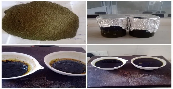
Fig.7: Maceration process — powdered leaf material, solvent maceration, and the concentrated semisolid extract obtained after filtration
5.7.3 Qualitative Chemical Tests
The ethanolic leaf extract was subjected to a standard battery of qualitative colour and precipitation tests for the major phytochemical classes, using the standard reagents and confirmatory reactions summarized in Table 5a.
|
Constituent |
Test(s) Applied |
Positive Indication |
|
Carbohydrates |
Fehling's, Benedict's, Iodine test |
Brick-red / reddish-brown precipitate; blue-black colour with starch |
|
Proteins |
Biuret, Xanthoproteic, Millon's, Ninhydrin test |
Violet/pink colour; yellow-orange precipitate; brick-red on boiling; deep blue/violet colour |
|
Alkaloids |
Wagner's, Dragendorff's, Mayer's, Hager's test |
Reddish-brown, orange, creamish-white or yellow precipitate, respectively |
|
Glycosides |
Keller–Kiliani, Legal's, Baljet's, Borntrager's, Foam test |
Reddish-brown, pink-red, yellow-orange colour or persistent foam, depending on test |
|
Flavonoids |
Shinoda test |
Red or orange colouration |
|
Tannins |
Ferric chloride test, Lead acetate test |
Blue-black/blue-green colour; precipitate formation |
|
Saponins |
Foam test on aqueous decoction |
Thick, persistent foam stable for several minutes |
Table 5a: Qualitative phytochemical tests applied and their positive indications
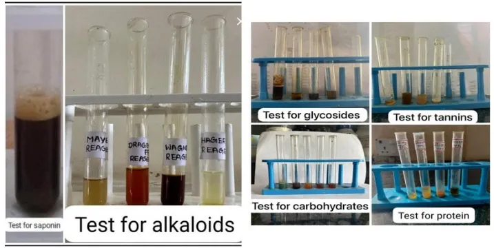
Fig. 7: Representative results of qualitative phytochemical screening tests — (left) test for saponins and alkaloids; (right) tests for glycosides, tannins, carbohydrates and proteins
5.8 In Vitro Pharmacological Screening
5.8.1 Anti-inflammatory Activity — Protein Denaturation Inhibition Assay
Anti-inflammatory potential was evaluated in vitro using the bovine serum albumin (BSA) protein-denaturation-inhibition assay, a widely used model founded on the principle that denaturation of tissue proteins is a recognized contributor to inflammatory and autoimmune pathology. Test solutions of the churnam extract were prepared at five ascending concentrations (10, 20, 30, 40 and 50 µl) and incubated with BSA under standardized conditions; aspirin was employed in parallel as the reference standard. Following incubation, the extent of protein denaturation in treated versus control samples was assessed spectrophotometrically, and the percentage inhibition of denaturation calculated for both the test sample and the standard at each concentration.
5.8.2 Antioxidant Activity — DPPH Free-Radical Scavenging Assay
Antioxidant capacity was assessed using the 2,2-diphenyl-1-picrylhydrazyl (DPPH) free-radical-scavenging assay, in which ascorbic acid served as the reference standard. The same concentration range (10–50 µl) was tested, and absorbance of the resulting solutions measured at 517 nm following a defined incubation period in the dark. Percentage radical-scavenging activity was calculated for both the test sample and the standard at each concentration, based on the reduction in absorbance relative to a DPPH-only control.
6. RESULTS AND DISCUSSION
The pharmacognostical and physicochemical standardization of Solanum nigrum leaf powder undertaken in this study provides a body of reference information relevant to the identification, authentication and quality control of the crude drug. Macroscopic and microscopic examination revealed features distinctive enough to support unambiguous identification of the plant material and to aid in the detection of adulteration.
6.1 Physicochemical Parameters
The physicochemical constants determined for the Solanum nigrum leaf churnam are summarized in Table 5, alongside the corresponding acceptable limits drawn from general pharmacopoeial guidance for herbal powders.
|
Parameter |
Value Obtained |
Acceptable Limit / Specification |
|
Moisture content (loss on drying) |
9.3% |
≤ 10% |
|
Total ash |
12.45% |
≤ 15% |
|
Acid-insoluble ash |
0.49% |
≤ 2% |
|
Water-soluble ash |
11% |
1–15% |
|
Chloroform-soluble extractive value |
3.5% |
1–5% |
|
Methanol-soluble extractive value |
12% |
10–20% |
|
Acetone-soluble extractive value |
9% |
5–10% |
|
Water-soluble extractive value |
22% |
15–30% |
|
Foaming index |
10 |
— |
|
Swelling index |
1.3 |
— |
Table 5: Physicochemical parameters of Solanum nigrum leaf churnam
All of the determined parameters fell comfortably within the acceptable ranges applied. The moisture content of 9.3% lies just below the upper safe limit of 10%, indicating a drying process adequate to minimize the risk of microbial spoilage while avoiding excessive desiccation that could otherwise compromise extractability. The low acid-insoluble ash value (0.49%, well under the 2% ceiling) points to minimal contamination with siliceous or earthy matter, supporting the purity of the processed material, while the total ash and water-soluble ash values reflect an inorganic mineral content consistent with expectations for a leafy crude drug of this type. The graded extractive values obtained in chloroform, methanol, acetone and water — rising from 3.5% in the relatively non-polar chloroform fraction to 22% in the highly polar aqueous fraction — indicate that the leaf churnam is comparatively rich in polar and moderately polar constituents, a pattern consistent with a phytochemical profile dominated by glycosides, flavonoids, tannins and related compounds rather than lipophilic constituents such as waxes or volatile oils. The foaming index of 10 corroborates the qualitative saponin-positive result obtained on phytochemical screening, while the low swelling index (1.3) indicates that the leaf material is not appreciably mucilaginous, distinguishing it from gum- or mucilage-rich botanical drugs.
6.2 Phytochemical Screening
Qualitative phytochemical screening of the ethanolic leaf extract, summarized in Table 6, confirmed the presence of all seven major phytoconstituent classes tested.
|
S. No. |
Phytochemical Test |
Ethanolic Leaf Extract |
|
1 |
Carbohydrates |
Positive |
|
2 |
Proteins |
Positive |
|
3 |
Alkaloids |
Positive |
|
4 |
Glycosides |
Positive |
|
5 |
Flavonoids |
Positive |
|
6 |
Tannins |
Positive |
|
7 |
Saponins |
Positive |
Table 6: Preliminary phytochemical screening of the ethanolic leaf extract of Solanum nigrum
The presence of this broad spectrum of secondary metabolites — most notably alkaloids, flavonoids, saponins and tannins — is consistent with earlier literature reports on the species and provides a plausible chemical basis for the antioxidant and anti-inflammatory activity subsequently observed in the biological assays.
6.3 Anti-inflammatory Activity (Protein Denaturation Inhibition Assay)
The results of the BSA protein-denaturation-inhibition assay, conducted with aspirin as the reference standard, are presented in Table 7 and Fig. 13.
|
Concentration |
% Inhibition — Standard (Aspirin) |
% Inhibition — Test Sample |
|
10 µl |
33.00% |
10.64% |
|
20 µl |
43.00% |
26.76% |
|
30 µl |
58.00% |
33.82% |
|
40 µl |
79.00% |
56.57% |
|
50 µl |
88.00% |
74.70% |
Table 7: In vitro anti-inflammatory activity (protein denaturation inhibition) of Solanum nigrum leaf churnam
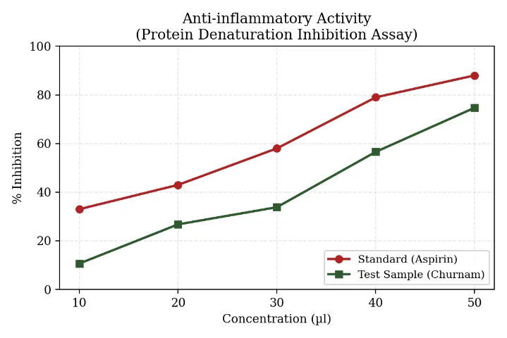
Fig. 8: Concentration-dependent inhibition of protein denaturation by Solanum nigrum leaf churnam compared with aspirin
Both the standard and the test sample exhibited a clear, concentration-dependent increase in percentage inhibition of protein denaturation, confirming a reproducible dose–response relationship. At the lowest concentration tested (10 µl), the churnam showed relatively modest activity (10.64%), whereas at the highest concentration (50 µl) inhibition rose markedly to 74.70%, representing close to 85% of the activity recorded for aspirin (88.00%) at the same concentration. Notably, the proportional gap between the test sample and the standard narrowed progressively with increasing concentration, suggesting that the active constituents of the churnam become increasingly effective at higher doses, possibly reflecting a saturation-dependent or synergistic mode of action among its constituent phytochemicals. By linear interpolation, the ICâ‚…â‚€ for the test sample was estimated at approximately 37.1 µl, compared with approximately 24.7 µl for aspirin; although the standard achieved half-maximal inhibition at a lower concentration, the sample's ability to approach 75% inhibition at 50 µl reflects a substantial degree of anti-inflammatory potential for a crude, unfractionated herbal preparation.
This activity is most plausibly attributable to the flavonoids, steroidal alkaloids and saponins identified during phytochemical screening, constituents known to stabilize protein tertiary structure and thereby inhibit the physical denaturation associated with the inflammatory cascade. The finding lends direct support to the traditional use of the plant in inflammatory gastric conditions, since local mucosal inflammation is recognized as a key contributor to the pathogenesis of peptic ulceration.
6.4 Antioxidant Activity (DPPH Radical Scavenging Assay)
Antioxidant potential, assessed via the DPPH free-radical-scavenging assay with ascorbic acid as the reference standard, is summarized in Table 8 and illustrated in Fig. 14.
|
Concentration |
% Inhibition — Ascorbic Acid |
% Inhibition — Test Sample |
|
10 µl |
34.91% |
25.49% |
|
20 µl |
45.61% |
41.10% |
|
30 µl |
68.82% |
49.47% |
|
40 µl |
74.34% |
57.66% |
|
50 µl |
81.50% |
65.57% |
Table 8: In vitro antioxidant activity (DPPH radical scavenging) of Solanum nigrum leaf churnam
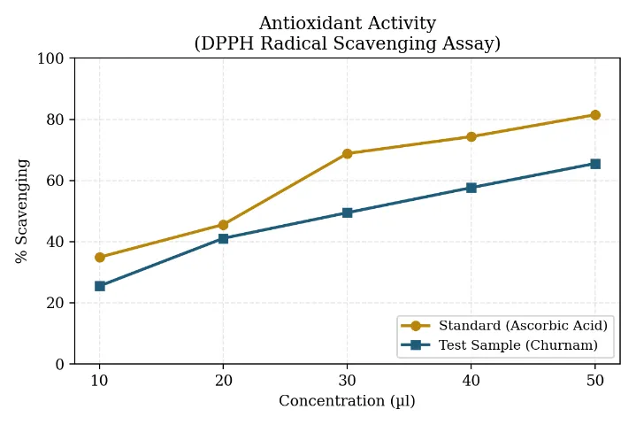
Fig. 8: Concentration-dependent DPPH radical-scavenging activity of Solanum nigrum leaf churnam compared with ascorbic acid
A similarly dose-dependent trend was evident in the antioxidant assay, with scavenging activity rising steadily from 25.49% at 10 µl to 65.57% at 50 µl. At the highest concentration tested, the churnam retained approximately 80.5% of the activity of ascorbic acid (65.57% versus 81.50%), indicating substantial free-radical-scavenging capacity. The ICâ‚…â‚€ for the test sample, interpolated between the 30 µl and 40 µl readings, was estimated at approximately 30.7 µl, compared with roughly 21.9 µl for ascorbic acid. Although the pure standard achieved half-maximal scavenging at a lower volume, the proximity of the sample's ICâ‚…â‚€ to that of the standard is noteworthy given that a crude plant extract typically contains a mixture of moderately active compounds rather than a single, highly potent antioxidant molecule.
The scavenging activity observed is consistent with the presence of flavonoids, phenolic compounds and steroidal saponins in the leaf extract, all recognized as effective hydrogen- or electron-donors capable of neutralizing the stable DPPH radical. This property is of direct relevance to the proposed anti-ulcer rationale of the study, since oxidative stress and excess reactive oxygen species generation at the gastric mucosal level are known to exacerbate mucosal injury and delay epithelial healing; a formulation exhibiting measurable free-radical-scavenging capacity may therefore contribute to gastroprotection by limiting oxidative damage to the mucosal lining.
6.5 Comparative Analysis of Anti-inflammatory and Antioxidant Findings
A side-by-side comparison of the two biological assays helps to place the observed activities in perspective and to draw out the shared mechanistic threads linking oxidative stress and inflammation in the context of gastric mucosal injury.
|
Evaluation Parameter |
Anti-inflammatory Activity |
Antioxidant Activity |
|
Experimental assay model |
BSA protein denaturation inhibition |
DPPH free-radical scavenging |
|
Reference standard drug |
Aspirin |
Ascorbic acid (Vitamin C) |
|
Maximum response at 50 µl (sample) |
74.70% inhibition |
65.57% scavenging |
|
Standard response at 50 µl |
88.00% |
81.50% |
|
Relative efficiency of sample vs. standard |
≈ 85% |
≈ 80.5% |
|
Estimated ICâ‚…â‚€ (sample vs. standard) |
37.1 µl vs. 24.7 µl |
30.7 µl vs. 21.9 µl |
|
Principal phytochemical contributors |
Steroidal alkaloids and saponins |
Flavonoids and phenolic compounds |
Table 9: Comparative summary of anti-inflammatory and antioxidant assay findings
Interpreted together, the lower ICâ‚…â‚€ recorded in the DPPH assay (30.7 µl) relative to the protein-denaturation assay (37.1 µl) suggests that free-radical neutralization is the more concentration-sensitive of the two activities. The anti-inflammatory assay, in turn, demonstrated that the crude churnam retains a substantial proportion (approximately 85%) of the relative efficiency of a well-established synthetic anti-inflammatory standard, a noteworthy result for an unrefined plant powder — an outcome plausibly reflecting the complementary, mutually reinforcing action of its antioxidant and anti-inflammatory phytoconstituents on oxidative-inflammatory tissue injury.
6.6 Proposed Label and Presentation of the Standardized Product
As a practical extension of the standardization exercise, an illustrative product label was designed for the finished Solanum nigrum leaf churnam, summarizing the composition, batch details, precautions and storage conditions expected of a properly quality-controlled herbal product (Fig. 15), consistent with regulatory expectations for traditional herbal products.
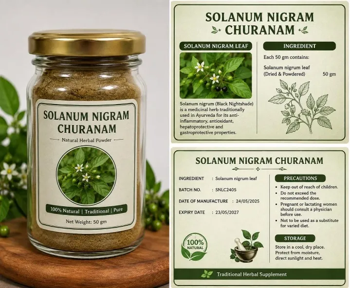
Fig. 9: Illustrative product label for standardized Solanum nigrum leaf churnam
7. DISCUSSION
The combined outcomes of the protein-denaturation-inhibition and DPPH radical-scavenging assays provide mutually reinforcing evidence for the anti-inflammatory and antioxidant potential of Solanum nigrum leaf churnam. Both assays demonstrated consistent, concentration-dependent activity, with the test sample achieving 74.70% anti-inflammatory inhibition and 65.57% antioxidant scavenging at the highest concentration examined (50 µl) — values that compare favourably, in proportional terms, with their respective reference standards despite the sample being a crude, unfractionated plant extract rather than an isolated, purified compound.
These observations carry particular mechanistic significance in the context of peptic ulcer disease, in which the gastric mucosa is subjected to a combined assault of local inflammation and oxidative stress arising from acid-pepsin aggression, H. pylori infection or NSAID-induced injury. The demonstrated capacity of the churnam to inhibit protein denaturation and scavenge free radicals in vitro lends preliminary scientific support to its traditional use and to its proposed role as a gastroprotective, anti-ulcer agent, and is consistent with the phytochemical profile — steroidal alkaloids, flavonoids and saponins — established during the standardization phase of this work.
It should be emphasized, however, that these findings rest on in vitro biochemical models, which, while useful as rapid, low-cost screening tools, cannot by themselves establish a direct causal link between the observed biochemical activity and actual protection of the gastric mucosa in a living organism. Further validation using established in vivo anti-ulcer models — for example, pylorus-ligation-induced or ethanol-induced gastric ulceration in rodents — together with histopathological assessment of the gastric mucosa, would be required before the gastroprotective potential of this churnam could be considered confirmed. Dose-ranging studies, assessment of acute and sub-chronic toxicity, and quantitative (rather than purely qualitative) phytochemical analysis — for instance by HPTLC or HPLC fingerprinting — would further strengthen the standardization profile established here and support any future move toward formulation development or regulatory submission.
CONCLUSION
The present investigation successfully carried out the pharmacognostical and physicochemical standardization of Solanum nigrum leaf powder (churnam) and explored its preliminary anti-inflammatory and antioxidant potential. Pharmacognostical evaluation yielded distinctive macroscopic and microscopic identification features that support the authentication and quality control of the powdered drug, while the physicochemical parameters determined — moisture content, ash values and extractive values — provide meaningful benchmarks against which the purity, quality and consistency of the formulation can be judged. Phytochemical screening confirmed the presence of alkaloids, flavonoids, saponins, tannins and other bioactive constituents likely to underlie the biological activities observed.
The bovine serum albumin protein-denaturation-inhibition assay revealed a clear, concentration-dependent anti-inflammatory response, with the test sample achieving 74.70% inhibition at 50 µl compared with 88.00% for aspirin, while the DPPH assay demonstrated a parallel concentration-dependent antioxidant effect, with 65.57% radical-scavenging activity at 50 µl against 81.50% for ascorbic acid. Taken together, these findings indicate that Solanum nigrum leaf churnam possesses promising antioxidant and anti-inflammatory activity that may plausibly support its traditional gastroprotective and anti-ulcer applications. As the present results derive solely from in vitro assays, further work — particularly employing established in vivo anti-ulcer models together with toxicological evaluation — is necessary to confirm the direct gastroprotective efficacy and overall safety of this standardized herbal preparation before it can be considered for translational or formulation development.
REFERENCES

A. Kavidha, Mohamed Shamnad K., Pragatheswaran A., Keerthikaran D., Pooja S.*, S. Nivetha, Pharmacognostical And Physio Chemical Standardization Of Solanum Nigrum Leaf Churnam, Int. J. Sci. R. Tech., 2026, 3 (9), 645-663. https://doi.org/10.5281/zenodo.23080422
 10.5281/zenodo.23080422