

We use cookies to ensure our website works properly and to personalise your experience. Cookies policy
Department of Microbiology and Biotechnology, Deogiri College, Chhatrapati Sambhajinagar - 431005, Maharashtra, India
Tridax procumbens L. is a valuable medicinal herbaceous plant that has been valued for centuries in Ayurvedic medicine. In this paper, the phytochemical composition of T. procumbens L. plant extract was studied for the presence of various biochemical compounds such as alkaloids, flavonoids, steroids, proteins and saponins. Also, the in vitro anticoagulant and anti-inflammatory activities of plant extracts were investigated in this paper. The qualitative phytochemical analysis of aqueous and methanolic plant extracts showed the presence of phytoconstituents such as tannins, alkaloids, saponins, flavonoids, phenols, steroids, anthocyanins, proteins and carbohydrates. The aqueous and methanolic extracts of T. procumbens L. were subjected to anticoagulant activity in human blood sample. Both extracts exhibited remarkable anticoagulant activity, suggesting their potential as natural anticoagulant agents. The in vitro anti-inflammatory activity for aqueous and methanolic extracts of T. procumbens L. was evaluated. The extract showed membrane stabilization activity. Therefore, the present study also supports the extraction, purification and the use of active constituents from T. procumbens L. in treating inflammatory disorders.
Since ancient times, plants have been utilized for both therapeutic and nutritional purposes, and many people rely on traditional medicine to meet their medical needs. Over the years, these herbal drugs have been shown to be effective. Around the world, a variety of plants and their parts are used to cure different illnesses [11]. According to the World Health Organization, more than 80% of the world’s population relies on traditional medicine for their primary healthcare needs [4].
Modern science has recognized several plant-derived drugs which possess either identified or unidentified chemical structures that are found to be clinically beneficial in various diseases. Many of the plant extracts are principally used in traditional medicines because they are readily accessible in rural areas and are relatively cheaper than modern medicines [9].
Plants have limitless ability to synthesize unique chemical substances that could be used in medicine and other fields. Alkaloids, steroids, tannins, glycosides, volatile oils, fixed oils, resins, phenols and flavonoids are among the active substances found in plants, which are deposited in specific areas such as leaves, flowers, bark, seeds, fruits and roots [4]. These substances serve as plant defence mechanisms against predation by microbes, insects, herbivores.
One such plant is Tridax procumbens L. (family Asteraceae), which is commonly called coat buttons. It is commonly used for medicinal purposes, because of its numerous pharmacological properties. These include analgesic, anti-anemic, anti-arthritic, anti-diabetic, antihypertensive, anti-inflammatory, antioxidant, antimicrobial, antipyretic, hepatoprotective, hypocholesterolemic and weight reducing properties [3].
Inflammation is the reaction of living tissues to injury, infection or irritation. Lysosomal enzymes released during inflammation produce a variety of disorders that lead to tissue injury by damaging the macromolecules and lipid peroxidation of membranes. This is assumed to be responsible for certain pathological conditions such as heart attacks, septic shocks and rheumatoid arthritis etc. The extracellular activity of these enzymes is said to be related to acute or chronic inflammation. Stabilization of lysosomal membrane is important in limiting the inflammatory response by inhibiting the release of lysosomal constituents of activated neutrophils such as bactericidal enzymes and proteases, which cause further tissue inflammation and damage upon extracellular release or by stabilizing the lysosomal membrane. Human red blood cell (HRBC) membrane or erythrocyte membrane is analogous to the lysosomal membrane and its stabilization implies that the extract may as well stabilize lysosomal membranes. Stabilization of HRBC membrane by hypotonicity induced membrane lysis can be taken as an in vitro measure of anti-inflammatory activity of the drugs or plant extracts [7].
The present study deals with the phytochemical screening, anticoagulation and in vitro anti-inflammatory activity of Tridax procumbens L., from the local area of Chhatrapati Sambhajinagar, Maharashtra (India).
MATERIAL AND METHOD
Collection of Plant Samples
The healthy, disease-free plants of T. procumbens L. were collected randomly from the local area of Chhatrapati Sambhajinagar, Maharashtra (India). It was authenticated from Botany Department, Deogiri College, Chhatrapati Sambhajinagar. The plant parts such as roots, leaves, and flowers were separated from the plant and washed thoroughly with distilled water. Further these cleaned plant materials were shade dried. After complete drying, the plant materials were ground into a fine powder using a mechanical grinder, and the resulting powdered samples were then kept in airtight containers.
Preparation of Plant Extract
The aqueous and methanolic extracts of T. procumbens L. were prepared using “Maceration method” by dissolving the powder in respective solvents. Following that, the container was sealed and left for three days under continuous stirring to ensure complete extraction. At the end of extraction, the extract was separated from the mixture by filtration. Subsequently, the solvents were evaporated by using a rotary evaporator.
Phytochemical Screening
Phytochemical screenings are the preliminary tests conducted to detect the presence of both primary and secondary metabolites in an extract. Following tests were performed to detect the presence of steroids, carbohydrates, proteins, anthocyanin, phenols, alkaloids, flavonoids, tannins, and saponins.
Detection of Steroids:
2 mL of extract was dissolved in 2 mL of chloroform, and shaken. Then a few drops of concentrated sulfuric acid were added into this mixture from the side of the test tubes. The formation of a red color in lower chloroform layer indicates the presence of steroids.
Detection of Carbohydrates (Molisch’s Test):
2 mL of the extract was treated with a few drops of Molisch’s reagent and the resulting mixture shaken properly. Few drops of concentrated H2SO4 were then added carefully down the side of the test tube. The formation of violet ring at the junction of two layers indicates the presence of carbohydrates.
Detection of Proteins (Ninhydrin Test):
2 mL of extract was taken in a test tube and treated with a few drops of ninhydrin reagent. The mixture was then placed in a water bath and allowed to boil for a few minutes. Formation of blue to purple color indicates the presence of proteins.
Detection of Anthocyanins:
2 mL of extract was taken in a test tube and treated with 2 mL of 2N HCl & NH3. The color change from pink-red to blue-violet indicates the presence of anthocyanins.
Detection of Phenols:
2 mL of extract was taken in a test tube and treated with a few drops of 10% alcoholic FeCl3 solution. The appearance of green, blue or violet color indicates the presence of phenolic compounds.
Detection of Tannins
2 mL of extract was taken in a test tube and treated with a few drops of 5% aqueous FeCl3. The formation of a green precipitate was an indication for the presence of tannins.
Detection of Alkaloids (Mayer’s Test):
2 mL of aqueous extract was mixed with 2 mL of 1% HCl on a steam bath and used for detection of alkaloids. The acidic mixture was treated with Mayer’s reagent (Potassium Mercuric Iodide). Formation of a yellow-colored precipitate indicates the presence of alkaloids.
Detection of Saponins (Foam Test):
5 mL of aqueous warmed extract was shaken vigorously. The formation of stable foam for at least ten minutes is an indication for the presence of saponins.
Detection of Flavonoids (Alkaline Reagent Test): 2 mL of extract was treated with 10% of NaOH solution. The formation of intense yellow color, which becomes colorless on addition of dilute acid, indicates the presence of flavonoids.
Anticoagulation activity:
Blood sample was collected from a healthy volunteer and transferred into three separate test tubes (2 mL each). 0.2 mL of methanolic extract was transferred into one test tube, 0.2 mL of methanol was transferred in second test tube and third test tube was considered as blank. The clotting activity of each tube was measured using a stopwatch and the clotting time was recorded.
Same procedure was followed for aqueous extract.
Anti-Inflammatory Activity:
Anti-inflammatory activity of plant extract was carried out using various in vitro models such as HRBC membrane stabilization activity, protein denaturation inhibitory activity and protease inhibitory activity. Standard drug was also used in order to compare the efficacy of anti-inflammatory activity of plant extract.
Membrane Stabilization
10 mL of fresh whole human blood was collected from healthy volunteers and transferred to centrifuge tubes. An equal volume of Alsever solution (2% dextrose, 0.8% sodium citrate, 0.5% citric acid and 0.42% NaCl) was mixed in the blood sample and centrifuged the mixture at 3,000 rpm for 5 min. After centrifugation, the pellet was washed with isotonic saline three times and made 10% suspension in isotonic saline.
Test Sample: The 0.5 mL containing 100μg of plant extract was taken and added in 0.5 mL of 10% RBCs suspension.
Only saline was added to the control tube instead of the test sample. Diclofenac was considered as a standard drug. All tubes were incubated in a water bath at 50°C for 30 min and after completion of incubation, cooled the tubes and added 0.5 mL of isotonic saline to reaction mixture. Centrifuged the reaction mixture at 3000 rpm for 5 min, supernatant of each mixture was collected and absorbance of each mixture was taken at 560 nm. The percent membrane stabilization activity was calculated using following formula:
% Protection = 100 - (Optical density of drug treated sample × 100)
Protein Denaturation Inhibitory Activity
0.5 mL of 1% aqueous solution of Bovine Serum Albumin (1% aqueous solution of Bovine Serum Albumin prepared in phosphate buffer saline of pH 6.4.) and 0.5 mL of plant extract (100μg) was taken in a test tube. 0.5 mL 1% aqueous solution of Bovine Serum Albumin was considered as a Control. The reaction mixture was incubated at 37°C for 20 min and then heated to 50°C for 20 min. Cooled the reaction mixture at room temperature and measured the turbidity spectrophotometrically at 660 nm. The percent inhibition was calculated by using following formula;
% Inhibition = [(Abs control - Abs sample) /Abs control] × 100
Protease Inhibitory Activity
0.06 g Trypsin, 0.5 mL 20mM Tris HCl Buffer and 100 µg/mL of plant extract was taken in a test tube. 20mM Tris HCl Buffer was considered as a blank. The mixture was incubated at 37°C for 5 min and then added 1 mL of 0.8% (W/V) casein. Again, incubated the mixture at 37°C for 20 min, then added 2 mL of 70% acetic acid to terminate the reaction. Centrifuged the mixture at 3000 rpm for 5 min, collected the supernatant and taken OD at 210 nm. The percent inhibition was calculated by using following formula;
% Inhibition = [(Abs control - Abs sample) /Abs control] × 100
RESULTS AND DISCUSSION
Preparation of Plant Extract:
The maceration of T. procumbens L. in aqueous and methanolic solvents yielded distinct crude extracts. Continuous stirring over three days maintained a concentration gradient that optimized metabolite extraction (Figure 1), while subsequent filtration and low-temperature rotary evaporation successfully isolated the final concentrates (Figure 2a and 2b).
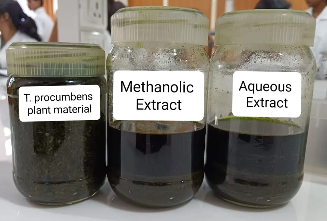
Figure 1: T. procumbens L. Plant Material and its Maceration Process
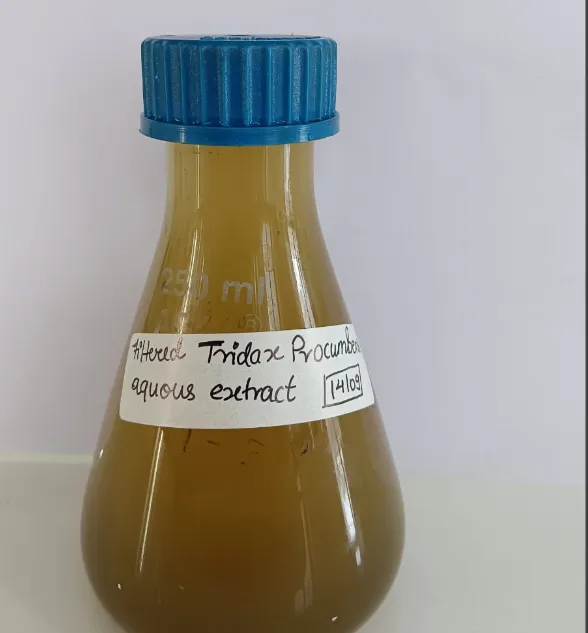
Figure 2a: Aqueous Extract
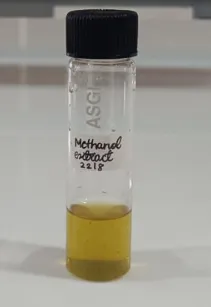
Figure 2b: Methanolic Extract
Phytochemical Screening:
Phytochemical screening of plant extract of T. procumbens L. showed the presence of the phytochemicals listed in the Table 1.
|
Sr. No. |
Phytoconstituent |
Methanolic extract |
Aqueous Extract |
|
1 |
Steroids |
Positive |
Positive |
|
2 |
Carbohydrates |
Positive |
Positive |
|
3 |
Proteins |
Positive |
Positive |
|
4 |
Anthocyanin |
Negative |
Positive |
|
5 |
Phenols |
Positive |
Positive |
|
6 |
Tannins |
Positive |
Negative |
|
7 |
Alkaloids |
Positive |
Positive |
|
8 |
Saponins |
Negative |
Positive |
|
9 |
Flavonoids |
Positive |
Positive |
Table 1: Phytochemical Screening Details of T. procumbens L. Plant Extracts
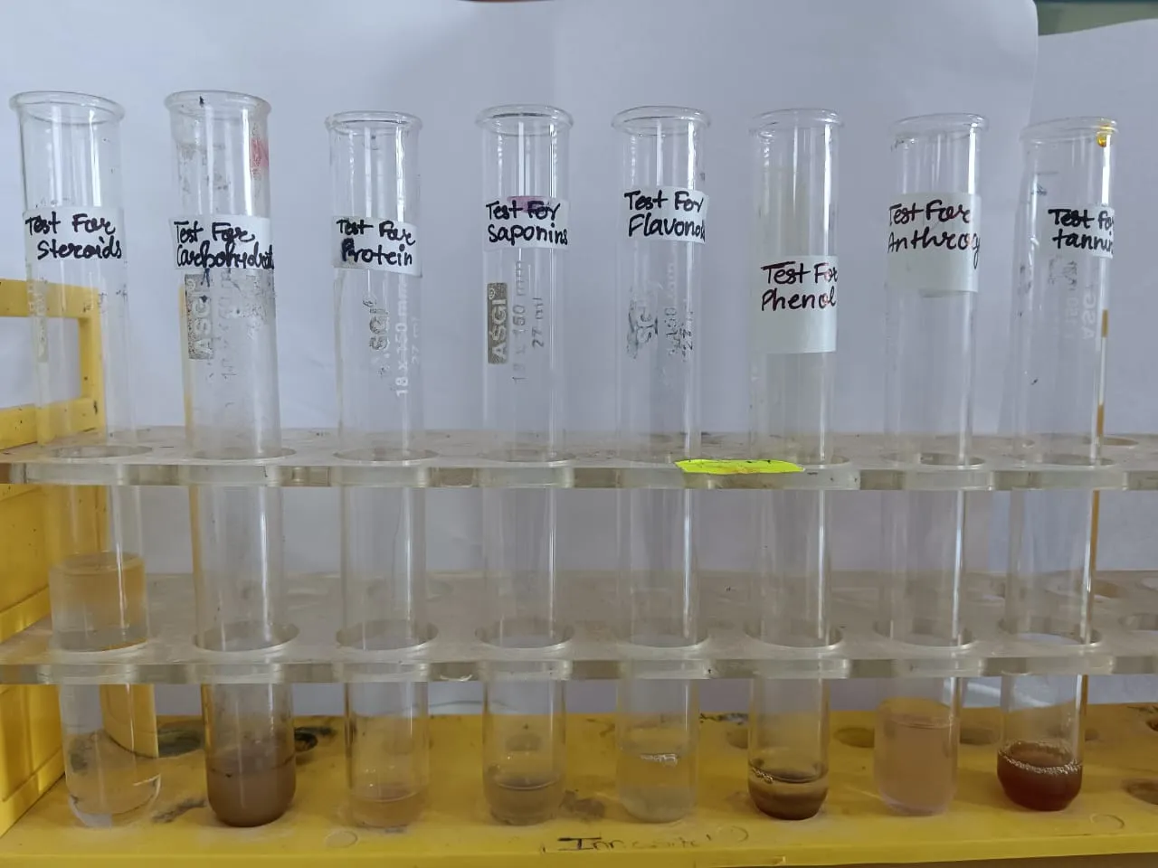
Figure 3: Representative Image of Phytochemical Screening of Plant Extract
Anticoagulation activity: The anticoagulation test results of T. procumbens L. presented in Table 2. The methanolic extract showed considerable anticoagulation activity with human blood when compared with blank and methanol.
|
Sr. No. |
Test Details |
Time in Minutes |
|
1 |
Methanolic Extract |
15 Min, 30 Sec |
|
2 |
Methanol |
10 Min, 15 Sec |
|
3 |
Blank |
05 Min, 00 Sec |
|
4 |
Time Difference |
10 Min, 30 Sec |
|
Sr. No. |
Test Details |
Time in Minutes |
|
1 |
Aqueous Extract |
12 Min, 45 Sec |
|
2 |
Distilled water |
02 Min, 30 Sec |
|
3 |
Blank |
05 Min, 00 Sec |
|
4 |
Time Difference |
07 Min, 45 Sec |
Table 2: Anticoagulation Activity Details
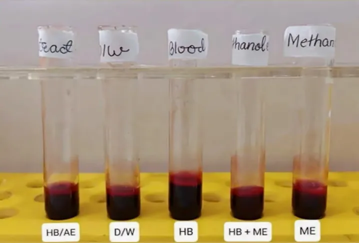
Figure 4: Anticoagulation Activity (HB: Human Blood, AE: Aqueous extract, D/W: Distilled Water, ME: Methanolic Extract)
Anti-Inflammatory activity:
The anti-inflammatory activity test results of T. procumbens L. presented in Figure 5. The aqueous extract showed significant anti-inflammatory activity when compared with blank and methanol.
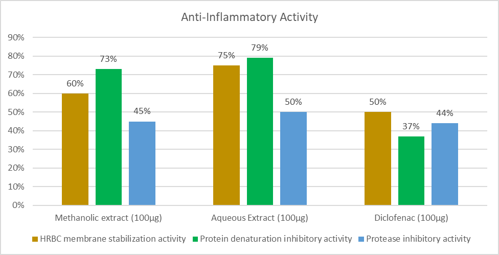
Figure 5: Graphical Representation of Anti-Inflammatory Activity
CONCLUSION
The obtained results suggest that both the aqueous and methanolic extracts of the aerial parts of Tridax procumbens L. exhibited significant anticoagulation and anti-inflammatory effects, likely due to the presence of phytochemicals like phenolics and flavonoids. This study provides preliminary evidence supporting the medicinal potential of Tridax procumbens L. in treating inflammatory disorders and preventing blood clot formation, thereby supporting its traditional medicinal use. However, further research is needed to isolate the bioactive components responsible for its anticoagulation and anti-inflammatory activities and their precise mechanisms of action.
REFERENCES



Sushmita S. Pawar*, Vaishnavi N. Kathar, Ankita M. Sahani, Phytochemical Screening, Anticoagulation And In Vitro Anti-Inflammatory Activity Of Tridax Procumbens L., Int. J. Sci. R. Tech., 2026, 3 (7), 998-1004. https://doi.org/10.5281/zenodo.21642426
 10.5281/zenodo.21642426