

We use cookies to ensure our website works properly and to personalise your experience. Cookies policy
Department of Pharmaceutical Chemistry, K.M. College of Pharmacy, Uthangudi, Madurai – 625107, Tamil Nadu, India
Tragia involucrata L. (Euphorbiaceae) is a medicinal plant with a well-documented history of traditional use in South Asia; its leaves have previously been associated with antioxidant and anti-inflammatory activity, although its neuropharmacological potential remains comparatively unexplored. The present study investigated the ethanolic leaf extract of T. involucrata, collected from Navinipatti, Melur Taluk, Madurai District, and authenticated by the Department of Botany, The American College, Madurai. Shade-dried, powdered leaves were subjected to exhaustive Soxhlet extraction with ethanol, and the concentrated extract was evaluated by preliminary qualitative phytochemical screening followed by Gas Chromatography–Mass Spectrometry (GC-MS) profiling to identify major volatile and semi-volatile constituents. Preliminary tests indicated the presence of alkaloids, flavonoids, tannins, cardiac glycosides, steroids/triterpenoids and phenolic compounds. Based on the GC-MS profile, five major phytoconstituents — squalene, phytol, neophytadiene, 9,12,15-octadecatrienoic acid ethyl ester, and hexadecanoic acid methyl ester — were selected and subjected to in silico molecular docking against human acetylcholinesterase (AChE) using the SwissDock web server, with visualization of the docked complexes in PyMOL. Squalene showed the most favourable predicted binding affinity toward AChE (?9.758 kcal/mol), followed by phytol (?8.156 kcal/mol), neophytadiene (?7.909 kcal/mol), 9,12,15-octadecatrienoic acid ethyl ester (?7.868 kcal/mol) and hexadecanoic acid methyl ester (?7.064 kcal/mol). These findings suggest that the ethanolic leaf extract of T. involucrata contains phytochemicals capable of favourable in silico interaction with AChE, supporting its preliminary candidacy for further target-based and cell-based neuropharmacological investigation.
Medicinal plants continue to play an important role in human healthcare and remain relevant to contemporary pharmaceutical research. Traditional medicine encompasses systems of knowledge that developed in diverse cultural settings and frequently employ plant-derived materials as remedies, creating an important interface between traditional knowledge, pharmacognosy, phytochemistry and modern pharmacology. The World Health Organization recognizes traditional, complementary and integrative medicine as an important component of health systems, provided its integration is evidence-based, safe and effective [1–3]. Natural products constitute an important reservoir of chemically diverse structures for drug discovery; historical examples such as morphine, quinine, paclitaxel and the vinca alkaloids illustrate how plant-derived molecules have contributed to modern therapeutics, either as final drugs or as lead compounds and pharmacophores for further optimization [4]. Systematic natural-product research typically proceeds through correct botanical identification, solvent extraction, preliminary phytochemical screening, chromatographic/spectrometric profiling, and biological or computational evaluation of the isolated or identified constituents [4,5].
Plants synthesize a wide range of secondary metabolites — alkaloids, flavonoids, phenolic acids, tannins, saponins, terpenoids and steroids — many of which possess biological activities of pharmaceutical interest. Flavonoids and phenolics are of interest for their interactions with reactive oxygen species and cell-signalling pathways, while terpenoids and sterols have been associated with antioxidant, anti-inflammatory and antimicrobial activity [5,6]. Qualitative phytochemical screening indicates only the probable presence of a class of compounds; more specific techniques such as GC-MS, LC-MS, HPLC and NMR are required to establish a detailed chemical profile, and in the present study preliminary screening and GC-MS profiling were used as complementary rather than interchangeable approaches [6].
Tragia involucrata L. (family Euphorbiaceae) is a climbing, herbaceous plant bearing characteristic stinging trichomes, widely distributed across the Indian subcontinent and documented in several South Asian traditional medicine systems [7,8]. Ethnopharmacological reviews describe its traditional use for inflammation, pain, respiratory ailments, skin disorders, gastrointestinal complaints and metabolic disorders [7,8]. Experimental studies have subsequently reported anti-inflammatory and analgesic activity of the root fraction [11], wound-healing activity in an excision-wound model [13], psychopharmacological (CNS-depressant) effects of the root extract [12], and antioxidant and anti-inflammatory activity of the leaf extract linked to its GC-MS-detectable phytochemical profile, including squalene [9,10]. Additional reports in the wider literature describe anticonvulsant, antitumor, antidiabetic, diuretic, antimicrobial and antifungal activities of extracts from different plant parts of T. involucrata, underscoring the chemical and pharmacological diversity of the species while also highlighting that this evidence base remains uneven across plant parts and biological endpoints [7,8]. Notably, detailed neuroprotective evaluation of the leaf extract using defined molecular targets or human neuronal cell models has received comparatively little attention, providing the rationale for the present investigation of the leaf rather than the more extensively studied root.
|
|
|
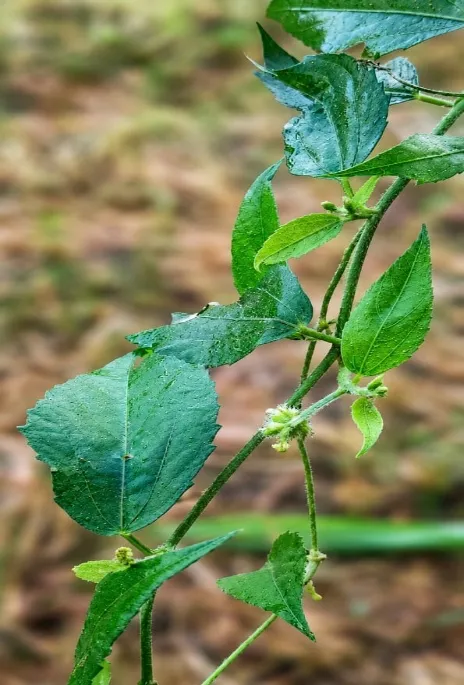
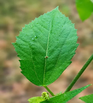
Fig. 1. Tragia involucrata
Neuroprotection refers broadly to strategies aimed at preserving neuronal structure and function against processes such as oxidative stress, mitochondrial dysfunction, protein misfolding and neuroinflammation that underlie neurodegenerative disorders including Alzheimer's disease, Parkinson's disease, Huntington's disease and amyotrophic lateral sclerosis [16,17]. Among these, the cholinergic hypothesis of Alzheimer's disease holds that degeneration of cholinergic neurons and reduced cholinergic transmission contribute to cognitive decline, providing the rationale for acetylcholinesterase (AChE) inhibition as a major symptomatic therapeutic strategy [19,20]. AChE, the enzyme responsible for hydrolysing acetylcholine at cholinergic synapses, therefore remains a widely used and structurally well-characterised target for both synthetic and natural-product-derived inhibitors [20,21]. Natural products are of particular interest as AChE-directed leads because individual phytochemicals can interact with the enzyme through diverse binding modes, and computational docking provides a rapid, resource-efficient means of prioritising candidates from a complex mixture such as a crude plant extract for subsequent experimental evaluation [15,18].
Molecular docking predicts the preferred orientation and relative binding affinity of a small-molecule ligand within a target protein's binding site and is widely used to rank candidate compounds ahead of experimental testing, although docking scores remain predictive rather than a direct experimental measure of inhibitory potency [15,18]. Building on the reported antioxidant and anti-inflammatory activity of T. involucrata leaves and on the well-established role of AChE in cholinergic neurotransmission, the present study combines preliminary qualitative phytochemical screening, GC-MS profiling and structure-based molecular docking to evaluate, for the first time, the potential of major constituents of the ethanolic leaf extract of T. involucrata to interact with human AChE. The specific objectives were: (i) to authenticate the plant material and prepare an ethanolic leaf extract by exhaustive Soxhlet extraction; (ii) to screen the extract qualitatively for major phytochemical classes; (iii) to identify its major volatile and semi-volatile constituents by GC-MS; and (iv) to evaluate the binding affinity of the most prominent GC-MS-identified constituents against human AChE using in silico molecular docking, thereby generating hypothesis-level evidence to guide future target-based and cell-based neuropharmacological investigation of this plant.
II. MATERIALS AND METHODS
A. Plant Material, Collection and Authentication
Fresh leaves of Tragia involucrata were collected from an uncultivated farmland at Navinipatti, Melur Taluk, Madurai District, Tamil Nadu, India. The plant material was identified and authenticated by Dr. D. Stephen, Associate Professor (Retd.), Department of Botany, The American College, Madurai.
B. Materials
Tragia involucrata leaves (shade-dried and coarsely powdered); ethanol (extraction solvent); Soxhlet apparatus with thimble; standard laboratory glassware (flask, condenser); and 1.5 mL airtight microcentrifuge tubes for storage of the concentrated extract [24].
C. Preparation of Powder
Collected leaves were washed with tap water followed by distilled water to remove surface debris, then shade-dried at 25–30 °C for 10–15 days until brittle (moisture content < 10%). The dried leaves were ground to a coarse powder (passing 40-mesh and retained on 60-mesh sieve). Thirty grams of powder were weighed into a Soxhlet thimble [25].
D. Ethanolic Soxhlet Extraction
The powdered material (30 g, defatted) was loosely packed into the thimble to two-thirds capacity to allow adequate solvent percolation. The Soxhlet apparatus was assembled with 150–300 mL of 80–95% ethanol in the receiving flask, the extractor loaded with the thimble, and a condenser supplied with continuous water flow (1–2 L/min). The assembly was heated gradually to ethanol reflux (~60 °C); vapourised solvent condensed and percolated through the powdered sample (~100–150 mL per siphon cycle). Extraction was continued for 8–18 hours (36–72 cycles) until the percolating solvent became pale and clear, with exhaustion confirmed by the absence of a positive phytochemical spot test (e.g., flavonoid reaction) on the final percolate [25].
E. Post-Extraction Processing
The apparatus was dismantled and the combined ethanolic extract filtered through Whatman No. 1 filter paper under vacuum. The filtrate was transferred to a pre-weighed Petri dish and the residual solvent removed by overnight lyophilisation. The resulting semi-solid extract was stored in an airtight 1.5 mL microcentrifuge container at 4 °C, protected from light, until further use [26].
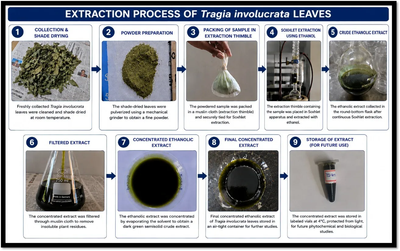
Fig. 2. Extraction Process for the Ethanolic Leaf Extract of Tragia involucrat
F. Preliminary Qualitative Phytochemical Screening
The concentrated ethanolic extract was subjected to standard qualitative colour and precipitation tests to detect the major phytochemical classes — alkaloids, flavonoids, tannins, cardiac glycosides, steroids/triterpenoids and phenolic compounds — following established protocols [27–31]; test conditions and inferences are summarised in Table I (Results).
G. GC-MS Profiling
Gas Chromatography–Mass Spectrometry couples chromatographic separation with mass-spectrometric detection, allowing tentative identification of volatile and semi-volatile constituents in a complex mixture based on retention behaviour and characteristic fragmentation patterns matched against a reference spectral library [32–39]. The instrument comprises a carrier-gas supply, injector, capillary separation column, ion source/mass analyser and detector (Fig. 3); the concentrated ethanolic leaf extract was injected and analysed under standard GC-MS operating conditions to generate a chromatographic profile from which major constituents were tentatively identified by comparison of their mass spectra with the NIST/library database.
GC–MS analysis of the sample was performed using an Agilent GC 8890 gas chromatograph coupled with an Agilent MS 5977C mass selective detector and an autosampler 7693A (Agilent Technologies, USA). Chromatographic separation was achieved using a DB-5ms capillary column (30 m × 0.25 mm i.d., 0.25 µm film thickness), with helium (99.999% purity) as the carrier gas at a constant flow rate of 1.0 mL/min. The injection port temperature was maintained at 250°C. The oven temperature was programmed from 50°C with an initial hold of 1 min, followed by an increase at a rate of 10°C/min to 300°C, with a final hold of 1 min. The mass spectrometer was operated over a mass scan range of m/z 0.6–1091. The obtained mass spectra were compared with those available in the NIST20 mass spectral library for compound identification, and data acquisition and processing were performed using MassHunter software.
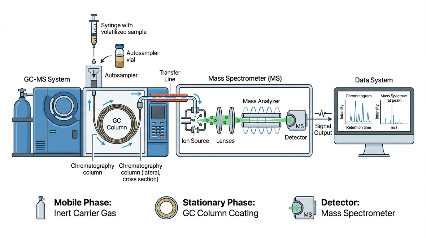
Fig. 3. Schematic of GC-MS Instrumentation
H. Molecular Docking
Based on prominence in the GC-MS profile, five major phytoconstituents — squalene, phytol, neophytadiene, 9,12,15-octadecatrienoic acid ethyl ester, and hexadecanoic acid methyl ester — were selected as ligands. The three-dimensional structure of human acetylcholinesterase (AChE) was obtained and prepared as the docking receptor, and the three-dimensional structures of the five ligands were prepared for docking. Docking calculations were performed using the SwissDock web server, which predicts favourable binding poses and estimates binding affinity (kcal/mol) for a small molecule within a defined target binding site using the EADock DSS algorithm [43]; more negative values indicate a more favourable predicted interaction within the scoring model used, though docking scores remain predictive rather than a direct experimental measurement of binding strength [15,18]. The resulting protein–ligand complexes were visualised in PyMOL, with the receptor displayed in cartoon representation and each docked ligand in stick representation, to examine the predicted binding orientation and spatial relationship with surrounding amino-acid residues.
III. RESULTS AND DISCUSSION
A. Preliminary Phytochemical Screening
Qualitative phytochemical screening of the ethanolic leaf extract of T. involucrata indicated the presence of alkaloids, flavonoids, tannins, cardiac glycosides, steroids/triterpenoids and phenolic compounds (Table I, Fig. 4). This phytochemical profile is broadly consistent with earlier reports on T. involucrata leaf and root extracts [9,10,14], and supports the rationale for further chromatographic and computational characterisation of the extract.
|
S. No. |
Test Performed |
Observation |
Inference |
|
1 |
Test for alkaloids: 1 mL extract + 2–3 drops Mayer's/Dragendorff's reagent |
Cream/orange precipitate |
Alkaloids present |
|
2 |
Flavonoids test: 1 mL extract + few drops lead acetate solution |
Yellow precipitate |
Flavonoids present |
|
3 |
Tannins test: 1 mL extract + few drops 5% ferric chloride |
Black-blue/greenish colour |
Tannins present |
|
4 |
Glycosides (Keller–Killiani) test: acetic acid + FeCl₃ + conc. Hâ‚‚SOâ‚„ |
Brown ring at interface |
Cardiac glycosides present |
|
5 |
Liebermann–Burchard test: acetic anhydride + conc. Hâ‚‚SOâ‚„ |
Blue/green/reddish colour |
Steroids/triterpenoids present |
|
6 |
Phenol test: 1 mL extract + 2 mL 5% ferric chloride |
Blue/green colour |
Phenolic compounds present |
Table I. Preliminary Qualitative Phytochemical Evaluation of the Ethanolic Leaf Extract of Tragia involucrata
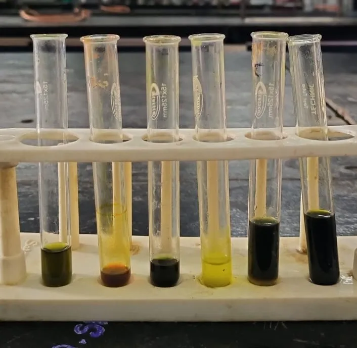
Fig. 4. Colour Reactions Observed in Preliminary Phytochemical Screening of the Ethanolic Leaf Extract of Tragia involucrata: (1) steroids/triterpenoids, (2) alkaloids, (3) tannins, (4) flavonoids, (5) glycosides, (6) phenols
B. GC-MS Identification of Major Phytoconstituents
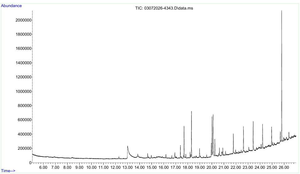
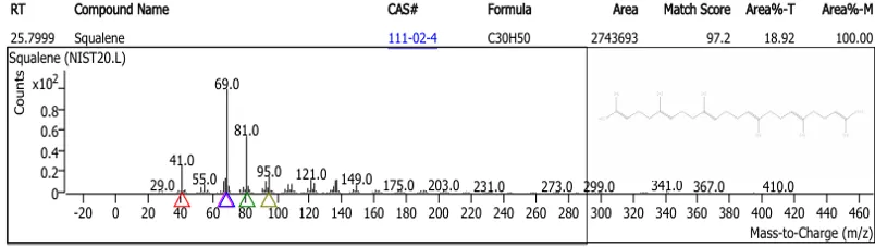
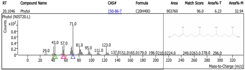
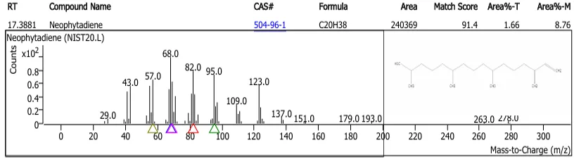
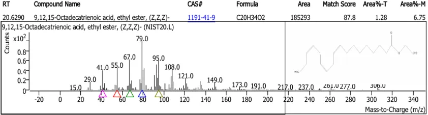
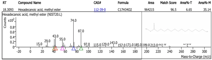
GC-MS analysis of the ethanolic leaf extract enabled tentative identification of its volatile and semi-volatile constituents by comparison of retention and fragmentation data with the spectral library. Five constituents were prominent in the profile and were prioritised for molecular docking based on their relative abundance and relevance to the present investigation: squalene, phytol, neophytadiene, 9,12,15-octadecatrienoic acid ethyl ester, and hexadecanoic acid methyl ester. This finding is consistent with earlier GC-MS work on T. involucrata leaf extracts, which similarly identified squalene among the major detectable constituents [10].
C. Molecular Docking Analysis
The five GC-MS-prioritised phytoconstituents were individually docked against human AChE using the SwissDock server; predicted binding affinities are summarised in Table II. Squalene showed the most favourable predicted binding affinity toward AChE (−9.758 kcal/mol), followed by phytol (−8.156 kcal/mol), neophytadiene (−7.909 kcal/mol), 9,12,15-octadecatrienoic acid ethyl ester (−7.868 kcal/mol) and hexadecanoic acid methyl ester (−7.064 kcal/mol), giving the predicted affinity order: squalene > phytol > neophytadiene > 9,12,15-octadecatrienoic acid ethyl ester > hexadecanoic acid methyl ester.
|
S. No. |
Ligand |
Binding Affinity (kcal/mol) |
|
1 |
Squalene |
−9.758 |
|
2 |
Phytol |
−8.156 |
|
3 |
Neophytadiene |
−7.909 |
|
4 |
9,12,15-Octadecatrienoic acid, ethyl ester |
−7.868 |
|
5 |
Hexadecanoic acid, methyl ester |
−7.064 |
Table II. Predicted Binding Affinity of Selected Phytoconstituents Against Human Acetylcholinesterase (SwissDock)
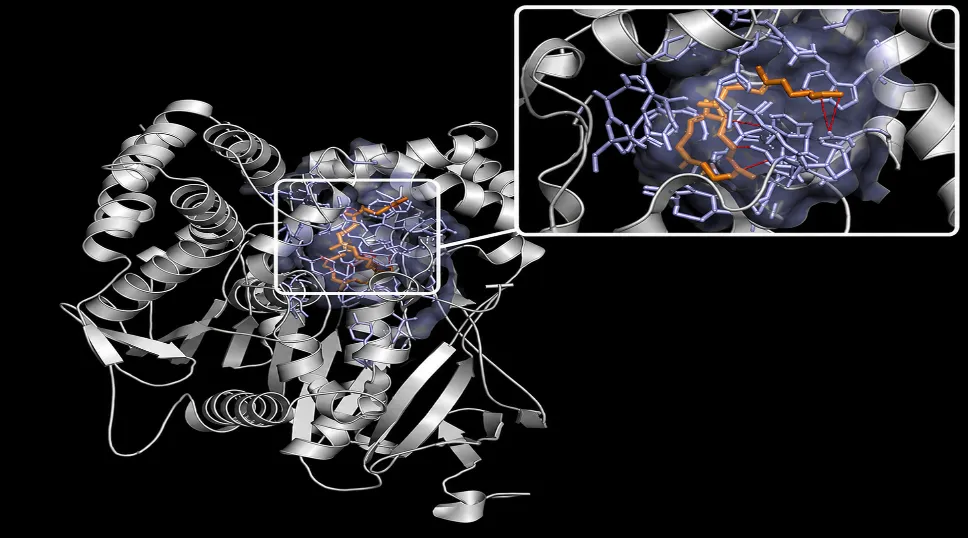
Fig. 5. Predicted Molecular Docking Interaction of Squalene with Acetylcholinesterase (AChE)
PyMOL-based visualisation of the docked complexes (Fig. 6) showed each ligand occupying the AChE binding region in stick representation within the cartoon-rendered protein, supporting the numerical docking results by illustrating the predicted spatial fit of the ligands within the binding site.
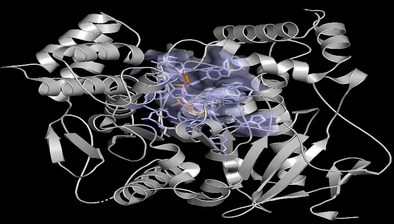
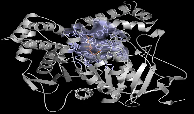
Fig. 6a–b. Predicted Docking Orientation of Phytol (left) and Neophytadiene (right) with AChE
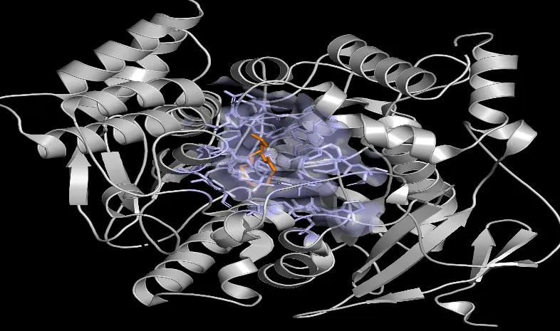
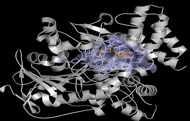
Fig. 6c–d. Predicted Docking Orientation of Hexadecanoic Acid, Methyl Ester (left) and 9,12,15-Octadecatrienoic Acid, Ethyl Ester (right) with AChE
The differences in predicted affinity among the five compounds may reflect differences in molecular size, shape, hydrophobicity and conformational flexibility that influence their ability to form favourable contacts within the AChE binding region. Squalene, a large triterpenoid hydrocarbon, and phytol, a diterpene alcohol, showed the two most favourable scores, consistent with previous reports linking squalene identified in T. involucrata leaf extracts to favourable in silico interaction with an inflammatory target [10], and with broader literature identifying phytol and related isoprenoids as candidates for AChE-directed natural-product screening [41,42]. However, a docking score alone cannot establish actual inhibitory activity; it should be interpreted as supporting, hypothesis-generating in silico evidence rather than confirmatory proof of enzyme inhibition [15,18], and requires corroboration through experimental AChE inhibition assays and, ideally, cytocompatibility evaluation in a relevant neuronal cell model such as SH-SY5Y cells [22,23] before any therapeutic inference can be drawn.
CONCLUSION
The present study provides an integrated preliminary chemical and in silico evaluation of the ethanolic leaf extract of Tragia involucrata L. Preliminary qualitative phytochemical screening confirmed the presence of alkaloids, flavonoids, tannins, cardiac glycosides, steroids/triterpenoids and phenolic compounds, consistent with earlier reports on this species. GC-MS profiling enabled tentative identification of the extract's major volatile and semi-volatile constituents, five of which — squalene, phytol, neophytadiene, 9,12,15-octadecatrienoic acid ethyl ester and hexadecanoic acid methyl ester — were prioritised for structure-based investigation. Molecular docking of these constituents against human acetylcholinesterase using the SwissDock server indicated that squalene possessed the most favourable predicted binding affinity (−9.758 kcal/mol), followed by phytol, neophytadiene, 9,12,15-octadecatrienoic acid ethyl ester and hexadecanoic acid methyl ester, with PyMOL-based visualisation structurally supporting these predicted interactions.
Taken together, the phytochemical, GC-MS and molecular docking findings indicate that the ethanolic leaf extract of T. involucrata contains constituents that are chemically diverse and computationally compatible with the AChE binding region, providing preliminary, hypothesis-generating evidence for further neuropharmacological exploration of this plant. As docking scores are predictive rather than confirmatory, these findings should be corroborated by experimental AChE inhibition assays and by cytocompatibility evaluation in a relevant neuronal cell model, such as SH-SY5Y cells, before any therapeutic inference is drawn. Subject to such validation, squalene and phytol in particular emerge as priority candidates for future isolation, quantification and mechanistic neuropharmacological study.
ACKNOWLEDGEMENT
The authors thank Dr. D. Stephen, Associate Professor (Retd.), Department of Botany, The American College, Madurai, for the identification and authentication of the plant material, and the Department of Pharmaceutical Chemistry, K.M. College of Pharmacy, Uthangudi, Madurai, for providing the laboratory facilities used in this work.
REFERENCES






Cholaraja K.*, Lavanya S., Logesh A., Madhu Bala S., Madhu Priya K.B.R., Manoj Kumar S., Phytochemical Screening, GC-MS Profiling And Molecular Docking Studies On The Ethanolic Leaf Extract Of Tragia Involucrata L. Against Acetylcholinesterase, Int. J. Sci. R. Tech., 2026, 3 (9), 532-543. https://doi.org/10.5281/zenodo.22958307
 10.5281/zenodo.22958307