

We use cookies to ensure our website works properly and to personalise your experience. Cookies policy
Govt. Degree and PG College, Siddipet (Autonomous), Central Research Lab
The structural behavior of DNA-protein complexes under altered physicochemical environments represents a critical area of investigation in understanding macromolecular organization beyond native cellular conditions. This study explores how controlled chemical disruption and externally applied mechanical forces influence the aggregation patterns of DNA-protein systems, providing insight into the dynamic nature of biomolecular assembly. In this work, cellular integrity was first disrupted using detergent-mediated solubilization, enabling the release of DNA-protein complexes into a free interactive environment. The system was further regulated through the addition of EDTA and sodium chloride, which respectively preserved DNA integrity by chelating divalent ions and promoted aggregation through electrostatic screening. These combined conditions created a tunable framework in which intermolecular interactions could be observed and manipulated. Such ionic modulation is consistent with established principles of polyelectrolyte behavior, where charge neutralization drives molecular condensation. Following chemical treatment, samples were subjected to hypergravitational conditions through centrifugation, introducing a directional force that significantly altered aggregation dynamics. This mechanical influence resulted in the formation of distinct structural patterns, including aligned compact assemblies and interconnected network-like aggregates. These observations highlight the role of non-equilibrium forces in shaping macromolecular architecture, a phenomenon supported by polymer dynamics under external stress. Microscopic examination revealed progressive transitions in structural organization, while spectrophotometric analysis using methylene blue confirmed the presence and accessibility of DNA within aggregated systems. The combined analytical approach allowed for both qualitative and quantitative assessment of aggregation behavior. Overall, the study demonstrates that DNA-protein aggregation is governed by a complex interplay of chemical, electrostatic, and mechanical factors. The findings provide a simplified yet powerful model for understanding biomolecular condensation, with potential implications in molecular biology, biomaterials development, and the study of phase-separated biological systems.
How biological macromolecules organize and behave when subjected to different physical stresses remains one of the more compelling puzzles in modern biophysics. Inside a living cell, DNA rarely exists in pure isolation. It’s constantly mingling with proteins, lipids, and various ions a bustling microenvironment that ultimately dictates its shape, stability, and functional accessibility. If we want to understand fundamental biology, or even just build better experimental techniques, we have to figure out how DNA behaves when forced out of this native comfort zone.
Because of its negatively charged phosphate backbone, cellular DNA essentially acts like a highly charged wire. Neighboring strands naturally repel each other, keeping the molecules from spontaneously clumping up under normal physiological conditions. But tweak that environment whether through chemical changes or physical force and that delicate balance shatters. The DNA might condense, aggregate, or completely collapse. These transitions aren't just random accidents; they are driven by strict physicochemical rules that control how large molecules assemble (Manning et al. 1969).
Proteins complicate this picture in fascinating ways. Normally, they act as the ultimate organizers, stabilizing chromatin and managing gene expression. Throw them into an experimental setup, however, and they can trigger wildly complex aggregation patterns. They effectively become viscoelastic scaffolds, bridging molecules together while simultaneously reacting to shifts in salt levels or mechanical tension. Because of this dual personality, DNA-protein systems act as quirky hybrids, blending the structural properties of polymers with those of colloids (de Gennes et al.1979).
Then there are the cellular lipids. They build the membrane walls and keep everything strictly compartmentalized. If you want to study what’s inside a cell, breaking those barriers is usually step one. That’s where amphipathic detergents come in. They easily slip into lipid bilayers and tear the architecture apart, forming mixed micelles and spilling the DNA-protein complexes out into the open. Suddenly moving from tight cellular confinement into a dispersed solution opens the door for entirely new, unexpected molecular interactions and clumping pathways to emerge (Helenius et al. 1975).
You also can't ignore the local ionic environment. Divalent metals like calcium and magnesium are double-edged swords: they stabilize nucleic acids, but they also wake up nucleases that can chew the DNA apart. Introducing a chelator like EDTA gives researchers a leash, selectively mopping up those ions to keep the DNA intact while blocking unwanted biochemical reactions. On the flip side, monovalent salts like sodium chloride work to mask the DNA's negative charge. This dampens the natural repulsion between strands, allowing them to get close enough to pack tightly together a classic phenomenon known as counterion condensation (Sigel et al., 1993).
Chemical shifts are only half the story, though; physical brute force matters just as much. Spin a sample at high relative centrifugal forces (RCF), and you introduce a strong directional pull that physically forces biomolecules to align, settle, and compact. Unlike gentle, equilibrium-driven processes, these force-induced changes yield non-equilibrium structures that bear the physical "scars" of the stress they just endured. Looking at these forced assemblies actually teaches us quite a bit about natural biological phenomena, like intense cellular crowding and phase separation (Rubinstein et al.2003).
Lately, the discovery of biomolecular condensates has completely changed how we view intracellular organization. We now know that genetics doesn't dictate everything; the surrounding physicochemical environment is pulling the strings too. These condensates are essentially membrane-free, liquid-like droplets born from multivalent interactions, and they are incredibly sensitive to salt, concentration limits, and physical force. Interestingly, the rules governing these natural systems look a whole lot like what we see in lab-controlled aggregation. This suggests that simplified experimental models are actually remarkably good at mimicking complex biology (Brangwynne et al. 2009, Banani et al. 2017).
That brings us to the core of the present study. We set out to systematically map exactly how DNA-protein complexes aggregate when hit with a combination of chemical destabilization and extreme gravitational force. By pulling specific experimental levers disrupting lipids with detergents, tweaking ion levels with EDTA and NaCl, and applying intense mechanical stress via centrifugation we can pause and observe transitional states that usually happen too fast to catch in nature. This approach lets us isolate individual variables while still seeing the big picture of how they gang up to reshape macromolecules.
What sets this investigation apart is how we look at the timeline of structural evolution. Aggregation isn't just a sudden, one-and-done endpoint. It's an entire continuum. We track the process from the initial, loose associations all the way to heavily condensed, directionally forced clumps. Each step along the way represents a real-time tug-of-war between entropy, intermolecular attraction, and outside physical input. Viewing aggregation as a moving picture rather than a static snapshot gives us a much deeper, nuanced understanding of how these assemblies actually form and stabilize.
To actually capture all of this, we combined microscopy with spectrophotometry, giving us a mix of visual proof and hard numbers. Under the microscope, we can watch the structural patterns unfold directly, pinpointing shifts in morphology, density, and connectivity. Meanwhile, spectrophotometry especially using DNA-binding dyes like methylene blue tells us exactly how accessible the molecules are and how they're interacting behind the scenes. Pairing the two methods gives us a comprehensive, 360-degree view of the aggregation process.
The ripple effects of this research go far beyond our specific lab setup. By detailing exactly how DNA-protein complexes react to environmental stress, this work helps pave the way for creating customizable biomaterials and cleaner nucleic acid extraction methods. It also peels back the curtain on the physics of molecular self-assembly. There are even broader implications for synthetic biology and prebiotic chemistry, fields that rely entirely on the spontaneous, organized formation of large molecules. Ultimately, this study frames DNA-protein aggregation not as a random biological mess, but as a highly dynamic, multi-factor process that we can purposefully control. By pulling together ideas from polymer physics, colloidal science, and molecular biology, we offer a fresh, unified framework for interpreting macromolecular behavior in both test tubes and living cells.
1.1 Previous research analysis
The way we look at DNA-protein aggregation hasn't stayed still; it’s a patchwork of molecular biology, polymer physics, and colloidal chemistry. Each field brings its own unique lens to how macromolecules behave when their environment shifts. Early on, scientists mostly treated DNA like an isolated vault of genetic code, hyper-focused on its role in heredity. Eventually, though, it became obvious that DNA is rarely flying solo. It’s almost always tangled up in complex assemblies with proteins and other biomolecules. That realization flipped the script. We stopped seeing DNA as just a static biological blueprint and started treating it as a dynamic physical system that physically shifts and transitions (Watson et al.1953).
Looking back, figuring out chromatin organization was a massive leap. Researchers saw how DNA wraps intimately around histone proteins to cram itself into the nucleus. A lot of this comes down to basic electrostatics: the positively charged histones cancel out the DNA's negatively charged backbone, which makes dense packing possible. This trick of charge neutralization eventually became the gold standard for understanding artificial DNA aggregation, where we use outside ions to force similar results in the lab (Kornberg et al.1974). While chromatin research was strictly biological, it planted an important seed. It proved that DNA condensation is entirely at the mercy of its chemical neighborhood.
Meanwhile, polymer scientists were building their own theoretical sandbox to explain DNA. They began modeling it as a semi-flexible polyelectrolyte a polymer governed by both its physical chain shape and its electrical charge. Studies in this arena proved that the tug-of-war between electrostatic repulsion and physical attraction dictates whether the DNA stretches out or collapses into a tight ball. When Manning's counterion condensation theory came along, we finally had the math to explain how soaking ions up in a solution lowers that repulsion and triggers clumping (Manning et al.1969). We lean heavily on these models to understand why DNA acts so erratically at different salt concentrations, which directly informs the experiments we are doing today.
Then there's the whole angle of membrane biology. Developing detergent-based extraction methods completely changed the game for isolating DNA and proteins from cells. We learned that detergents essentially hack into lipid bilayers, tear them apart to form micelles, and dump the cell's contents out. Sure, it makes extraction easier, but it also totally rewires the physical space where these biomolecules interact. Take away the lipid walls, and molecules start moving faster and clumping together in totally new ways (Helenius et al. 1974).
You also can't talk about nucleic acid stability without getting into metal ions and chelating agents. We’ve known for a long time that divalent ions like magnesium (Mg²âº) keep DNA structured, but they also act like a green light for nucleases to come in and chew that DNA apart. That's why molecular biologists obsessively use EDTA. It works like a sponge, soaking up those metal ions to paralyze the enzymes. But previous work proves EDTA does more than just play bodyguard; it actively tweaks the surrounding ionic environment, which indirectly shifts how things aggregate. It's a perfect example of how ionic control both protects and manipulates a system (Sigel et al.1993).
Lately, the spotlight has swung toward non-equilibrium systems specifically, what happens when you hit biomolecules with raw physical force. We’ve always used centrifugation to separate things, but researchers are finally paying attention to how it physically molds molecular structures. When you subject a polymer to force, it aligns, stretches, and compacts. The resulting shapes look nothing like what you get under calm, equilibrium conditions. This is a big deal for DNA-protein systems, where mechanical stress and chemical signals combine to create incredibly complex, tangled aggregates (Rubinstein et al.2003).
Another huge modern shift is the focus on biomolecular phase separation. Think of it as proteins and nucleic acids spontaneously organizing into their own distinct compartments, entirely without membranes. These condensates rely on multivalent interactions and will drastically change shape if you mess with the temperature, salt, or concentration. Because these natural droplets behave so much like the artificial aggregates we make in the lab, scientists have started using simplified experimental models to decode the underlying rules. It's a brilliant way to bridge the gap between test-tube chemistry and actual living biology (Brangwynne et al.2009, Banani et al. 2017).
Even with all this progress, we’re still missing a crucial piece of the puzzle. Nobody has really figured out how to integrate chemical destabilization, ionic regulation, and mechanical force into one cohesive experiment. Most studies isolate these variables, which means we miss out on seeing how they amplify or cancel each other out. This current work fills that exact gap. We are throwing everything at the system at once: shredding membranes with detergents, manipulating ions with EDTA and NaCl, and cranking up the hypergravitational stress via centrifugation. Doing it this way catches all those fleeting, transitional states that separate, isolated studies usually miss.
To sum it up, past research gives us a rock-solid baseline for understanding how DNA and proteins interact, both biologically and physically. But these systems are messy and complex. They demand a holistic approach. By standing on the shoulders of established theories and mixing up the experimental conditions, this study aims to build a much clearer, comprehensive picture of exactly why and how macromolecules aggregate.
Genetic material aggregation depends on various factors which involves globule polymer aggregation to ion induced aggregation and sometimes protein adhesion aggregation. Here to study the structural aggregation all these types of aggregation can be taken into consideration while drawing conclusion.
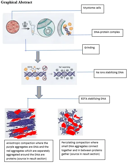
Muscle tissue from the organism is taken which is first crushed using a grinding apparatus which breaks down the structural complexity of the protein. Which frees up the following tissue sample for further breakdown. Small sample can then be taken and the layers of cell apparatus which are bound together should be broken down by using various compounds which help better isolate the DNA-protein complex.
2.1 Lipid breakdown
Lipids exist in cell membrane which constitutes phospholipids + cholesterol + glycolipids. Intramyocellular lipid droplets are energy reservoirs for sustained energy production which contain triglycerides + cholesteryl esters. Mitochondria is another lipid heavy structure which includes phospholipids + cardiolipin. (Alberts, B. et al.2014) Sarcoplasmic reticulum also contains lipid reserves. Transverse tubules which is present in sarcolemma and lipid bilayer structure and conduct electrical signals. Nuclear envelope which is double layer around nucleus which also is in phospholipid bilayer. Cytosolic lipid binding complexes. Which is protein bound fatty acids. Peroxisomes which contain lipids involved in beta oxidation of very long chain fatty acids and membrane bound organelles.
These lipids which are bound can be solubilized by using detergent which can easily remove the lipids present in the cells.
2.2 Mechanism
2.3 EDTA
This is a very important compound which helps in protecting DNA which is a major component needed in this experiment which helps us to carry out the separation and aggregation without damaging the DNA molecule. Its major role is as a chelating agent which binds magnesium ions and calcium ions and divalent metal ions.
2.3.1 Mechanism
2.4 NaCl
This is a major component which allows the negative charge to be neutralized and DNA to be aggregated.
2.4.1 Mechanism
2.4.1.1 Procedure breakdown:
3.1 Before Aggregation
As seen in fig 1, the DNA-protein structure complexes are observed where the internal dense DNA material is surrounded by globular protein. These are DNA rich aggregates which are taken from the surrounding supernatant of the visible material structure formed from the supernatant. These are also mixed with denatured proteins and lipid remnants.
These complexes irregular and non-uniform shaped with dense core and filamentous edges. These are not clear long structures due to the extraction being aimed from the supernatant free-floating particles rather than the visible cluster of DNA precipitate. this sample also consist of protein debris and cell fragments and other debris.
In this observation, the visible complex of structures is in such way due to the NaCl, EDTA, ethanol and mechanical handling done in the process of extracting which in turn changed its phase to being globular. This is called polymer physics phenomenon.
Each of these following images show various types of patters which can be further elaborated as follows based on the numbering given in the description of the image.
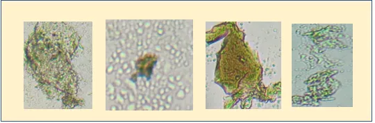
Fig 1: preliminary structural showcase of isolated DNA-protein complex which shows 4 main structures were left to right can be denoted as 1, 2, 3, 4 respectively
Image 1
This type of complexes is more commonly seen throughout the initial analysis which shows a globular complex of DNA-protein complex where the bonds between the molecules are more tightly woven and when zoomed in for a closer examination as seen in fig 2 we can see the structural format of this complex where slightly filamentous structures can be seen in the darker DNA rich areas and more globular protein structures in the transparent regions.
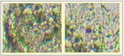
Fig 2: closer look on the first image of the DNA-protein complex
This can be described as core shell composite structure with differential density distribution. The outer region which is transparent protein is more optically diffused and it has higher scattering which shows that its hydrated and less compact. This is also likely a viscoelastic matrix which is protein and residual lipids. The inner dark zone of DNA rich area has high optical density and indicates localized concentration of polymer chains. DNA is not fully collapsed here and is dispersed and surrounded by a soft protein network. This structure is not uniform because of change in densities and different hydration levels along with salt concentration, this creates microphase separation between DNA rich and protein rich domains. The boundaries of this structure are less defined and slight irregularities appear this indicates the structure is free floating and not confined within a bigger matrix. This could be due to internal pressure of polymer entropy which is working against external matrix constraints.
Image 2
As seen in the fig 1 second image the central dark DNA structure is much more defined optically dense structure with sharper boundary and minimal filamentous extensions. Unlike first structure this has less internal granularity visible and more uniform optical density. This implies reduced internal mobility and lower structural integrity this due to the addition of NaCl transitioned from heterogenous to quasi homogenous condensed state. Here, we can observe the region around the structure which is slightly less dense in particles this is a local depletion zone which occurs when molecules like protein fragments around the DNA. These structures are compact and relatively smooth compared to before image which implies interfacial energy is dominating the chain entropy which means minimizing the surface area and has condensing behavior basically due to the external factor of NaCl. The first image to the second image shows the difference between polymer system of DNA-protein complex to colloidal like object. This can be basically stated like having high macromolecular density around the DNA which is also loosely packed. This also shows the reduced hydration in these types of cases.
Image 3
This is similarly to the second image is a very condensed DNA which is not surrounded by protein which shows the denaturation of surrounding protein. This structure is compact and bundled and appears like a single dense particle. But the DNA molecular patter does not seem to be heavily aggregated together when we have a closer look at the structure, as seen in the fig 3.
This also doesn’t show any internal differentiation which means this went to collapse due to the addition of NaCl faster than reorganization. Here, we can observe no visible connections to surrounding material and no bridging structures. This can also be treated as a isolated DNA domain. This structure behaves like a colloidal particle rather than a polymer assembly. This is a terminal fragmentation state with no further structural reorganization, which is not ideal to the goal of this experiment.
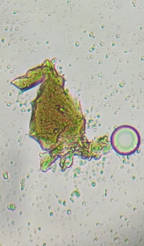
Fig 3: slightly magnified 3rd image which does not show heavy aggregation
Image 4
This structure when compared to others and analyzed appears to be similar to the first image structure when it comes to the basis of structural organization and properties. This is important due to the fact that these types of structures are important for the assessment of aggregation patterns of DNA-protein matrixes.
Before the aggregation studies the immediately separated DNA-protein matrix is first analyzed using this device. For accurate results we can add methylene blue to both the control medium which is distilled water and then the supernatant, where DNA can form electrostatic binds to methylene blue due to the negative charge property of DNA and positive charge property of methylene blue. This can hold the DNA and can check the absorbance at 660nm wavelength (Green, F. J. 1990) which can prove several factors such as,
4.1 Absorbance and transmittance analysis
Sample absorbance (Aâ‚›) = 1.163
Blank/control absorbance (Aᵦ) = 0.301
The corrected absorbance is 0.862.
This shows the high absorbance and scattering rate of DNA bound with methylene blue in spectrophotometer. The error in the control blank due to the methylene blue is corrected and normalized in the corrected absorbance section. And the reading in the spectrophotometer is shown in fig 4.
|
Parameter |
Value |
Unit/Remarks |
|
Wavelength |
660 |
nm |
|
Sample Absorbance (As) |
1.163 |
Raw absorbance of DNA–methylene blue complex |
|
Blank/Control Absorbance (Ab) |
0.301 |
Distilled water + methylene blue |
|
Corrected Absorbance |
0.862 |
As − Ab = 1.163 − 0.301 |
|
Transmittance |
13.7 |
% |
|
Dye Used |
Methylene Blue |
DNA-binding dye |
|
Sample Type |
DNA–Protein Complex |
Supernatant fraction |
|
Interpretation |
High DNA-associated absorbance |
Indicates DNA presence and accessibility |
5.1 Relative centrifugal force calculation
Rotation speed of centrifuge = 4000 RPM
Radius of the sample container = 5cm
RCF =1.118 ×10⁻⁵ × r × (RPM) × 2 (CRC Handbook of Chemistry and Physics)
RCF =1.118×10−5×5 × (4000)2
RCF = 894.4.
This can also be denoted as 894 × g, which is around 894 times the gravity which is being applied upon the sample which can be classified as a hypergravitational condition, which is more than sufficient for macromolecular aggregation and sedimentation.
5.1.2 After aggregation
5.1.2.1 Primary observations
The DNA-protein complex visibly is settled below the sample tube which is expected for this experiment.
5.1.3 Structural observation
Throughout the analysis there are 2 types of aggregation which are common and most prevalent.
5.1.3.1 Anisotropic compaction
As seen in fig 5, this type of aggregation is the most prevalent and visible which is a very clear indication of aggregation pattern. This is also called anisotropic aggregation where; the structure is compressed in a directional band. The RCF imposed a directional force gradient causing macromolecules to align and compact along the radial axis. Protein around this also is compressed but still behaves like a viscoelastic medium.
The DNA-protein complex moved radially outward where local concertation is increased. During the process the DNA-protein complex underwent stretch, overlap and intertwining and went from disentangling to frozen topology.
This structure has dense core region with slightly diffused edges this indicates the micro stratification under RCF. This can occur due to different mass of DNA-protein complex and different sedimentation coefficients.
This is not a structure of equilibrium; this is a condensed phase structure with indications of clear applied force and non-relaxed network. This can be further interpreted as force-based polymer collapse. The protein also has scaffold type structures and constraint. This can further prove the aggregation changes under RCF.
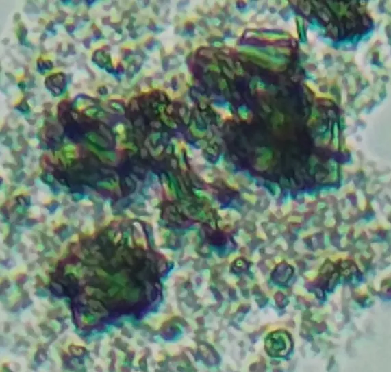
Fig 5: Anisotropic compaction
5.1.4 Percolating network complex
This is another type of aggregation observed in this where there is a continuity in the mass with no clear internal boundaries apart from the DNA to protein difference. The interior is granular and non-uniform pockets. Here we can observe the DNA-protein complex has come very much together with very low gaps which overlap one another, here we can observe a granular or sometimes globular structure different from the previous filamentous structure. The surface is like sub-clusters fused together rather than smooth bodies. This occurs due to the smaller DNA-protein structures form first and then stick upon each other as seen in fig 6.
This forms a network like structure which forms places with darker in color and then lighter in color as it goes outward. This shows the non-uniform crosslinking of DNA-protein complexes. The wrinkling structure is due to the local dehydration during the RCF process, which can create capillary compaction which gives it the visible structure.
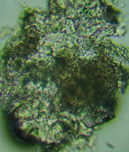
Fig 6: Percolating network complex
This study offers a deep structural look into how DNA-protein complexes aggregate when subjected to chemical destabilization and extreme gravitational forces. What we observed is a clear, step-by-step transition. The system shifts from loosely tangled polymeric networks into heavily condensed, force-driven architectures, perfectly illustrating how physicochemical parameters ultimately dictate macromolecular behavior.
When we examined the extracted DNA-protein complexes, they didn't look uniform at all. Instead, they formed messy, irregular shapes featuring a densely packed DNA core surrounded by a fuzzy, diffuse protein matrix. This kind of organization is a classic hallmark of a polymer-colloid hybrid. Essentially, the DNA acts as a semi-flexible polyelectrolyte trapped inside a stretchy, viscoelastic protein gel (de Gennes et al.1979). We also noticed wispy filamentous extensions and uneven density gradients, which tells us the structural collapse isn't total. There is a continuous, dynamic tug-of-war happening between the macromolecules. It's the exact kind of intermediate state you usually see during phase separation, where the chaotic push of entropy constantly battles against the condensing pull of enthalpy (Brangwynne et al.2009).
None of this structural exposure would be possible without detergents tearing apart the cellular lipids. By driving amphipathic molecules into the phospholipid bilayers, the detergents force the membranes to curve, break, and form micelles (Helenius et al. 1975). This effectively demolishes the compartmental barriers, releasing the DNA-protein complexes into a free-floating, highly interactive state. Interestingly, the leftover lipid fragments don't just vanish. They likely stick around in the surrounding matrix, adding to its overall viscoelasticity and subtly altering the kinetics of the aggregation process.
Next, the introduction of EDTA acts as a crucial stabilizer. It works by hunting down and chelating divalent ions like Mg²âº. By doing so, it essentially paralyzes DNA-degrading nucleases and stops unwanted electrostatic bridges from forming too early (Sigel et al.1993). Because of this, the DNA avoids random, messy precipitation. Instead, it remains structurally accessible and primed to participate in a highly controlled aggregation sequence. (manning et,al. 1969).
Sodium chloride (NaCl) is the real engine driving the aggregation here, working through electrostatic screening. If you look at the progression from the clumpy, heterogeneous structures in Image 1 to the tightly packed, uniform entities in Image 2, you are seeing counterion condensation theory in action. The sodium ions step in to mask the DNA's negatively charged phosphate backbone, effectively killing the natural repulsion between the strands (Manning et al.). Once that barrier falls, the molecules pack tightly together, seeking a more energetically stable, condensed state. As the boundaries smooth out and the internal graininess fades, it becomes clear that minimizing surface energy has overpowered configurational entropy. The entire system practically transforms into a colloidal-like phase.
But there is a limit. Image 3 reveals isolated, ultra-dense particles, pointing to a kinetically trapped state. Basically, the ionic collapse happened so incredibly fast that the structures never had a chance to properly reorganize or link up. Because aggregation outpaced network formation, the system produced these dead-end, discrete blobs. This is a textbook example of diffusion-limited aggregation, which frequently happens when polymers collapse under extreme ionic strength (Lin et al.1989). It highlights a critical boundary in the experimental setup: push the salt concentration too high, and you completely block the formation of functional, interconnected macromolecular networks.
Our spectrophotometric data backs all of this up perfectly. We recorded a high corrected absorbance of 0.862 at 660 nm, confirming that the methylene blue dye is heavily interacting with the DNA. This proves the genetic material is both present and physically accessible within the complexes. On the flip side, the transmittance was quite low (roughly 13.7%). That sharp drop is caused by intense light scattering, which is exactly what you would expect from massive, aggregated structures blocking the light path (Lakowicz et al.2006). Seeing both high absorption and heavy scattering reinforces the conclusion that we are dealing with a incredibly dense, uneven system filled with differently sized particles.
The most dramatic transformations, however, happened when we cranked up the hypergravitational stress to roughly 894 × g. Applying that kind of external force imposes strict directional rules on the system. We observed distinct anisotropic compaction meaning the mechanical pressure actually overpowered the molecules' natural chemical interactions, forcing the DNA-protein complexes to align and compress along a single radial axis. This is a fantastic real-world example of force-induced polymer collapse. The macromolecules get shoved into non-equilibrium states, locking them into frozen, stressed topologies (Rubinstein et al.2003). On top of that, the visible stratification and density gradients suggest that heavier, denser structures settled out differently than the lighter ones.
We also found percolating network complexes, pointing to a secondary aggregation stage driven by cluster-cluster interactions. These structures look granular and are made of fused subunits, bearing a strong resemblance to the fractal aggregation patterns typically seen in colloidal chemistry (Meakin et al.). Furthermore, the structures showed signs of dehydration-induced wrinkling and capillary compaction. This suggests that as water was forced out during centrifugation, solvent dynamics actively helped cement the final architecture.
Ultimately, this study proves that DNA-protein aggregation isn't just a simple, single-step event. It's a complex, multi-stage transformation dictated by a delicate blend of chemical, electrostatic, and mechanical forces. By carefully balancing detergent liberation, EDTA stabilization, salt-driven condensation, and centrifugal alignment, we created a highly varied landscape of structural outcomes. More importantly, it shows that manipulating these specific parameters allows us to steer aggregation toward exact, desired architectures. That level of control has massive potential for designing biomaterials, advancing molecular biology, and even modeling prebiotic chemistry.
What we are seeing here mirrors intracellular phase separation and the way biomolecular condensates form inside living cells, guided by the exact same nanoscale forces (Banani et al. 2017). Because of this, our work goes far beyond being a simple extraction protocol. It serves as a robust model system for studying force-modulated macromolecular assembly, paving the way for the next generation of tunable biological materials and synthetic cellular systems.
CONCLUSION
This study demonstrates that DNA-protein aggregation isn't just a messy byproduct of extraction it's a highly tunable physicochemical process. By exposing these macromolecules to specific chemical destabilizers and intense physical forces, we can actively steer them into distinct structural states.
The process starts when detergents break open the cell. Rather than simply spilling the contents, this creates a transitional space where biomolecules can freely reorganize, mirroring how amphipathic agents reshape lipid walls.
From there, EDTA steps in as a vital stabilizer. By sequestering divalent ions, it shuts down degrading enzymes and prevents random clumping. This ensures the aggregation follows precise electrostatic rules, resulting in authentic structural transitions rather than degraded artifacts. Adding NaCl then shifts the system entirely. Salt masks the DNA's electrical charge, neutralizing natural repulsion and forcing tight molecular packing. But balance is everything: push the salt levels too high and the system becomes kinetically trapped, freezing before diverse networks can form. Finding that ionic sweet spot is crucial.
Perhaps the most exciting results came from the hypergravitational experiments. Spinning the samples at roughly 894 × g completely rewrites the rules. Instead of settling into chemical equilibrium, this extreme directional force compresses and aligns the molecules. It proves that raw mechanical power can override natural entropy, making aggregation as much an engineering challenge as a chemical one. Alongside these dense, anisotropic structures, we also observed massive, interconnected fractal-like webs, highlighting the incredible versatility of these assemblies.
Crucially, our methylene blue spectrophotometric data confirmed that even highly compressed DNA remains chemically active and easily detectable. This functional accessibility is vital for any downstream applications.
Ultimately, we've built a working model for how biological polymers respond to simultaneous chemical and physical stress. Because these artificial aggregates closely mimic natural biomolecular condensates, this research does more than just improve basic extraction mechanics. It lays the groundwork for better understanding cellular phase separation and helps pave the way for next-generation, force-responsive biomaterials.
REFERENCES


Kummari Sujay*, T. Jagadeshwarachari, P. AyodhyaReddy, Preliminary Analysis Of Structural Aggregation In DNA-Protein Complex Under High Relative Centrifugal Force In Myotome Cells In NCBI Taxonomic ID: 36718 Organisms, Int. J. Sci. R. Tech., 2026, 3 (8), 196-208. https://doi.org/10.5281/zenodo.21806306
 10.5281/zenodo.21806306