View Article

Abstract

Electroceuticals are a hi-tech type of medical therapy, which is drug-free, and involves a specialised electrical stimulation of the neural circuits to treat a disease and create physiological balance in the body. According to the understanding that the nervous system plays a significant role in the regulation of vital body functions, electroceuticals manipulate some neural pathway to influence organ activity in a way that it has never been heard before. Comparing to traditional drugs, which often affect multiple systems and produce a vast number of adverse effects, electroceuticals provide precise, targeted interventions with minimal off-target effects. Recent advances in developmental bioelectricity and bioelectronic medicine also mean that this technology can now be applied to electrically active non-neuronal cells, new therapeutic opportunities in diseases such as cancer, inflammation, metabolic disorders, neurodegeneration, and congenital abnormalities have been opened. Electroceuticals can potentially change the future of medicine by making the lives of billions of people healthier, and reconsider the treatment of complex conditions in a more precise, personalized, and side-effect-free manner, as the number of chronic conditions continues to grow across the globe.

Keywords

Electroceuticals, Drug-free, Nerves, bioelectric therapy, Devices.

Introduction

× Popup Image

Electroceuticals is the use of devices to meddle with neural signalling in medicine, a portmanteau of the terms electronic and pharmaceutical.  The endocrine system is controlled by the central nervous system in a number of complex feedback systems. Moreover, most of the drug’s act via endocrine or neuronal final-receptors. Nonetheless, these effects cannot be strictly localised to the injured region or organ, hence, all familiar drugs, not to mention surgeries and non-surgeries, have their own side effects. In this connection, imagine a day when the electricity will substitute pharmaceuticals with the major aspect of medical care. Consequently, physicians may use the electroceuticals which target definite nerve fibers or certain brain circuits and can treat any disease, instead of medications or more complex procedures. In other words, the neuronal impulses within the body will be re-programmed to get the lost functionality back and a healthy balance. Consequently, they were able to regulate a large number of physiological actions, such as food intake, heartbeat, pancreatic functioning, and liver, kidney and spleen activities. They might possibly lower the inflammation and revert multiple diseases, such as heart failure, diabetes and obesity, high blood pressure and pulmonary and cerebrovascular diseases.

In 20 years, the electroceutical is expected to be a routine part of medical practice, treating up to 2 billion chronically ill or 25 percent of the global population. [1]

Considering that the nervous system plays a crucial role in regulating homeostatic and functional processes of the body, electroceuticals that regulate the nervous system have enormous potential in the management of diseases (Figure 1). This observation has been proved to play a significant role in the phenotypes and functions of cells and tissues at various stages according to developmental bioelectricity. Recent research has gone further to extend the role of ion-mediated electrical transmission to other types of cells other than neurones. This opens up possibilities to be used in a range of areas such as metabolic disorders, neurodegeneration, cancer, and birth abnormalities.  [2-4]

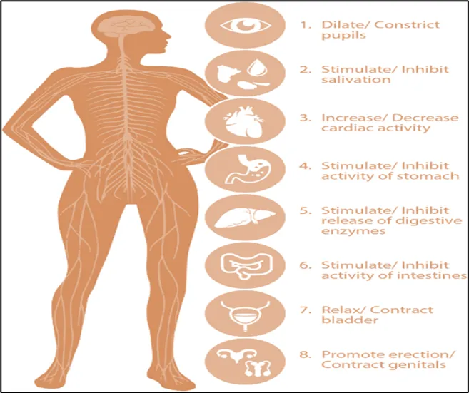
Figure 1. Anatomy showing nervous system control of key organs.

The nervous system is central to controlling the function of all major organs and homeostatic processes in the body and mediated through electric signaling. Some of the body functions controlled by the human nervous system include food intake, dilation of airways, cardiac activity, pancreatic activity, brain activity, bladder control, liver, kidney, and spleen functions, and inflammation. Studies in other model systems have shown that the central nervous system also controls some aspects of embryogenesis [5]and regeneration. Adapted with permission.[6]

  • MECHANISM:

Bioelectric signaling operates at four different levels within the body:

  1. Cellular level bioelectric signaling:

The study of electrical signaling between and within cells that is mediated by changes in the resting membrane potential (Vmem), which is regulated by the movement of ions (K+, Na+, Ca2+, and Cl−) across the plasma membrane, is known as cellular level bioelectric signaling.[7]

Important aspects of cell behaviour, including migration, death, differentiation, and proliferation, are altered by ion channels, which provide bioelectrical signals that are transformed into secondary messenger responses. A fascinating subset of bioelectric signaling at the cellular level is that of organelles and molecules. This electrical signaling, regulated by intracellular signaling pathways, plays a crucial role in preserving homeostasis within organelles such as the nuclear envelope, mitochondria, endoplasmic reticulum, phagosomes, and lysosomes.[8] For example, researchers have identified two separate groups of mitochondria with unique mitochondrial membrane potentials (Δ𝜓m) in both mouse brain stem cells and human dermal stem cells.[9] While high Δ𝜓m stem cells are more likely to differentiate into neurones, low Δ𝜓m cells are quiescent stem cells needed to maintain a neural precursor pool. By only adjusting mitochondrial potential, this could offer an alternative to the present protocols for neural development. The absence of instruments for measuring the voltage gradient across organelle membranes, an area of active method development, contributes to the immature research status of organelle level bioelectric signalling. [10] Through an electro-Fenton effect produced by electric-field-sensitized magnetite, it has been demonstrated that alternating electric fields can break down magnetite-bound protein aggregates in Alzheimer's disease at the molecular level. [11,12] Bioelectric signalling at the cellular level has many uses, such as metabolism, quorum sensing, biofilm development in bacteria, and cancer. [13]

  • APPLICATIONS CELLULAR LEVEL BIOELECTRIC SIGNALING:
  1. Stem Cells:

Recent research on the function of bioelectric signalling in stem cells has shown that endogenous electrical signalling plays a functional role in promoting differentiation and sustaining proliferative states. [14-16] or example, research on the gut of Drosophila has found conserved pathways (like mGluR) that connect intestinal stem cell proliferation to bioelectric regulators of Ca2+, Na+, and Cl−. [17, 18] The large-conductance potassium (BK) channel plays a crucial role in regulating the proliferation, self-renewal, and hyperpolarization-driven differentiation of human mesenchymal and cardiac stem cells. Additionally, recent studies have highlighted the importance of calcium signaling, initiated by mechanosensitive ion channels, in controlling stem cell migration, differentiation, and growth. It has been shown that Vmem promotes the escape from pluripotency and the initiation of germ layer differentiation (cell fate commitment) during embryonic development by means of the calcium and mammalian target of rapamycin (mTOR) signaling pathways. Under the new terminology, therapies that modify this endogenous cellular bioelectric signaling are categorized as cellular bioelectroceuticals. [19,20]

  1. Cancer:

Ion channel dysregulation has long been associated with cancer. [21] The function of resting membrane potential has been investigated more recently, though, and variations in Vmem have been linked to enhanced cell invasion, proliferation, and metastasis [22] (and generally, loss of cooperativity towards normal morphostasis). [23] Individual cancer cells' bioelectrical characteristics are very different from those of normal cells; they have dysregulated ion-channel expression and activity, and their Vmem values are significantly depolarised (−5 to −40 mV compared to −40 to −90 mV in normal epithelial cells). [24] Currently, this research is being expanded to include breast cancer and human glioblastoma (GBM).[25] Cone's classic study has demonstrated a correlation between mitosis and Vmem in somatic cells, with terminally differentiated cells undergoing proliferation due to prolonged depolarisation. [26]

Research involving Xenopus has also influenced advancements in human cell applications. For instance, existing ion-channel drugs have been adapted to modulate the bioelectrical characteristics of GBM cells, leading to suppressed tumor growth and promoting a more differentiated and senescent state in these cells.[27] The Djamgoz lab introduced the "Celex (cellular excitability) hypothesis" of metastasis, suggesting that increased electrical excitability enhances the invasiveness, activity, and disruptive behavior of cancer cells, ultimately contributing to their metastatic potential.[28]

  1. Tissue Level Bioelectric Signaling:

Electrical impulses in the majority of tissues vary depending on whether the tissue is growing, wounded, infected, or benign. For instance, electric currents have been recorded in tumour tissue, wound healing [29], bacterial infections of the gut epithelium [30], and tissue growth. [31]

  • APPLICATIONS OF TISSUE LEVEL BIOELECTRIC SIGNALING:
  1. Wound Healing:

An endogenous electrical field is created across damaged tissue in wounds; the strength of this field varies depending on the kind of tissue. [32, 33] The transepithelial membrane potential collapses to zero at the wound site, in contrast to the unwounded epidermis. This creates an electrical gradient that acts as a signal for directed cell movement into the area of damage, or electrotaxis. [34] The importance of this bioelectric cue is shown by its ability to suppress other directional cues for cell migration in wound healing. [35] When administered to the wound, a redox reaction creates wireless microcurrents that resemble the physiological electric field of the skin, which aids in wound healing and has antibacterial properties. [36, 37]

  1. Tumor Tissue:

Electrical fields known as tumor-treating electrical fields (TTFields) are being introduced in cancer treatment and are locally applied to tumor tissue to prevent the growth of cancer cells and cause them to die. [38,39]  Tumor Treating Fields (TTFields) represent mid-strength alternating electric fields, usually ranging between 1 and 3 volts per centimeter and at mid-range frequencies of between 100 and 300 kHz (100,000 and 300,000 cycles per second). These disciplines are precisely targeted to interfere with the cancer cell functioning without affecting inactive and non-dividing cells. Two theories address the effect that TTFields has on cancerous cells. [40, 41] First, TTFields disrupt the folding of the polar cellular networks, such as tubulin and septin, and affect the correct formation of the mitotic spindle during the metaphase. Subsequently the nonuniform intracellular electric field generated by TTFields when the cell goes cytocinesis modulates polarisable structures such as microtubules, organelles, ions, proteins and DNA. The structural disruption takes place due to the interrupted cell division. Novocure has been working on Tumor Treating Fields (TTFields) since 2000, which have been approved by the FDA as a form of treatment of mesothelioma, newly diagnosed glioblastoma (GBM) and recurrent GBM under the brand names Optune and Optune Lua. As of 2017, Novocure is in clinical trials of combining TTFields with anti-PD-1 antibody KEYTRUDA (pembrolizumab) to treat non-small cell lung cancer as part of the KEYNOTE-B36 clinical trial. [42]

  1. Organ Level Bioelectric Signaling:

Communication between organs is facilitated by electrical signals transmitted through peripheral nerves, which link the organs to the central nervous system and to one another.[43] In order to fix malfunctions in such electric signaling and neuronal circuits, organ level BEMs use electronic devices (such implants or wearables) to deliver corrected electric doses or fields that target specific biochemical systems. As the processes behind diseases are more known, the specificity of BEMs' actions on target pathways actually lies on a continuum that is constantly shifting. Nonetheless, we suggest that these interventions could be categorized as either "broad" or "precise," based on whether they specifically address the neural and molecular mechanisms underlying the disease or simply alleviate its symptoms.[44]

  • Organ Level Bioelectric Signaling Is of Two Types:
    1. Organ Level Broad BEMS:

Broad BEMs are devices that emit non-specific electrical signals to modulate nerve activity, aiming to control symptoms in long-term conditions such as pain and tremors.[45] Broad BEMs, also known as neuromodulatory, neurostimulatory, or nerve stimulation devices, have been on the market for a long time due to their extensive history of altering neuronal activity. Instead of dynamically altering on the millisecond scale, which is essential for achieving precise therapeutic outcomes, broad BEMs usually block or stimulate using stable, simple waveforms. Furthermore, the mechanism of action (MoA) of a number of these treatments is unknown, and there is contradictory information regarding their effectiveness.[46]

    1. Organ Level Precise BEMS:

Precise BEMs are devices that deliver carefully directed electrical pulses to stimulate specific biological pathways. This targeted approach allows for more accurate therapeutic outcomes and may reduce side effects by limiting the activity to a localized area. For instance, the inflammatory reflex, which uses the Vagus nerve to transfer neural information, recognises and reacts to inflammation and cytokine production in order to preserve immune system homeostasis. [47]This neurological reaction can be modulated, inflammatory cytokines can be suppressed, and immunological homeostasis may be restored by sending precise electrical pulses down the Vagus nerve at particular points.[48] It is anticipated that specifically targeting the inflammatory reflex will be safer and less immunosuppressive than systemic anti-inflammatory drugs. [49] To address inflammation, certain BEMs focus on stimulating the auricular, cervical, splenic, or pancreatic branches of the vagus nerve, which are involved in regulating biological pathways that influence inflammation and cytokine production in chronic autoimmune disorders. For instance, SetPoint Medical Inc. is conducting a Phase 2 pilot study for rheumatoid arthritis and a proof-of-concept trial for Crohn’s disease, both under FDA breakthrough designation. [50,51] Galvani Bioelectronics is conducting proof of concept research in RA that focusses on the splenic nerve. [52] Electrical stimulation of cardiovascular reflexes is another application for precise BEMs. One example is treating hypertension by focussing on The baroreflex, which is sensitive to blood pressure (Kyushu University).[53] as well as cardiac failure (CVRx). [54,55]

  1. Systems Level Bioelectric Signaling:

Voltage gradients define details like axial polarity, patterning, and organ identification (size and shape) at the organism level. [56] and are essential for embryonic growth, remodelling, and regeneration. [57,58] It has been suggested that these system-level patterning effects are governed by the morphogenetic code, which operates above the genetic and molecular levels. This code directs gene expression and cellular behavior in a top-down manner, guiding the development of specific organ-level structures and function.[59]

  • EVOLUTION OF ELECTROCEUTICALS:

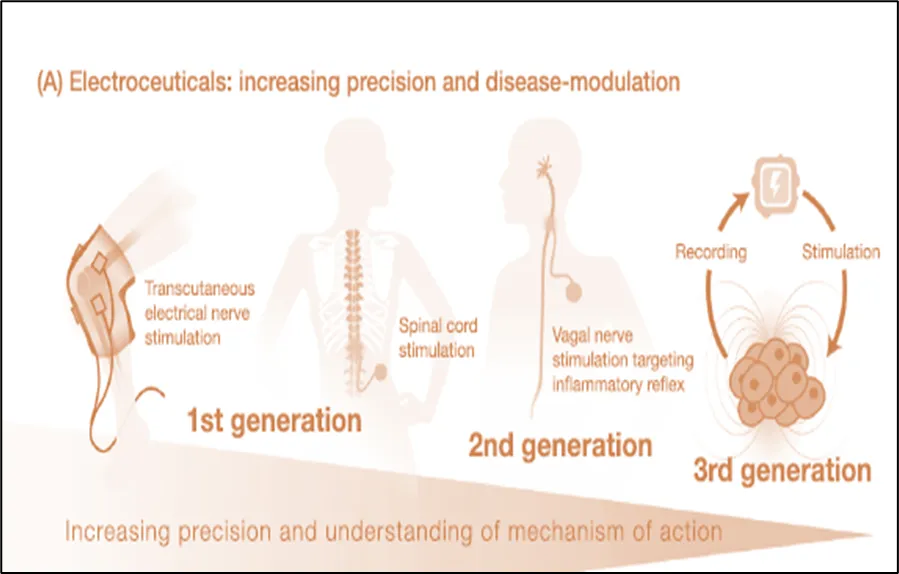
Figure 2. Evolution of electroceuticals.

  1. First Generation Electroceuticals:

It includes a set of well-known medical treatments that use electric current to create an effect, such as deep brain stimulation, transcutaneous electrical nerve stimulation, cardiac defibrillators, cardiac pacemakers, cochlear and retinal implants, and spinal cord stimulation.[60] Although these treatments are therapeutically effective, they are comparatively broad-acting and focus on excitable tissues rather than particular nerve fibres or bundles. They employ basic waveforms that are not specifically designed according to the mechanism of action, aiming to generally reduce symptoms without a complete understanding of the biological processes involved.[61]

  1. Second Generation Electroceuticals:

This is aimed at altering disease with more targeted electro modulation. An instance of this with more knowledge of the principles of biology is the utilisation of miniature devices that aim a section of nerve fibbers or bundles [62] The clinical trials relating to the scope of these devices extend beyond neurological issues and is more on the similarity to the pharmaceutical drug development. [62] A good example of this genre is the splenic stimulation equipment of Galvani [63] and the Microregulators system by Setpoint [62].  Although vague nerve stimulation (VNS) is not a new technique (the VNS devices first emerged more than 30 years ago) and some of the VNS devices (like transcutaneous vagus nerve stimulators) are considered first-generation devices, the two devices address the vagus nerve with increasing specificity and are based on a better understanding of the neural molecular inflammatory pathways. [62,63] The experimental discovery of Kevin Tracey and Paul Peter Tak into the vagal inflammatory reflex gave the first indication that the nervous system is actively and reflexively regulating inflammation when it takes place [64] Genovese et al. validated this in research controlled by sham that they conducted and they showed that the likelihood of a response to treatment. [65]  In order to address some of these challenges, however, new modalities of therapeutic guiding are currently being studied, these encompass closed-loop and synchronized electrical stimulation, neuronal and non-neuronal biomarker use. Should there be positive findings out of these next-generation device trials properly powered, it is hoped that it will provide essential clinical validation in the future. [60,66]

  1. Third Generation Electroceuticals:

More precisely, third-generation electroceuticals use a technique to target individual cells or particular nerve fibers.[60] Third-generation electroceuticals are wireless, closed-loop, and frequently biodegradable or miniature bioelectronic systems that stimulate neuronal and non-neural tissues precisely and in real time, allowing for disease-modifying treatments that go beyond symptom management. [66]

  • THE PROSPECTS OF BIOELECTRIC THERAPY:

A Comprehensively Adaptive Method

The future of personalised bioelectric therapy is hinted at by today's closed-loop devices, which will employ gathered data to apply neural stimulations more precisely in order to produce therapeutic results. [67]

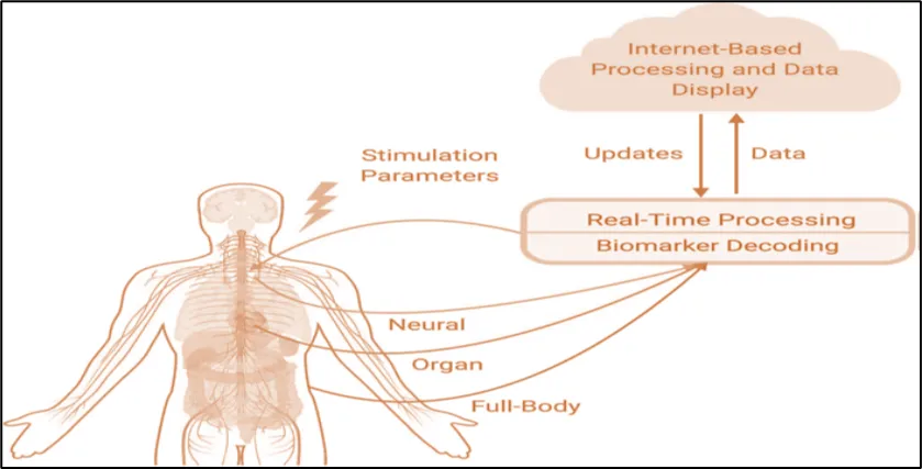
Figure 3. Actual and longer-term processing of neuronal, organ, and full-body state data will be used in personalized bioelectric therapy in the future to adjust stimulation levels for the best possible therapeutic results.[68]

Devices will be able to generate a consistent and customized therapeutic response when stimulation parameters are controlled by combining neuronal, organ, and full-body state data. Devices that convert collected data into comprehensible biomarkers and patient status evaluations that control given stimulation are still being developed. In order to achieve this, there are many obstacles related to implantable and Computer-based technologies, biomedical analysis of neural signals, and the engineering of medical devices. These devices must be able to record, decode, and control neurological, physiological, and state biomarkers over an extended period of time. In an effort to reach this future generation, various commercial and scientific organisations are tackling these issues from various perspectives. A configurable approach to system design research is used by the Dynamic Neuro-Modulator (DyNeuMo) is an adaptable brain stimulation device developed through a partnership between Bioinduction, Imperial College, and Oxford University. This system enables adjustment of stimulation settings based on multiple layers of patient data by combining population-level neural signals with broader contextual information, such as circadian cycles or movement patterns detected by accelerometers.[69] Companies such as Neuralink have invested heavily in developing advanced implantable technologies. Neuralink has engineered a high-density electrode array along with a specialized surgical robot to facilitate accurate, scalable, and long-term high-bandwidth implantation. Their objective is to build brain–machine interfaces with high data transfer capacity, suitable for both medical and non-medical uses.[70]

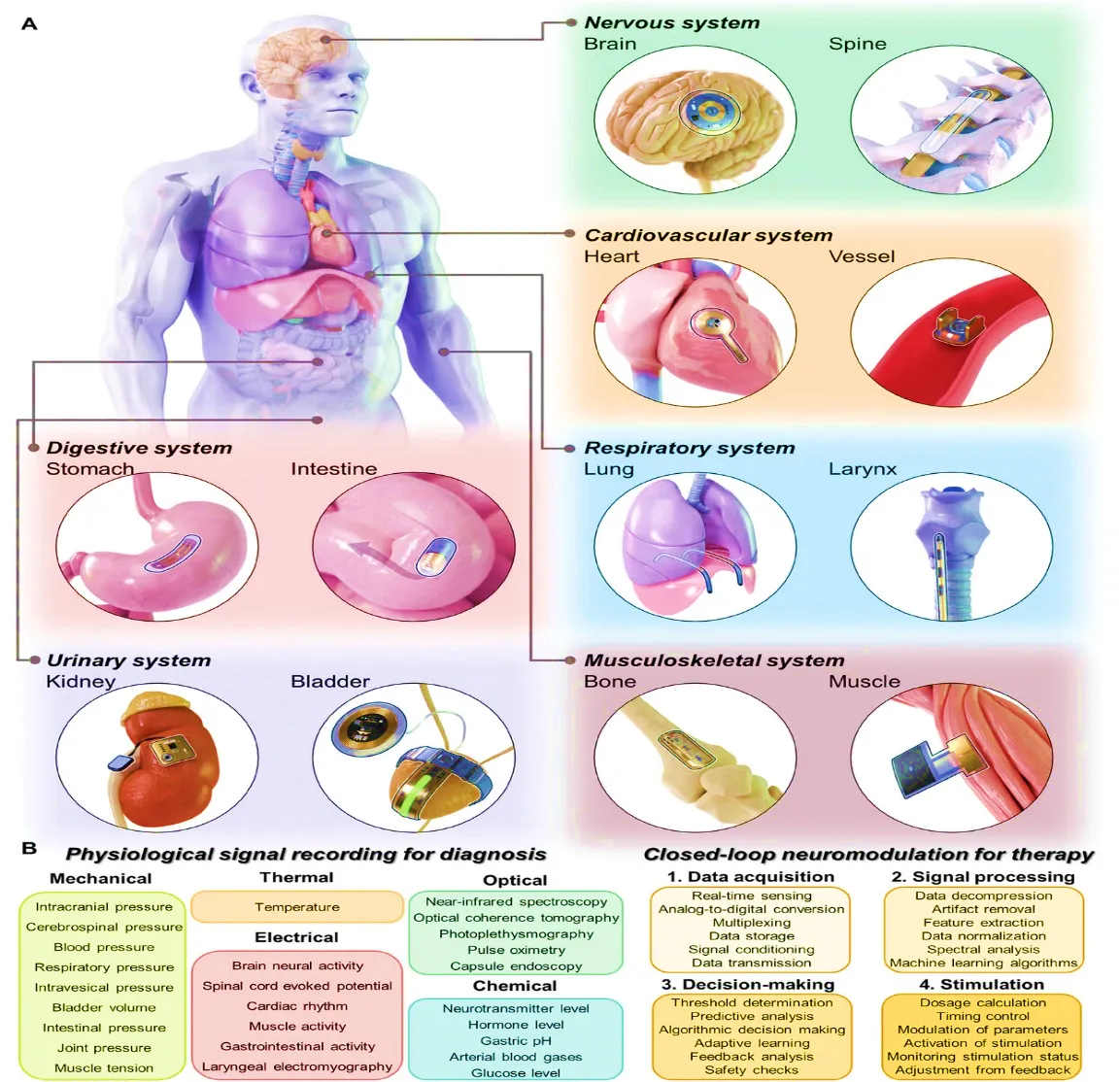
Figure. 4 Implanted bioelectronic devices tailored to a particular organ.

Categorization of implantable monitoring and therapeutic devices for the neurological, cardiovascular, respiratory, digestive, urinary, and musculoskeletal systems, among other organ systems.[71]

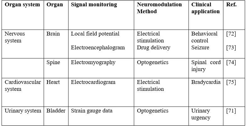
Table 1. Clinical applications for implantable bioelectronic devices with closed loops.

  • Therapeutic Implantable Neuromodulation Devices:

Figure 5. Devices for therapeutic closed-loop neuromodulation that are implanted. A) Diagram illustrating open-loop systems, conventional closed-loop systems, and AI-driven closed-loop systems.[75

  1. Closed Loop:

The implementation sequence is a multi-phase, intricate process that includes a number of technological and computational elements to guarantee the system's efficacy and flexibility. First, data collecting entails physiological signal sensing in real time, which is essential for closed-loop systems. Analog-to-digital conversion is the next step in transforming the obtained analogue signals into digital form. Signal conditioning is used to improve the signals for precise interpretation, and multiplexing is used to manage many signals. [76] These signals have finally been analysed, stored, and sent for additional examination. [77] The next crucial step is signal processing, which involves thoroughly processing raw data. [78] Data decompression is used to control data complexity and size. By removing noise and unnecessary information, artefact removal preserves the accuracy of the data. To find particular aspects of the gathered signals that are necessary for precise analysis, feature extraction is carried out. By standardising the data, data normalisation enables cross-session or cross-time comparability. [79] Spectral analysis aids in the comprehension of the signals' frequency components, and machine learning algorithms are used to analyse and learn from the data, improving the system's capacity for decision-making and prediction. The next step is decision-making, where the system makes therapeutic decisions by interpreting the data it has analysed. Setting the parameters or boundaries for stimulation is known as threshold determination. The efficient operation of a closed loop neuromodulation system depends on the intricate connections between each of these phases. [78] Choi et al. provided examples of advanced conventional closed-loop systems, including a transient closed-loop system that combines a bioresorbable pacemaker for autonomous electrotherapy with a network of wireless body-integrated devices. Studies involving the hearts of rats, canines, and humans have shown that this system is capable of autonomous rate-adaptive pacing in cardiac pacing. [75]

  1. Open Loop System:

An open-loop system consists of sensors that detect physiological signals and stimulators that deliver neuromodulation without feedback control. These systems lack the ability to adapt or provide feedback in real time and function according to preset parameters. The foundation for more sophisticated neuromodulation methods has been established by open-loop systems. By adding a feedback mechanism, conventional closed-loop systems offer a substantial improvement over open -loop systems. In these systems, a processor processes physiological signals that are picked up by sensors. A controller receives the feedback and stimulation parameters that are set using the processed data. The system may adapt its stimulation in response to real-time physiological data thanks to this controller. This feedback loop is essential for increasing the system's adaptability since it enables the stimulation parameters to be changed in response to shifting physiological demands. [71,74]

  1. AI-Based Closed-Loop Systems:

Artificial intelligence (AI) greatly enhances closed-loop systems' performance for effective and secure neuromodulation treatment. Following initial signal sensing and processing, the data helps build big data in AI-based closed-loop systems. AI systems learn from this data and continuously improve the system's reaction, frequently using neural networks. Through this process, the system is able to make better decisions and optimise the controller's output. A level of accuracy and customisation that was previously impossible is made possible by AI's capacity to evaluate enormous datasets and learn from intricate patterns. With the combination of multidisciplinary science and engineering, each of these evolutionary stages shows a stronger integration of technology. The transition from static, preset systems to dynamic, learning systems highlights how technology can be used to treat and control a variety of diseases in an adaptive manner in addition to interacting with biological systems. [80,81,82]

  • TYPES OF ELECTROCEUTICAL AND BIOELECTRIC THERAPY:
  1. Electroconvulsive Therapy (ECT):

Through electrodes positioned at certain points on the anaesthetised patient's head, ECT creates a sequence of electrical currents that cause a seizure. ECT is often administered twice or three times per week, with a total of six to nine depression therapy sessions. [75,76]and ten to twenty schizophrenia sessions [77,78].More than 70% of individuals receiving ECT are diagnosed with severe depression and usually do not respond to standard treatments for the condition.[79]Acute mania, malignant catatonia, and schizophrenia are further neuropsychiatric disorders that may be treated with ECT.[80]According to earlier research examining the mechanism of ECT, patients exhibited a) an elevated seizure threshold during or following ECT therapy,[81]as well as b) the hypothalamic-pituitary-adrenal (HPA) axis being downregulated,[82]which, respectively, are neurophysiological alterations linked to the effects of antidepressants and anticonvulsants. A meta-analysis of seven controlled clinical trials conducted between 1956 and 2003 found consistent evidence that ECT is more effective than other treatments, including placebos and antidepressant medications. The analysis revealed that patients were about 11 times more likely to respond to ECT compared to a placebo (OR = 11.08, 95% CI 3.10–39.65) and approximately four times more likely to respond than those receiving antidepressant drugs (OR = 3.72, 95% CI 2.60–5.32).[83]

  • Advantages Of Electroconvulsive Therapy:
  1. The primary therapeutic advantage of electroconvulsive therapy (ECT) is that patients typically experience rapid symptom alleviation. In 1982, Gangadhar and associates [84] compared the effectiveness of imipramine and ECT in treating endogenous depression in a randomised controlled trial.
  2. Although ECT had fewer side effects and had a significantly faster effect, both therapies were equally effective at reducing depressed symptoms. Another study that looked into the effectiveness of ECT for psychotic severe depression found that 95% of patients experienced remission after just 10 to 14 days after starting treatment.[85]
  3. Given that severe psychotic symptoms of sadness sometimes result in potentially fatal situations like increased suicidal thoughts or food refusal,[86]
  4. For severe or acute mental illnesses that require immediate intervention to alleviate symptoms, electroconvulsive therapy (ECT) may be helpful as a first-line treatment. Furthermore, people who experience negative side effects from any medicine or who are unable to take specific medications (such as nursing mothers) may find ECT to be a helpful alternative. This may also hold true for people with depression who are physically unwell and who are more likely to suffer from deteriorating somatic symptoms as a side effect of antidepressant drugs.[87]
  5. ECT is typically advised as a monotherapy in situations where the side effects of psychopharmacological therapies are unbearable.[88]
  1. Vagus Nerve Stimulation (VNS):

The term "VNS" describes a treatment method that applies intermittent electrical pulses to the vagus nerve, a mixed parasympathetic nerve composed of approximately 80% sensory (afferent) fibers and 20% motor (efferent) fibers, which is located on both sides of the neck. [89,90]Through its afferent fibers, the VNS primarily transmits bodily inputs, including visceral sensory and gustatory information, either directly or indirectly to brain regions such the orbitofrontal cortex, amygdala, and hypothalamus. [91,92]Since changes in these brain regions are often suggestive of a range of neuropsychiatric disorders, stimulating these circuits has been suggested as an alternative treatment for patients with refractory epilepsy and treatment-resistant depression. in 1997 as well as 2005. In order to stimulate the left vagus nerve via a wire, a pulse generator is often implanted on the patient's left upper chest during an invasive VNS procedure.[93]Applying electrical stimulation to the left cymba conchae of the outer ear (transcutaneous auricular vagus nerve stimulation, or ta-VNS) or the neck (non-invasive vagus nerve stimulation, or n-VNS) is another non-invasive technique for VNS.[94] The cymba conchae area receives the vagus nerve's auricular branch.[95] When compared to the effects of the conventional invasive form of VNS, previous research has shown that ta-VNS has a similar effect on the brain. [96,97]

  • Advantages Of VNS:
  1. The main advantage of ta-VNS is that it provides a non-invasive method to deliver VNS by specifically stimulating the auricular branch of the vagus nerve (ABVN), which supplies the outer ear and is easily accessible. Additionally, while mild skin irritation or ulceration are the most common side effects, ta-VNS treatment has not been associated with any serious adverse effects.[98]
  2. The fact that VNS can be used for a variety of conditions other than depression and epilepsy is another benefit. Previous research has demonstrated that VNS has an anti-inflammatory impact by Inhibiting the secretion of proinflammatory cytokines like tumor necrosis factor-alpha (TNF-α) and interleukin-6 (IL-6).[99-101]
  3.  Consequently, VNS may be used therapeutically to treat a range of inflammatory disorders, such as stroke, diabetes, sepsis, obesity, and pain management.[102]
  4. Deep Brain Stimulation (DBS):

Deep Brain Stimulation (DBS) is a surgical technique in which electrode leads are implanted into targeted areas of the brain through small openings in the skull, guided by neuroimaging and stereotactic methods. These electrodes are connected to an implanted pulse generator (IPG) placed under the skin, which delivers electrical stimulation to the brain.The IPG's stimulation parameters, which are normally set at 60–130 Hz, 2–10 V, and 60–200 ms pulse width, can be externally changed by clinicians.[103] When it comes to treating Parkinson's disease, DBS has received FDA approval and has largely taken the place of ablative neurosurgery in patients with movement abnormalities such essential tremor and dystonia.[104] DBS has also shown promise in treating depression that is resistant to therapy.[105,106] and OCD (obsessive-compulsive disorder) [107,108] For the treatment of Parkinson's disease, the subthalamic nucleus (STN) and the internal segment of the globus pallidus (GPi), both components of the basal ganglia, are commonly targeted areas in the brain. [109,110]

  • Advantages Of DBS:
  1. Because DBS has the benefits of reversibility and adaptability, it has surpassed surgery in treating lesioning for movement disorders.[111]
  2. After surgical implantation, stimulation parameters including location, intensity, and current field shape can be altered to suit specific requirements (e.g., medication adjustments).[112]
  3. Through monitoring, clinicians can quickly detect any adverse effects that may have arisen from the inadvertent activation of brain regions around the target area. These DBS features make it feasible to gradually lessen potential side effects or improve therapeutic efficacy.[113]
  • LIMITATIONS:
  1. Muscle discomfort, nausea, and postictal headache are the most often reported physical side effects of electroconvulsive therapy. Up to 45% of ECT patients report having headaches. [114]
  2. Cognitive impairments, such as temporary confusion immediately following ECT treatment due to the effects of seizures and anaesthesia, and retrograde memory loss, which is characterised by a severe, ongoing inability to date back previous information, are occasionally described in addition to physical side effects. [115]
  3. Headaches, dyspnea, lower facial paralysis, coughing, and voice alterations are often reported side effects during stimulation. The high upfront expenditures of hospitalization, implantation surgery, and device procurement are another disadvantage of invasive VNS procedures. [116,117]
  4. Intracerebral haemorrhages, haematomas, paralysis, infection, electrode displacement, and skin erosion are among the problems associated with surgery and hardwires.[118]

Although they are less severe than those caused by surgery, stimulation-induced side effects like headaches, ocular deviations, muscular contractions, discomfort, and dysarthria are more frequently described. Frequent follow-up visits, the high expense of the implantation surgery, and battery maintenance are additional concerns that frequently strain patients receiving DBS treatment. [119]

CONCLUSION

The growing complexity of electroceutical technologies introduces distinct challenges from a drug development perspective and marks a significant shift in therapeutic approaches. To advance this field, there is a strong need for researchers to deepen their understanding of the natural bioelectric code that influences tissue and organ-level organization, and to develop more effective methods for modulating both neural and non-neural bioelectric states in humans and preclinical models.

Looking ahead, electroceuticals hold great potential to accurately diagnose and treat a wide range of diseases, promote tissue and organ regeneration, and possibly reduce the impact of aging. Their long-term commercial success will rely on targeting conditions with significant unmet medical needs, where electroceuticals can clearly outperform existing treatments in terms of durability, safety, and effectiveness while also addressing factors like ease of use, intellectual property protection, scalability, cost, and reimbursement strategies.

Realizing this vision requires a more comprehensive understanding of the foundational principles of bioelectric function and how it conveys meaningful biological information across different levels of physiological organization, as outlined in the referenced ontology, along with the interconnections between those levels.

REFERENCES

  1. Mishra S. Electroceuticals in medicine–The brave new future. Indian heart journal. 2017 Sep 1;69(5):685-6.
  2. Levin M, Selberg J, Rolandi M. Endogenous bioelectrics in development, cancer, and regeneration: drugs and bioelectronic devices as electroceuticals for regenerative medicine. Iscience. 2019 Dec 20;22:519-33.
  3. Pai VP, Cervera J, Mafe S, Willocq V, Lederer EK, Levin M. HCN2 channel-induced rescue of brain teratogenesis via local and long-range bioelectric repair. Frontiers in Cellular Neuroscience. 2020 May 26;14:136.
  4. Nuccitelli R. Application of pulsed electric fields to cancer therapy. Bioelectricity. 2019 Mar 1;1(1):30-4.
  5. Herrera-Rincon C, Levin M. Booting up the organism during development: Pre-behavioral functions of the vertebrate brain in guiding body morphogenesis. Communicative & integrative biology. 2018 Jan 2;11(1):e1433440.
  6. Kumar A, Brockes JP. Nerve dependence in tissue, organ, and appendage regeneration. Trends in neurosciences. 2012 Nov 1;35(11):691-9.
  7. Klier PE, Gest AM, Martin JG, Roo R, Navarro MX, Lesiak L, Deal PE, Dadina N, Tyson J, Schepartz A, Miller EW. Bioorthogonal, fluorogenic targeting of voltage-sensitive fluorophores for visualizing membrane potential dynamics in cellular organelles. Journal of the American Chemical Society. 2022 Jul 1;144(27):12138-46.
  8. Liu H, He Z, April SL, Trefny MP, Rougier JS, Salemi S, Olariu R, Widmer HR, Simon HU. Biochemical re-programming of human dermal stem cells to neurons by increasing mitochondrial membrane potential. Cell Death & Differentiation. 2019 Jun;26(6):1048-61.
  9. Matamala E, Castillo C, Vivar JP, Rojas PA, Brauchi SE. Imaging the electrical activity of organelles in living cells. Communications biology. 2021 Mar 23;4(1):389.
  10. Choi Y, Lee WS, Lee J, Park SH, Kim S, Kim KH, Park S, Kim EH, Kim JK. Capacitive electrode-based electric field treatments on redox-toxic iron deposits in transgenic AD mouse models: the electroceutical targeting of Alzheimer’s disease feasibility study. International journal of molecular sciences. 2023 May 31;24(11):9552.
  11. Bok J, Ha J, Ahn BJ, Jang Y. Disease-modifying effects of non-invasive electroceuticals on β-amyloid plaques and tau tangles for Alzheimer’s disease. International Journal of Molecular Sciences. 2022 Dec 30;24(1):679.
  12. Robinson AJ, Jain A, Sherman HG, Hague RJ, Rahman R, Sanjuan‐Alberte P, Rawson FJ. Toward hijacking bioelectricity in cancer to develop new bioelectronic medicine. Advanced Therapeutics. 2021 Mar;4(3):2000248.
  13. Bhavsar MB, Leppik L, Costa Oliveira KM, Barker JH. Role of bioelectricity during cell proliferation in different cell types. Frontiers in bioengineering and biotechnology. 2020 Jul 2;8:603.
  14. Sundelacruz S, Moody AT, Levin M, Kaplan DL. Membrane potential depolarization alters calcium flux and phosphate signaling during osteogenic differentiation of human mesenchymal stem cells. Bioelectricity. 2019 Mar 1;1(1):56-66.
  15. Fennelly C, Soker S. Bioelectric properties of myogenic progenitor cells. Bioelectricity. 2019 Mar 1;1(1):35-45.
  16. Petsakou A, Perrimon N. Bioelectric regulation of intestinal stem cells. Trends in cell biology. 2023 Jul 1;33(7):555-67.
  17. Deng H, Gerencser AA, Jasper H. Signal integration by Ca2+ regulates intestinal stem-cell activity. Nature. 2015 Dec 10;528(7581):212-7.
  18. Pchelintseva E, Djamgoz MB. Mesenchymal stem cell differentiation: Control by calcium‐activated potassium channels. Journal of Cellular Physiology. 2018 May;233(5):3755-68.
  19. Sempou E, Kostiuk V, Zhu J, Cecilia Guerra M, Tyan L, Hwang W, Camacho-Aguilar E, Caplan MJ, Zenisek D, Warmflash A, Owens ND. Membrane potential drives the exit from pluripotency and cell fate commitment via calcium and mTOR. Nature communications. 2022 Nov 5;13(1):6681.
  20. Litan A, Langhans SA. Cancer as a channelopathy: ion channels and pumps in tumor development and progression. Frontiers in cellular neuroscience. 2015 Mar 17;9:86.
  21. Ribeiro M, Elghajiji A, Fraser SP, Burke ZD, Tosh D, Djamgoz MB, Rocha PR. Human breast cancer cells demonstrate electrical excitability. Frontiers in neuroscience. 2020 Apr 30;14:404.
  22. Levin M. Bioelectrical approaches to cancer as a problem of the scaling of the cellular self. Progress in biophysics and molecular biology. 2021 Oct 1;165:102-13.
  23. Levin M. Molecular bioelectricity in developmental biology: new tools and recent discoveries: control of cell behavior and pattern formation by transmembrane potential gradients. Bioessays. 2012 Mar;34(3):205-17.
  24. Payne SL, Ram P, Srinivasan DH, Le TT, Levin M, Oudin MJ. Potassium channel-driven bioelectric signalling regulates metastasis in triple-negative breast cancer. EBioMedicine. 2022 Jan 1;75.
  25. Cone Jr CD. Unified theory on the basic mechanism of normal mitotic control and oncogenesis. Journal of Theoretical Biology. 1971 Jan 1;30(1):151-81.
  26. Mathews J, Kuchling F, Baez-Nieto D, Diberardinis M, Pan JQ, Levin M. Ion channel drugs suppress cancer phenotype in NG108-15 and U87 cells: toward novel electroceuticals for glioblastoma. Cancers. 2022 Mar 15;14(6):1499.
  27. Djamgoz MB, Fraser SP, Brackenbury WJ. In vivo evidence for voltage-gated sodium channel expression in carcinomas and potentiation of metastasis. Cancers. 2019 Oct 28;11(11):1675.
  28. Reid B, Zhao M. The electrical response to injury: molecular mechanisms and wound healing. Advances in wound care. 2014 Feb 1;3(2):184-201.
  29. Sun Y, Reid B, Ferreira F, Luxardi G, Ma L, Lokken KL, Zhu K, Xu G, Sun Y, Ryzhuk V, Guo BP. Infection-generated electric field in gut epithelium drives bidirectional migration of macrophages. PLoS biology. 2019 Apr 9;17(4):e3000044.
  30. Zhu K, Hum NR, Reid B, Sun Q, Loots GG, Zhao M. Electric Fields at Breast Cancer and Cancer Cell Collective Galvanotaxis. Sci. Rep. 10, 8712 [Internet]. 2020
  31. Jaffe LF, Vanable Jr JW. Electric fields and wound healing. Clinics in dermatology. 1984 Jul 1;2(3):34-44.
  32. Zhao M, Chalmers L, Cao L, Vieira AC, Mannis M, Reid B. Electrical signaling in control of ocular cell behaviors. Progress in retinal and eye research. 2012 Jan 1;31(1):65-88.
  33. Zhao Y, Garcia BA. Cold Spring Harbor Perspect. Biol. 2015;7:a025064.
  34. Zhao M. Electrical fields in wound healing—an overriding signal that directs cell migration. InSeminars in cell & developmental biology 2009 Aug 1 (Vol. 20, No. 6, pp. 674-682). Academic Press.
  35. Banerjee J, Das Ghatak P, Roy S, Khanna S, Sequin EK, Bellman K, Dickinson BC, Suri P, Subramaniam VV, Chang CJ, Sen CK. Improvement of human keratinocyte migration by a redox active bioelectric dressing. PLoS One. 2014 Mar 3;9(3):e89239.
  36. Banerjee J, Ghatak PD, Roy S, Khanna S, Hemann C, Deng B, Das A, Zweier JL, Wozniak D, Sen CK. Silver-zinc redox-coupled electroceutical wound dressing disrupts bacterial biofilm. PloS one. 2015 Mar 24;10(3):e0119531.
  37. Kirson ED, Gurvich Z, Schneiderman R, Dekel E, Itzhaki A, Wasserman Y, Schatzberger R, Palti Y. Disruption of cancer cell replication by alternating electric fields. Cancer research. 2004 May 1;64(9):3288-95.
  38. Kirson ED, Dbalý V, Tovaryš F, Vymazal J, Soustiel JF, Itzhaki A, Mordechovich D, Steinberg-Shapira S, Gurvich Z, Schneiderman R, Wasserman Y. Alternating electric fields arrest cell proliferation in animal tumor models and human brain tumors. Proceedings of the National Academy of Sciences. 2007 Jun 12;104(24):10152-7.
  39. Carrieri FA, Smack C, Siddiqui I, Kleinberg LR, Tran PT. Tumor treating fields: at the crossroads between physics and biology for cancer treatment. Frontiers in oncology. 2020 Oct 30;10:575992.
  40. Tuszynski JA, Wenger C, Friesen DE, Preto J. An overview of sub-cellular mechanisms involved in the action of TTFields. International journal of environmental research and public health. 2016 Nov;13(11):1128.
  41. Novocure, Novocure Announces Clinical Trial Collaboration with MSD to Evaluate Tumor Treating Fields Together with KEYTRUDA (pembrolizumab) in Non-Small Cell Lung Cancer, Press Release, 2020.
  42. Horn CC, Ardell JL, Fisher LE. Electroceutical targeting of the autonomic nervous system. Physiology. 2019 Mar 1;34(2):150-62.
  43. Cracchiolo M, Ottaviani MM, Panarese A, Strauss I, Vallone F, Mazzoni A, Micera S. Bioelectronic medicine for the autonomic nervous system: clinical applications and perspectives. Journal of Neural Engineering. 2021 Mar 17;18(4):041002.
  44. Knotkova H, Hamani C, Sivanesan E, Le Beuffe MF, Moon JY, Cohen SP, Huntoon MA. Neuromodulation for chronic pain. The Lancet. 2021 May 29;397(10289):2111-24.
  45. Jensen MP, Brownstone RM. Mechanisms of spinal cord stimulation for the treatment of pain: still in the dark after 50 years. European journal of pain. 2019 Apr;23(4):652-9.
  46. Koopman FA, Schuurman PR, Vervoordeldonk MJ, Tak PP. Vagus nerve stimulation: a new bioelectronics approach to treat rheumatoid arthritis?. Best practice & research Clinical rheumatology. 2014 Aug 1;28(4):625-35.
  47. Pavlov VA, Chavan SS, Tracey KJ. Bioelectronic medicine: from preclinical studies on the inflammatory reflex to new approaches in disease diagnosis and treatment. Cold Spring Harbor perspectives in medicine. 2020 Mar 1;10(3):a034140.
  48. Toffa DH, Touma L, El Meskine T, Bouthillier A, Nguyen DK. Learnings from 30 years of reported efficacy and safety of vagus nerve stimulation (VNS) for epilepsy treatment: a critical review. Seizure. 2020 Dec 1;83:104-23.
  49. Koopman FA, Chavan SS, Miljko S, Grazio S, Sokolovic S, Schuurman PR, Mehta AD, Levine YA, Faltys M, Zitnik R, Tracey KJ. Vagus nerve stimulation inhibits cytokine production and attenuates disease severity in rheumatoid arthritis. Proceedings of the National Academy of Sciences. 2016 Jul 19;113(29):8284-9.
  50. Levine YA, Faltys M, Chernoff D. Harnessing the inflammatory reflex for the treatment of inflammation-mediated diseases. Cold Spring Harbor Perspectives in Medicine. 2020 Jan 1;10(1):a034330.
  51. Sokal DM, McSloy A, Donegà M, Kirk J, Colas RA, Dolezalova N, Gomez EA, Gupta I, Fjordbakk CT, Ouchouche S, Matteucci PB. Splenic nerve neuromodulation reduces inflammation and promotes resolution in chronically implanted pigs. Frontiers in immunology. 2021 Mar 29;12:649786.
  52. Abraham WT, Zile MR, Weaver FA, Butter C, Ducharme A, Halbach M, Klug D, Lovett EG, Müller-Ehmsen J, Schafer JE, Senni M. Baroreflex activation therapy for the treatment of heart failure with a reduced ejection fraction. JACC: Heart Failure. 2015 Jun;3(6):487-96.
  53. Stavrakis S, Stoner JA, Humphrey MB, Morris L, Filiberti A, Reynolds JC, Elkholey K, Javed I, Twidale N, Riha P, Varahan S. TREAT AF (transcutaneous electrical Vagus nerve stimulation to suppress atrial fibrillation) a randomized clinical trial. Clinical Electrophysiology. 2020 Mar 1;6(3):282-91.
  54. Lanni JS, Peal D, Ekstrom L, Chen H, Stanclift C, Bowen ME, Mercado A, Gamba G, Kahle KT, Harris MP. Integrated K+ channel and K+ Cl-cotransporter functions are required for the coordination of size and proportion during development. Developmental biology. 2019 Dec 15;456(2):164-78.
  55. Harris MP. Bioelectric signaling as a unique regulator of development and regeneration. Development. 2021 May 15;148(10):dev180794.
  56. Bates E. Ion channels in development and cancer. Annual review of cell and developmental biology. 2015 Nov 13;31(1):231-47.
  57. Levin M, Martyniuk CJ. The bioelectric code: An ancient computational medium for dynamic control of growth and form. Biosystems. 2018 Feb 1;164:76-93.
  58. Lee SK, Jeakins GS, Tukiainen A, Hewage E, Armitage OE. Next-generation bioelectric medicine: harnessing the therapeutic potential of neural implants. Bioelectricity. 2020 Dec 1;2(4):321-7.
  59. Knotkova H, Hamani C, Sivanesan E, Le Beuffe MF, Moon JY, Cohen SP, Huntoon MA. Neuromodulation for chronic pain. The Lancet. 2021 May 29;397(10289):2111-24.
  60. Genovese MC, Gaylis NB, Sikes D, Kivitz A, Horowitz DL, Peterfy C, Glass EV, Levine YA, Chernoff D. Safety and efficacy of neurostimulation with a miniaturised vagus nerve stimulation device in patients with multidrug-refractory rheumatoid arthritis: a two-stage multicentre, randomised pilot study. The Lancet Rheumatology. 2020 Sep 1;2(9):e527-38.
  61. Sokal DM, McSloy A, Donegà M, Kirk J, Colas RA, Dolezalova N, Gomez EA, Gupta I, Fjordbakk CT, Ouchouche S, Matteucci PB. Splenic nerve neuromodulation reduces inflammation and promotes resolution in chronically implanted pigs. Frontiers in immunology. 2021 Mar 29;12:649786.
  62. Tracey KJ. The inflammatory reflex. Nature. 2002 Dec 19;420(6917):853-9.
  63. Ramadi KB, Srinivasan SS, Traverso G. Electroceuticals in the gastrointestinal tract. Trends in pharmacological sciences. 2020 Dec 1;41(12):960-76.
  64. Li S, Zhu H, Chen JD. Intestinal electrical stimulation synchronized with intestinal slow wave ameliorates glucagon-induced hyperglycemia in rats. Neuromodulation: Technology at the Neural Interface. 2024 Feb 1;27(2):312-20.
  65. Ardell JL, Nier H, Hammer M, Southerland EM, Ardell CL, Beaumont E, KenKnight BH, Armour JA. Defining the neural fulcrum for chronic vagus nerve stimulation: implications for integrated cardiac control. The Journal of physiology. 2017 Nov 15;595(22):6887-903.
  66. Lee SK, Jeakins GS, Tukiainen A, Hewage E, Armitage OE. Next-generation bioelectric medicine: harnessing the therapeutic potential of neural implants. Bioelectricity. 2020 Dec 1;2(4):321-7.
  67. Zamora M, Toth R, Morgante F, Ottaway J, Gillbe T, Martin S, Lamb G, Noone T, Benjaber M, Nairac Z, Sehgal D. DyNeuMo Mk-1: Design and pilot validation of an investigational motion-adaptive neurostimulator with integrated chronotherapy. Experimental neurology. 2022 May 1;351:113977.
  68. Musk E. An integrated brain-machine interface platform with thousands of channels. Journal of medical Internet research. 2019 Oct 31;21(10):e16194.
  69. Mickle AD, Won SM, Noh KN, Yoon J, Meacham KW, Xue Y, McIlvried LA, Copits BA, Samineni VK, Crawford KE, Kim DH. A wireless closed-loop system for optogenetic peripheral neuromodulation. Nature. 2019 Jan 17;565(7739):361-5.
  70. Zhou A, Santacruz SR, Johnson BC, Alexandrov G, Moin A, Burghardt FL, Rabaey JM, Carmena JM, Muller R. A wireless and artefact-free 128-channel neuromodulation device for closed-loop stimulation and recording in non-human primates. Nature biomedical engineering. 2019 Jan;3(1):15-26.
  71. Ouyang W, Lu W, Zhang Y, Liu Y, Kim JU, Shen H, Wu Y, Luan H, Kilner K, Lee SP, Lu Y. A wireless and battery-less implant for multimodal closed-loop neuromodulation in small animals. Nature Biomedical Engineering. 2023 Oct;7(10):1252-69.
  72. Kathe C, Michoud F, Schönle P, Rowald A, Brun N, Ravier J, Furfaro I, Paggi V, Kim K, Soloukey S, Asboth L. Wireless closed-loop optogenetics across the entire dorsoventral spinal cord in mice. Nature biotechnology. 2022 Feb;40(2):198-208.
  73. Y. S. Choi, H. Jeong, R. T. Yin, R. Avila, A. Pfenniger, J. Yoo, J. Y. Lee, A. Tzavelis, Y. J. Lee, S. W. Chen, H. S. Knight, S. Kim, H. Y. Ahn, G. Wickerson, A. Vazquez-Guardado, E. Higbee-Dempsey, B. A. Russo, M. A. Napolitano, T. J. Holleran, L. A. Razzak, A. N. Miniovich, G. Lee, B. Geist, B. Kim, S. Han, J. A. Brennan, K. Aras, S. S. Kwak, J. Kim, E. A. Waters, et al., Science 2022, 376, 1006.
  74. Zheng J, Ki WH, Tsui CY. A fully integrated analog front end for biopotential signal sensing. IEEE Transactions on Circuits and Systems I: Regular Papers. 2018 Aug 16;65(11):3800-9
  75. Jiang Y, Trotsyuk AA, Niu S, Henn D, Chen K, Shih CC, Larson MR, Mermin-Bunnell AM, Mittal S, Lai JC, Saberi A. Wireless, closed-loop, smart bandage with integrated sensors and stimulators for advanced wound care and accelerated healing. Nature biotechnology. 2023 May;41(5):652-62.
  76. Kwon S, Kim HS, Kwon K, Kim H, Kim YS, Lee SH, Kwon YT, Jeong JW, Trotti LM, Duarte A, Yeo WH. At-home wireless sleep monitoring patches for the clinical assessment of sleep quality and sleep apnea. Science Advances. 2023 May 24;9(21):eadg9671.
  77. Kim J, Kim C, Han HB, Cho CJ, Yeom W, Lee SQ, Choi JH. A bird’s-eye view of brain activity in socially interacting mice through mobile edge computing (MEC). Science Advances. 2020 Dec 2;6(49):eabb9841.
  78. Topalovic U, Barclay S, Ling C, Alzuhair A, Yu W, Hokhikyan V, Chandrakumar H, Rozgic D, Jiang W, Basir-Kazeruni S, Maoz SL. A wearable platform for closed-loop stimulation and recording of single-neuron and local field potential activity in freely moving humans. Nature neuroscience. 2023 Mar;26(3):517-27.
  79. Afshar P, Khambhati A, Stanslaski S, Carlson D, Jensen R, Linde D, Dani S, Lazarewicz M, Cong P, Giftakis J, Stypulkowski P. A translational platform for prototyping closed-loop neuromodulation systems. Frontiers in neural circuits. 2013 Jan 22;6:117.
  80. Yoo J, Shoaran M. Neural interface systems with on-device computing: machine learning and neuromorphic architectures. Current opinion in biotechnology. 2021 Dec 1;72:95-101.
  81. Pagnin D, de Queiroz V, Pini S, Cassano GB. Efficacy of ECT in depression: a meta-analytic review. Focus. 2008 Jan;6(1):155-62.
  82. Gangadhar BN, Kapur RL, Kalyanasundaram S. Comparison of electroconvulsive therapy with imipramine in endogenous depression: a double blind study. Br J Psychiatry. 1982;141:367–371. doi: 10.1192/bjp.141.4.367.
  83. Husain MM, Rush AJ, Fink M, Knapp R, Petrides G, Rummans T, Biggs MM, O'Connor K, Rasmussen K, Litle M, Zhao W. Speed of response and remission in major depressive disorder with acute electroconvulsive therapy (ECT): a Consortium for Research in ECT (CORE) report. The Journal of clinical psychiatry. 2004 Apr 1;65(4):19907.
  84. Parker G, Hadzi-Pavlovic D, Hickie I, Mitchell P, Wilhelm K, Brodaty H, Boyce P, Eyers K, Pedic F. Psychotic depression: a review and clinical experience. Australian & New Zealand Journal of Psychiatry. 1991 Jun;25(2):169-80.
  85. Franco-Bronson K. The management of treatment-resistant depression in the medically ill. Psychiatric Clinics of North America. 1996 Jun 1;19(2):329-50.
  86. Baghai TC, Möller HJ. Electroconvulsive therapy and its different indications. Dialogues in clinical neuroscience. 2008 Mar 31;10(1):105-17.
  87. Browning KN, Travagli RA. Central nervous system control of gastrointestinal motility and secretion and modulation of gastrointestinal functions. Comprehensive physiology. 2014 Oct 17;4(4):1339-68.
  88. Yu ZJ, Weller RA, Sandidge K, Weller EB. Vagus nerve stimulation: can it be used in adolescents or children with treatment-resistant depression?. Current Psychiatry Reports. 2008 Apr;10(2):116-22.
  89. Foley JO, DuBois FS. Quantitative studies of the vagus nerve in the cat: I. The ratio of sensory to motor fibers. The Journal of Nervous and Mental Disease. 1937 Nov 1;86(5):587.
  90. Zagon A. Does the vagus nerve mediate the sixth sense?. Trends in neurosciences. 2001 Nov 1;24(11):671-3.
  91. [93] Terry RS, Tarver WB, Zabara J. The implantable neurocybernetic prosthesis system. Pacing and Clinical Electrophysiology. 1991 Jan;14(1):86-93.
  92. Van Leusden JW, Sellaro R, Colzato LS. Transcutaneous Vagal Nerve Stimulation (tVNS): a new neuromodulation tool in healthy humans?. Frontiers in psychology. 2015 Feb 10;6:102.
  93. Busch V, Zeman F, Heckel A, Menne F, Ellrich J, Eichhammer P. The effect of transcutaneous vagus nerve stimulation on pain perception–an experimental study. Brain stimulation. 2013 Mar 1;6(2):202-9.
  94. Dietrich, S., Smith, J., Scherzinger, C., Hofmann-Preiß, K., Freitag, T., Eisenkolb, A. and Ringler, R., 2008. A novel transcutaneous vagus nerve stimulation leads to brainstem and cerebral activations measured by functional MRI/Funktionelle Magnetresonanztomographie zeigt Aktivierungen des Hirnstamms und weiterer zerebraler Strukturen unter transkutaner Vagusnervstimulation.
  95. Kraus T, Hösl K, Kiess O, Schanze A, Kornhuber J, Forster CB. BOLD fMRI deactivation of limbic and temporal brain structures and mood enhancing effect by transcutaneous vagus nerve stimulation. Journal of neural transmission. 2007 Nov;114(11):1485-93.
  96. He W, Jing X, Wang X, Rong P, Li L, Shi H, Shang H, Wang Y, Zhang J, Zhu B. Transcutaneous auricular vagus nerve stimulation as a complementary therapy for pediatric epilepsy: a pilot trial. Epilepsy & Behavior. 2013 Sep 1;28(3):343-6.
  97. Bernik TR, Friedman SG, Ochani M, DiRaimo R, Ulloa L, Yang H, Sudan S, Czura CJ, Ivanova SM, Tracey KJ. Pharmacological stimulation of the cholinergic antiinflammatory pathway. The Journal of experimental medicine. 2002 Mar 18;195(6):781-8.
  98. Das UN. Vagus nerve stimulation, depression, and inflammation. Neuropsychopharmacology. 2007 Sep;32(9):2053-4.
  99. Guarini S, Altavilla D, Cainazzo MM, Giuliani D, Bigiani A, Marini H, Squadrito G, Minutoli L, Bertolini A, Marini R, Adamo EB. Efferent vagal fibre stimulation blunts nuclear factor-κB activation and protects against hypovolemic hemorrhagic shock. Circulation. 2003 Mar 4;107(8):1189-94.
  100. Johnson RL, Wilson CG. A review of vagus nerve stimulation as a therapeutic intervention. Journal of inflammation research. 2018 May 16:203-13.
  101. Holtzheimer PE, Mayberg HS. Deep brain stimulation for psychiatric disorders. Annual review of neuroscience. 2011 Jul 21;34(1):289-307.
  102. Wichmann T, DeLong MR. Deep brain stimulation for neurologic and neuropsychiatric disorders. Neuron. 2006 Oct 5;52(1):197-204.
  103. Bewernick BH, Hurlemann R, Matusch A, Kayser S, Grubert C, Hadrysiewicz B, Axmacher N, Lemke M, Cooper-Mahkorn D, Cohen MX, Brockmann H. Nucleus accumbens deep brain stimulation decreases ratings of depression and anxiety in treatment-resistant depression. Biological psychiatry. 2010 Jan 15;67(2):110-6.
  104. Mayberg HS, Lozano AM, Voon V, McNeely HE, Seminowicz D, Hamani C, Schwalb JM, Kennedy SH. Deep brain stimulation for treatment-resistant depression. Neuron. 2005 Mar 3;45(5):651-60.
  105. Abelson JL, Curtis GC, Sagher O, Albucher RC, Harrigan M, Taylor SF, Martis B, Giordani B. Deep brain stimulation for refractory obsessive-compulsive disorder. Biological psychiatry. 2005 Mar 1;57(5):510-6.
  106. Nuttin B, Cosyns P, Demeulemeester H, Gybels J, Meyerson B. Electrical stimulation in anterior limbs of internal capsules in patients with obsessive-compulsive disorder. Lancet. 1999 Oct 30;354(9189):1526.
  107. Aziz TZ, Peggs D, Sambrook MA, Crossman AR. Lesion of the subthalamic nucleus for the alleviation of 1‐methyl‐4‐phenyl‐1, 2, 3, 6‐tetrahydropyridine (MPTP)‐induced parkinsonism in the primate. Movement disorders: official journal of the Movement Disorder Society. 1991;6(4):288-92.
  108. Bergman H, Wichmann T, DeLong MR. Reversal of experimental parkinsonism by lesions of the subthalamic nucleus. Science. 1990 Sep 21;249(4975):1436-8.
  109. Ghika J, Villemure JG, Fankhauser H, Favre J, Assal G, Ghika-Schmid F. Efficiency and safety of bilateral contemporaneous pallidal stimulation (deep brain stimulation) in levodopa-responsive patients with Parkinson's disease with severe motor fluctuations: a 2-year follow-up review. Journal of neurosurgery. 1998 Nov 1;89(5):713-8.
  110. Hickey P, Stacy M. Deep brain stimulation: a paradigm shifting approach to treat Parkinson's disease. Frontiers in neuroscience. 2016 Apr 28;10:173.
  111. de Koning PP, Figee M, van den Munckhof P, Schuurman PR, Denys D. Current status of deep brain stimulation for obsessive-compulsive disorder: a clinical review of different targets. Current psychiatry reports. 2011 Aug;13(4):274-82.
  112. Jaffe R. The practice of electroconvulsive therapy: recommendations for treatment, training, and privileging: a task force report of the American Psychiatric Association. American Journal of Psychiatry. 2002 Feb 1;159(2):331-.
  113. Ghaziuddin N, Laughrin D, Giordani B. Cognitive side effects of electroconvulsive therapy in adolescents. Journal of Child and adolescent Psychopharmacology. 2000;10(4):269-76.
  114. Ben‐Menachem E, Manon‐Espaillat R, Ristanovic R, Wilder BJ, Stefan H, Mirza W, Tarver WB, Wernicke JF, First International Vagus Nerve Stimulation Study Group. Vagus nerve stimulation for treatment of partial seizures: 1. A controlled study of effect on seizures. Epilepsia. 1994 May;35(3):616-26.
  115. Handforth A, DeGiorgio CM, Schachter SC, Uthman BM, Naritoku DK, Tecoma ES, Henry TR, Collins SD, Vaughn BV, Gilmartin RC, Labar DR. Vagus nerve stimulation therapy for partial-onset seizures: a randomized active-control trial. Neurology. 1998 Jul;51(1):48-55.
  116. Hariz MI. Complications of deep brain stimulation surgery. Movement disorders: official journal of the Movement Disorder Society. 2002 Mar;17(S3):S162-6.
  117. Grill WM. Safety considerations for deep brain stimulation: review and analysis. Expert review of medical devices. 2005 Jul 1;2(4):409-20.

Reference

  1. Mishra S. Electroceuticals in medicine–The brave new future. Indian heart journal. 2017 Sep 1;69(5):685-6.
  2. Levin M, Selberg J, Rolandi M. Endogenous bioelectrics in development, cancer, and regeneration: drugs and bioelectronic devices as electroceuticals for regenerative medicine. Iscience. 2019 Dec 20;22:519-33.
  3. Pai VP, Cervera J, Mafe S, Willocq V, Lederer EK, Levin M. HCN2 channel-induced rescue of brain teratogenesis via local and long-range bioelectric repair. Frontiers in Cellular Neuroscience. 2020 May 26;14:136.
  4. Nuccitelli R. Application of pulsed electric fields to cancer therapy. Bioelectricity. 2019 Mar 1;1(1):30-4.
  5. Herrera-Rincon C, Levin M. Booting up the organism during development: Pre-behavioral functions of the vertebrate brain in guiding body morphogenesis. Communicative & integrative biology. 2018 Jan 2;11(1):e1433440.
  6. Kumar A, Brockes JP. Nerve dependence in tissue, organ, and appendage regeneration. Trends in neurosciences. 2012 Nov 1;35(11):691-9.
  7. Klier PE, Gest AM, Martin JG, Roo R, Navarro MX, Lesiak L, Deal PE, Dadina N, Tyson J, Schepartz A, Miller EW. Bioorthogonal, fluorogenic targeting of voltage-sensitive fluorophores for visualizing membrane potential dynamics in cellular organelles. Journal of the American Chemical Society. 2022 Jul 1;144(27):12138-46.
  8. Liu H, He Z, April SL, Trefny MP, Rougier JS, Salemi S, Olariu R, Widmer HR, Simon HU. Biochemical re-programming of human dermal stem cells to neurons by increasing mitochondrial membrane potential. Cell Death & Differentiation. 2019 Jun;26(6):1048-61.
  9. Matamala E, Castillo C, Vivar JP, Rojas PA, Brauchi SE. Imaging the electrical activity of organelles in living cells. Communications biology. 2021 Mar 23;4(1):389.
  10. Choi Y, Lee WS, Lee J, Park SH, Kim S, Kim KH, Park S, Kim EH, Kim JK. Capacitive electrode-based electric field treatments on redox-toxic iron deposits in transgenic AD mouse models: the electroceutical targeting of Alzheimer’s disease feasibility study. International journal of molecular sciences. 2023 May 31;24(11):9552.
  11. Bok J, Ha J, Ahn BJ, Jang Y. Disease-modifying effects of non-invasive electroceuticals on β-amyloid plaques and tau tangles for Alzheimer’s disease. International Journal of Molecular Sciences. 2022 Dec 30;24(1):679.
  12. Robinson AJ, Jain A, Sherman HG, Hague RJ, Rahman R, Sanjuan‐Alberte P, Rawson FJ. Toward hijacking bioelectricity in cancer to develop new bioelectronic medicine. Advanced Therapeutics. 2021 Mar;4(3):2000248.
  13. Bhavsar MB, Leppik L, Costa Oliveira KM, Barker JH. Role of bioelectricity during cell proliferation in different cell types. Frontiers in bioengineering and biotechnology. 2020 Jul 2;8:603.
  14. Sundelacruz S, Moody AT, Levin M, Kaplan DL. Membrane potential depolarization alters calcium flux and phosphate signaling during osteogenic differentiation of human mesenchymal stem cells. Bioelectricity. 2019 Mar 1;1(1):56-66.
  15. Fennelly C, Soker S. Bioelectric properties of myogenic progenitor cells. Bioelectricity. 2019 Mar 1;1(1):35-45.
  16. Petsakou A, Perrimon N. Bioelectric regulation of intestinal stem cells. Trends in cell biology. 2023 Jul 1;33(7):555-67.
  17. Deng H, Gerencser AA, Jasper H. Signal integration by Ca2+ regulates intestinal stem-cell activity. Nature. 2015 Dec 10;528(7581):212-7.
  18. Pchelintseva E, Djamgoz MB. Mesenchymal stem cell differentiation: Control by calcium‐activated potassium channels. Journal of Cellular Physiology. 2018 May;233(5):3755-68.
  19. Sempou E, Kostiuk V, Zhu J, Cecilia Guerra M, Tyan L, Hwang W, Camacho-Aguilar E, Caplan MJ, Zenisek D, Warmflash A, Owens ND. Membrane potential drives the exit from pluripotency and cell fate commitment via calcium and mTOR. Nature communications. 2022 Nov 5;13(1):6681.
  20. Litan A, Langhans SA. Cancer as a channelopathy: ion channels and pumps in tumor development and progression. Frontiers in cellular neuroscience. 2015 Mar 17;9:86.
  21. Ribeiro M, Elghajiji A, Fraser SP, Burke ZD, Tosh D, Djamgoz MB, Rocha PR. Human breast cancer cells demonstrate electrical excitability. Frontiers in neuroscience. 2020 Apr 30;14:404.
  22. Levin M. Bioelectrical approaches to cancer as a problem of the scaling of the cellular self. Progress in biophysics and molecular biology. 2021 Oct 1;165:102-13.
  23. Levin M. Molecular bioelectricity in developmental biology: new tools and recent discoveries: control of cell behavior and pattern formation by transmembrane potential gradients. Bioessays. 2012 Mar;34(3):205-17.
  24. Payne SL, Ram P, Srinivasan DH, Le TT, Levin M, Oudin MJ. Potassium channel-driven bioelectric signalling regulates metastasis in triple-negative breast cancer. EBioMedicine. 2022 Jan 1;75.
  25. Cone Jr CD. Unified theory on the basic mechanism of normal mitotic control and oncogenesis. Journal of Theoretical Biology. 1971 Jan 1;30(1):151-81.
  26. Mathews J, Kuchling F, Baez-Nieto D, Diberardinis M, Pan JQ, Levin M. Ion channel drugs suppress cancer phenotype in NG108-15 and U87 cells: toward novel electroceuticals for glioblastoma. Cancers. 2022 Mar 15;14(6):1499.
  27. Djamgoz MB, Fraser SP, Brackenbury WJ. In vivo evidence for voltage-gated sodium channel expression in carcinomas and potentiation of metastasis. Cancers. 2019 Oct 28;11(11):1675.
  28. Reid B, Zhao M. The electrical response to injury: molecular mechanisms and wound healing. Advances in wound care. 2014 Feb 1;3(2):184-201.
  29. Sun Y, Reid B, Ferreira F, Luxardi G, Ma L, Lokken KL, Zhu K, Xu G, Sun Y, Ryzhuk V, Guo BP. Infection-generated electric field in gut epithelium drives bidirectional migration of macrophages. PLoS biology. 2019 Apr 9;17(4):e3000044.
  30. Zhu K, Hum NR, Reid B, Sun Q, Loots GG, Zhao M. Electric Fields at Breast Cancer and Cancer Cell Collective Galvanotaxis. Sci. Rep. 10, 8712 [Internet]. 2020
  31. Jaffe LF, Vanable Jr JW. Electric fields and wound healing. Clinics in dermatology. 1984 Jul 1;2(3):34-44.
  32. Zhao M, Chalmers L, Cao L, Vieira AC, Mannis M, Reid B. Electrical signaling in control of ocular cell behaviors. Progress in retinal and eye research. 2012 Jan 1;31(1):65-88.
  33. Zhao Y, Garcia BA. Cold Spring Harbor Perspect. Biol. 2015;7:a025064.
  34. Zhao M. Electrical fields in wound healing—an overriding signal that directs cell migration. InSeminars in cell & developmental biology 2009 Aug 1 (Vol. 20, No. 6, pp. 674-682). Academic Press.
  35. Banerjee J, Das Ghatak P, Roy S, Khanna S, Sequin EK, Bellman K, Dickinson BC, Suri P, Subramaniam VV, Chang CJ, Sen CK. Improvement of human keratinocyte migration by a redox active bioelectric dressing. PLoS One. 2014 Mar 3;9(3):e89239.
  36. Banerjee J, Ghatak PD, Roy S, Khanna S, Hemann C, Deng B, Das A, Zweier JL, Wozniak D, Sen CK. Silver-zinc redox-coupled electroceutical wound dressing disrupts bacterial biofilm. PloS one. 2015 Mar 24;10(3):e0119531.
  37. Kirson ED, Gurvich Z, Schneiderman R, Dekel E, Itzhaki A, Wasserman Y, Schatzberger R, Palti Y. Disruption of cancer cell replication by alternating electric fields. Cancer research. 2004 May 1;64(9):3288-95.
  38. Kirson ED, Dbalý V, Tovaryš F, Vymazal J, Soustiel JF, Itzhaki A, Mordechovich D, Steinberg-Shapira S, Gurvich Z, Schneiderman R, Wasserman Y. Alternating electric fields arrest cell proliferation in animal tumor models and human brain tumors. Proceedings of the National Academy of Sciences. 2007 Jun 12;104(24):10152-7.
  39. Carrieri FA, Smack C, Siddiqui I, Kleinberg LR, Tran PT. Tumor treating fields: at the crossroads between physics and biology for cancer treatment. Frontiers in oncology. 2020 Oct 30;10:575992.
  40. Tuszynski JA, Wenger C, Friesen DE, Preto J. An overview of sub-cellular mechanisms involved in the action of TTFields. International journal of environmental research and public health. 2016 Nov;13(11):1128.
  41. Novocure, Novocure Announces Clinical Trial Collaboration with MSD to Evaluate Tumor Treating Fields Together with KEYTRUDA (pembrolizumab) in Non-Small Cell Lung Cancer, Press Release, 2020.
  42. Horn CC, Ardell JL, Fisher LE. Electroceutical targeting of the autonomic nervous system. Physiology. 2019 Mar 1;34(2):150-62.
  43. Cracchiolo M, Ottaviani MM, Panarese A, Strauss I, Vallone F, Mazzoni A, Micera S. Bioelectronic medicine for the autonomic nervous system: clinical applications and perspectives. Journal of Neural Engineering. 2021 Mar 17;18(4):041002.
  44. Knotkova H, Hamani C, Sivanesan E, Le Beuffe MF, Moon JY, Cohen SP, Huntoon MA. Neuromodulation for chronic pain. The Lancet. 2021 May 29;397(10289):2111-24.
  45. Jensen MP, Brownstone RM. Mechanisms of spinal cord stimulation for the treatment of pain: still in the dark after 50 years. European journal of pain. 2019 Apr;23(4):652-9.
  46. Koopman FA, Schuurman PR, Vervoordeldonk MJ, Tak PP. Vagus nerve stimulation: a new bioelectronics approach to treat rheumatoid arthritis?. Best practice & research Clinical rheumatology. 2014 Aug 1;28(4):625-35.
  47. Pavlov VA, Chavan SS, Tracey KJ. Bioelectronic medicine: from preclinical studies on the inflammatory reflex to new approaches in disease diagnosis and treatment. Cold Spring Harbor perspectives in medicine. 2020 Mar 1;10(3):a034140.
  48. Toffa DH, Touma L, El Meskine T, Bouthillier A, Nguyen DK. Learnings from 30 years of reported efficacy and safety of vagus nerve stimulation (VNS) for epilepsy treatment: a critical review. Seizure. 2020 Dec 1;83:104-23.
  49. Koopman FA, Chavan SS, Miljko S, Grazio S, Sokolovic S, Schuurman PR, Mehta AD, Levine YA, Faltys M, Zitnik R, Tracey KJ. Vagus nerve stimulation inhibits cytokine production and attenuates disease severity in rheumatoid arthritis. Proceedings of the National Academy of Sciences. 2016 Jul 19;113(29):8284-9.
  50. Levine YA, Faltys M, Chernoff D. Harnessing the inflammatory reflex for the treatment of inflammation-mediated diseases. Cold Spring Harbor Perspectives in Medicine. 2020 Jan 1;10(1):a034330.
  51. Sokal DM, McSloy A, Donegà M, Kirk J, Colas RA, Dolezalova N, Gomez EA, Gupta I, Fjordbakk CT, Ouchouche S, Matteucci PB. Splenic nerve neuromodulation reduces inflammation and promotes resolution in chronically implanted pigs. Frontiers in immunology. 2021 Mar 29;12:649786.
  52. Abraham WT, Zile MR, Weaver FA, Butter C, Ducharme A, Halbach M, Klug D, Lovett EG, Müller-Ehmsen J, Schafer JE, Senni M. Baroreflex activation therapy for the treatment of heart failure with a reduced ejection fraction. JACC: Heart Failure. 2015 Jun;3(6):487-96.
  53. Stavrakis S, Stoner JA, Humphrey MB, Morris L, Filiberti A, Reynolds JC, Elkholey K, Javed I, Twidale N, Riha P, Varahan S. TREAT AF (transcutaneous electrical Vagus nerve stimulation to suppress atrial fibrillation) a randomized clinical trial. Clinical Electrophysiology. 2020 Mar 1;6(3):282-91.
  54. Lanni JS, Peal D, Ekstrom L, Chen H, Stanclift C, Bowen ME, Mercado A, Gamba G, Kahle KT, Harris MP. Integrated K+ channel and K+ Cl-cotransporter functions are required for the coordination of size and proportion during development. Developmental biology. 2019 Dec 15;456(2):164-78.
  55. Harris MP. Bioelectric signaling as a unique regulator of development and regeneration. Development. 2021 May 15;148(10):dev180794.
  56. Bates E. Ion channels in development and cancer. Annual review of cell and developmental biology. 2015 Nov 13;31(1):231-47.
  57. Levin M, Martyniuk CJ. The bioelectric code: An ancient computational medium for dynamic control of growth and form. Biosystems. 2018 Feb 1;164:76-93.
  58. Lee SK, Jeakins GS, Tukiainen A, Hewage E, Armitage OE. Next-generation bioelectric medicine: harnessing the therapeutic potential of neural implants. Bioelectricity. 2020 Dec 1;2(4):321-7.
  59. Knotkova H, Hamani C, Sivanesan E, Le Beuffe MF, Moon JY, Cohen SP, Huntoon MA. Neuromodulation for chronic pain. The Lancet. 2021 May 29;397(10289):2111-24.
  60. Genovese MC, Gaylis NB, Sikes D, Kivitz A, Horowitz DL, Peterfy C, Glass EV, Levine YA, Chernoff D. Safety and efficacy of neurostimulation with a miniaturised vagus nerve stimulation device in patients with multidrug-refractory rheumatoid arthritis: a two-stage multicentre, randomised pilot study. The Lancet Rheumatology. 2020 Sep 1;2(9):e527-38.
  61. Sokal DM, McSloy A, Donegà M, Kirk J, Colas RA, Dolezalova N, Gomez EA, Gupta I, Fjordbakk CT, Ouchouche S, Matteucci PB. Splenic nerve neuromodulation reduces inflammation and promotes resolution in chronically implanted pigs. Frontiers in immunology. 2021 Mar 29;12:649786.
  62. Tracey KJ. The inflammatory reflex. Nature. 2002 Dec 19;420(6917):853-9.
  63. Ramadi KB, Srinivasan SS, Traverso G. Electroceuticals in the gastrointestinal tract. Trends in pharmacological sciences. 2020 Dec 1;41(12):960-76.
  64. Li S, Zhu H, Chen JD. Intestinal electrical stimulation synchronized with intestinal slow wave ameliorates glucagon-induced hyperglycemia in rats. Neuromodulation: Technology at the Neural Interface. 2024 Feb 1;27(2):312-20.
  65. Ardell JL, Nier H, Hammer M, Southerland EM, Ardell CL, Beaumont E, KenKnight BH, Armour JA. Defining the neural fulcrum for chronic vagus nerve stimulation: implications for integrated cardiac control. The Journal of physiology. 2017 Nov 15;595(22):6887-903.
  66. Lee SK, Jeakins GS, Tukiainen A, Hewage E, Armitage OE. Next-generation bioelectric medicine: harnessing the therapeutic potential of neural implants. Bioelectricity. 2020 Dec 1;2(4):321-7.
  67. Zamora M, Toth R, Morgante F, Ottaway J, Gillbe T, Martin S, Lamb G, Noone T, Benjaber M, Nairac Z, Sehgal D. DyNeuMo Mk-1: Design and pilot validation of an investigational motion-adaptive neurostimulator with integrated chronotherapy. Experimental neurology. 2022 May 1;351:113977.
  68. Musk E. An integrated brain-machine interface platform with thousands of channels. Journal of medical Internet research. 2019 Oct 31;21(10):e16194.
  69. Mickle AD, Won SM, Noh KN, Yoon J, Meacham KW, Xue Y, McIlvried LA, Copits BA, Samineni VK, Crawford KE, Kim DH. A wireless closed-loop system for optogenetic peripheral neuromodulation. Nature. 2019 Jan 17;565(7739):361-5.
  70. Zhou A, Santacruz SR, Johnson BC, Alexandrov G, Moin A, Burghardt FL, Rabaey JM, Carmena JM, Muller R. A wireless and artefact-free 128-channel neuromodulation device for closed-loop stimulation and recording in non-human primates. Nature biomedical engineering. 2019 Jan;3(1):15-26.
  71. Ouyang W, Lu W, Zhang Y, Liu Y, Kim JU, Shen H, Wu Y, Luan H, Kilner K, Lee SP, Lu Y. A wireless and battery-less implant for multimodal closed-loop neuromodulation in small animals. Nature Biomedical Engineering. 2023 Oct;7(10):1252-69.
  72. Kathe C, Michoud F, Schönle P, Rowald A, Brun N, Ravier J, Furfaro I, Paggi V, Kim K, Soloukey S, Asboth L. Wireless closed-loop optogenetics across the entire dorsoventral spinal cord in mice. Nature biotechnology. 2022 Feb;40(2):198-208.
  73. Y. S. Choi, H. Jeong, R. T. Yin, R. Avila, A. Pfenniger, J. Yoo, J. Y. Lee, A. Tzavelis, Y. J. Lee, S. W. Chen, H. S. Knight, S. Kim, H. Y. Ahn, G. Wickerson, A. Vazquez-Guardado, E. Higbee-Dempsey, B. A. Russo, M. A. Napolitano, T. J. Holleran, L. A. Razzak, A. N. Miniovich, G. Lee, B. Geist, B. Kim, S. Han, J. A. Brennan, K. Aras, S. S. Kwak, J. Kim, E. A. Waters, et al., Science 2022, 376, 1006.
  74. Zheng J, Ki WH, Tsui CY. A fully integrated analog front end for biopotential signal sensing. IEEE Transactions on Circuits and Systems I: Regular Papers. 2018 Aug 16;65(11):3800-9
  75. Jiang Y, Trotsyuk AA, Niu S, Henn D, Chen K, Shih CC, Larson MR, Mermin-Bunnell AM, Mittal S, Lai JC, Saberi A. Wireless, closed-loop, smart bandage with integrated sensors and stimulators for advanced wound care and accelerated healing. Nature biotechnology. 2023 May;41(5):652-62.
  76. Kwon S, Kim HS, Kwon K, Kim H, Kim YS, Lee SH, Kwon YT, Jeong JW, Trotti LM, Duarte A, Yeo WH. At-home wireless sleep monitoring patches for the clinical assessment of sleep quality and sleep apnea. Science Advances. 2023 May 24;9(21):eadg9671.
  77. Kim J, Kim C, Han HB, Cho CJ, Yeom W, Lee SQ, Choi JH. A bird’s-eye view of brain activity in socially interacting mice through mobile edge computing (MEC). Science Advances. 2020 Dec 2;6(49):eabb9841.
  78. Topalovic U, Barclay S, Ling C, Alzuhair A, Yu W, Hokhikyan V, Chandrakumar H, Rozgic D, Jiang W, Basir-Kazeruni S, Maoz SL. A wearable platform for closed-loop stimulation and recording of single-neuron and local field potential activity in freely moving humans. Nature neuroscience. 2023 Mar;26(3):517-27.
  79. Afshar P, Khambhati A, Stanslaski S, Carlson D, Jensen R, Linde D, Dani S, Lazarewicz M, Cong P, Giftakis J, Stypulkowski P. A translational platform for prototyping closed-loop neuromodulation systems. Frontiers in neural circuits. 2013 Jan 22;6:117.
  80. Yoo J, Shoaran M. Neural interface systems with on-device computing: machine learning and neuromorphic architectures. Current opinion in biotechnology. 2021 Dec 1;72:95-101.
  81. Pagnin D, de Queiroz V, Pini S, Cassano GB. Efficacy of ECT in depression: a meta-analytic review. Focus. 2008 Jan;6(1):155-62.
  82. Gangadhar BN, Kapur RL, Kalyanasundaram S. Comparison of electroconvulsive therapy with imipramine in endogenous depression: a double blind study. Br J Psychiatry. 1982;141:367–371. doi: 10.1192/bjp.141.4.367.
  83. Husain MM, Rush AJ, Fink M, Knapp R, Petrides G, Rummans T, Biggs MM, O'Connor K, Rasmussen K, Litle M, Zhao W. Speed of response and remission in major depressive disorder with acute electroconvulsive therapy (ECT): a Consortium for Research in ECT (CORE) report. The Journal of clinical psychiatry. 2004 Apr 1;65(4):19907.
  84. Parker G, Hadzi-Pavlovic D, Hickie I, Mitchell P, Wilhelm K, Brodaty H, Boyce P, Eyers K, Pedic F. Psychotic depression: a review and clinical experience. Australian & New Zealand Journal of Psychiatry. 1991 Jun;25(2):169-80.
  85. Franco-Bronson K. The management of treatment-resistant depression in the medically ill. Psychiatric Clinics of North America. 1996 Jun 1;19(2):329-50.
  86. Baghai TC, Möller HJ. Electroconvulsive therapy and its different indications. Dialogues in clinical neuroscience. 2008 Mar 31;10(1):105-17.
  87. Browning KN, Travagli RA. Central nervous system control of gastrointestinal motility and secretion and modulation of gastrointestinal functions. Comprehensive physiology. 2014 Oct 17;4(4):1339-68.
  88. Yu ZJ, Weller RA, Sandidge K, Weller EB. Vagus nerve stimulation: can it be used in adolescents or children with treatment-resistant depression?. Current Psychiatry Reports. 2008 Apr;10(2):116-22.
  89. Foley JO, DuBois FS. Quantitative studies of the vagus nerve in the cat: I. The ratio of sensory to motor fibers. The Journal of Nervous and Mental Disease. 1937 Nov 1;86(5):587.
  90. Zagon A. Does the vagus nerve mediate the sixth sense?. Trends in neurosciences. 2001 Nov 1;24(11):671-3.
  91. [93] Terry RS, Tarver WB, Zabara J. The implantable neurocybernetic prosthesis system. Pacing and Clinical Electrophysiology. 1991 Jan;14(1):86-93.
  92. Van Leusden JW, Sellaro R, Colzato LS. Transcutaneous Vagal Nerve Stimulation (tVNS): a new neuromodulation tool in healthy humans?. Frontiers in psychology. 2015 Feb 10;6:102.
  93. Busch V, Zeman F, Heckel A, Menne F, Ellrich J, Eichhammer P. The effect of transcutaneous vagus nerve stimulation on pain perception–an experimental study. Brain stimulation. 2013 Mar 1;6(2):202-9.
  94. Dietrich, S., Smith, J., Scherzinger, C., Hofmann-Preiß, K., Freitag, T., Eisenkolb, A. and Ringler, R., 2008. A novel transcutaneous vagus nerve stimulation leads to brainstem and cerebral activations measured by functional MRI/Funktionelle Magnetresonanztomographie zeigt Aktivierungen des Hirnstamms und weiterer zerebraler Strukturen unter transkutaner Vagusnervstimulation.
  95. Kraus T, Hösl K, Kiess O, Schanze A, Kornhuber J, Forster CB. BOLD fMRI deactivation of limbic and temporal brain structures and mood enhancing effect by transcutaneous vagus nerve stimulation. Journal of neural transmission. 2007 Nov;114(11):1485-93.
  96. He W, Jing X, Wang X, Rong P, Li L, Shi H, Shang H, Wang Y, Zhang J, Zhu B. Transcutaneous auricular vagus nerve stimulation as a complementary therapy for pediatric epilepsy: a pilot trial. Epilepsy & Behavior. 2013 Sep 1;28(3):343-6.
  97. Bernik TR, Friedman SG, Ochani M, DiRaimo R, Ulloa L, Yang H, Sudan S, Czura CJ, Ivanova SM, Tracey KJ. Pharmacological stimulation of the cholinergic antiinflammatory pathway. The Journal of experimental medicine. 2002 Mar 18;195(6):781-8.
  98. Das UN. Vagus nerve stimulation, depression, and inflammation. Neuropsychopharmacology. 2007 Sep;32(9):2053-4.
  99. Guarini S, Altavilla D, Cainazzo MM, Giuliani D, Bigiani A, Marini H, Squadrito G, Minutoli L, Bertolini A, Marini R, Adamo EB. Efferent vagal fibre stimulation blunts nuclear factor-κB activation and protects against hypovolemic hemorrhagic shock. Circulation. 2003 Mar 4;107(8):1189-94.
  100. Johnson RL, Wilson CG. A review of vagus nerve stimulation as a therapeutic intervention. Journal of inflammation research. 2018 May 16:203-13.
  101. Holtzheimer PE, Mayberg HS. Deep brain stimulation for psychiatric disorders. Annual review of neuroscience. 2011 Jul 21;34(1):289-307.
  102. Wichmann T, DeLong MR. Deep brain stimulation for neurologic and neuropsychiatric disorders. Neuron. 2006 Oct 5;52(1):197-204.
  103. Bewernick BH, Hurlemann R, Matusch A, Kayser S, Grubert C, Hadrysiewicz B, Axmacher N, Lemke M, Cooper-Mahkorn D, Cohen MX, Brockmann H. Nucleus accumbens deep brain stimulation decreases ratings of depression and anxiety in treatment-resistant depression. Biological psychiatry. 2010 Jan 15;67(2):110-6.
  104. Mayberg HS, Lozano AM, Voon V, McNeely HE, Seminowicz D, Hamani C, Schwalb JM, Kennedy SH. Deep brain stimulation for treatment-resistant depression. Neuron. 2005 Mar 3;45(5):651-60.
  105. Abelson JL, Curtis GC, Sagher O, Albucher RC, Harrigan M, Taylor SF, Martis B, Giordani B. Deep brain stimulation for refractory obsessive-compulsive disorder. Biological psychiatry. 2005 Mar 1;57(5):510-6.
  106. Nuttin B, Cosyns P, Demeulemeester H, Gybels J, Meyerson B. Electrical stimulation in anterior limbs of internal capsules in patients with obsessive-compulsive disorder. Lancet. 1999 Oct 30;354(9189):1526.
  107. Aziz TZ, Peggs D, Sambrook MA, Crossman AR. Lesion of the subthalamic nucleus for the alleviation of 1‐methyl‐4‐phenyl‐1, 2, 3, 6‐tetrahydropyridine (MPTP)‐induced parkinsonism in the primate. Movement disorders: official journal of the Movement Disorder Society. 1991;6(4):288-92.
  108. Bergman H, Wichmann T, DeLong MR. Reversal of experimental parkinsonism by lesions of the subthalamic nucleus. Science. 1990 Sep 21;249(4975):1436-8.
  109. Ghika J, Villemure JG, Fankhauser H, Favre J, Assal G, Ghika-Schmid F. Efficiency and safety of bilateral contemporaneous pallidal stimulation (deep brain stimulation) in levodopa-responsive patients with Parkinson's disease with severe motor fluctuations: a 2-year follow-up review. Journal of neurosurgery. 1998 Nov 1;89(5):713-8.
  110. Hickey P, Stacy M. Deep brain stimulation: a paradigm shifting approach to treat Parkinson's disease. Frontiers in neuroscience. 2016 Apr 28;10:173.
  111. de Koning PP, Figee M, van den Munckhof P, Schuurman PR, Denys D. Current status of deep brain stimulation for obsessive-compulsive disorder: a clinical review of different targets. Current psychiatry reports. 2011 Aug;13(4):274-82.
  112. Jaffe R. The practice of electroconvulsive therapy: recommendations for treatment, training, and privileging: a task force report of the American Psychiatric Association. American Journal of Psychiatry. 2002 Feb 1;159(2):331-.
  113. Ghaziuddin N, Laughrin D, Giordani B. Cognitive side effects of electroconvulsive therapy in adolescents. Journal of Child and adolescent Psychopharmacology. 2000;10(4):269-76.
  114. Ben‐Menachem E, Manon‐Espaillat R, Ristanovic R, Wilder BJ, Stefan H, Mirza W, Tarver WB, Wernicke JF, First International Vagus Nerve Stimulation Study Group. Vagus nerve stimulation for treatment of partial seizures: 1. A controlled study of effect on seizures. Epilepsia. 1994 May;35(3):616-26.
  115. Handforth A, DeGiorgio CM, Schachter SC, Uthman BM, Naritoku DK, Tecoma ES, Henry TR, Collins SD, Vaughn BV, Gilmartin RC, Labar DR. Vagus nerve stimulation therapy for partial-onset seizures: a randomized active-control trial. Neurology. 1998 Jul;51(1):48-55.
  116. Hariz MI. Complications of deep brain stimulation surgery. Movement disorders: official journal of the Movement Disorder Society. 2002 Mar;17(S3):S162-6.
  117. Grill WM. Safety considerations for deep brain stimulation: review and analysis. Expert review of medical devices. 2005 Jul 1;2(4):409-20.

Photo
Sakshi Wadgaonkar
Corresponding author

Womens College Of Pharmacy, Peth Vadgaon, Kolhapur, Maharashrta

Photo
Sayali Patil
Co-author

Womens College Of Pharmacy, Peth Vadgaon, Kolhapur, Maharashrta

Photo
Ankita Patil
Co-author

Womens College Of Pharmacy, Peth Vadgaon, Kolhapur, Maharashrta

Photo
Sanika Mali
Co-author

Womens College Of Pharmacy, Peth Vadgaon, Kolhapur, Maharashrta

Photo
Shradha Lokare
Co-author

Womens College Of Pharmacy, Peth Vadgaon, Kolhapur, Maharashrta

Photo
Dhanraj Jadge
Co-author

Womens College Of Pharmacy, Peth Vadgaon, Kolhapur, Maharashrta

Sakshi Wadgaonkar*, Sayali Patil, Ankita Patil, Sanika Mali, Shradha Lokare, Dhanraj Jadge, Electroceuticals: The Future Of Drug Free Therapy, Int. J. Sci. R. Tech., 2026, 3 (6), 220-236. https://doi.org/10.5281/zenodo.20526658

More related articles
Comparative Analysis of Free Radical Scavenging in...
Nisha Shri Chengamaraju, M. Ganga Raju, NVL. Suvarchala Reddy, M....
The Impact of Artificial Intelligence in Gene Ther...
Vemula Greeshma, Dr. Venu Gopal, Dr. C. Bhuvaneswar Rao...
Gene Therapy on Type 1 Diabetes Mellitus: An Overview of Contemporary Treatment ...
Nikita Choudhary , Maggie Jo Alex, Prerana Nyaynit, Khushi Malve , Saujanya Rokade , Jhanvi Yadav ...
The Role Of Biochemical Parameters In Thyroid Disorders...
Sujatha Gundeboiyna, Prasad Namburi , Mallikarjuna Allam, Suneetha Jarugumalli, Kande Madhavi, Tanz...
Related Articles
Gene Therapy: Current Status and Future Perspectives...
Satyajeet Pawar, Gauri Bhange, Ashlesha Chandale, Dr. Vijaykumar Kale, Snehal Kadbhane...
Recent Advancements in Gene Therapy in the Treatment of Triple-Negative Breast C...
Sanchari Bhattacharya, Prasiddha Das, Samriddha Chakravorty , Arthita Koley...
Comparative Analysis of Free Radical Scavenging in Moringa oleifera, Sauropus an...
Nisha Shri Chengamaraju, M. Ganga Raju, NVL. Suvarchala Reddy, M. Lakshmi Madhuri, Chong Man Ning...
More related articles
Comparative Analysis of Free Radical Scavenging in Moringa oleifera, Sauropus an...
Nisha Shri Chengamaraju, M. Ganga Raju, NVL. Suvarchala Reddy, M. Lakshmi Madhuri, Chong Man Ning...
The Impact of Artificial Intelligence in Gene Therapy...
Vemula Greeshma, Dr. Venu Gopal, Dr. C. Bhuvaneswar Rao...