

We use cookies to ensure our website works properly and to personalise your experience. Cookies policy
Regional Forensic Science Laboratory, Chhatrapati Sambhajinagar, Maharashtra.
In forensic serology, the absorption elution method is one of the most adapted methods to determine the ABO blood group from the biological evidence. Blood groups are classified based on the presence or absence of a specific antigen or antibody. Blood group can be determined from a human’s body fluids, such as blood, semen, saliva and others. Determination of blood groups from forensic exhibits is a crucial step in forensic investigation, as it links the crime, crime scene, victim, and suspect. In this paper, we will study how the different variations of washes given to the blood stains and the different dilution of Anti- serum affects the agglutination reaction of ABO blood grouping. Freshly prepared stains of known blood groups were taken and they were studied for the gradation of agglutination reaction with the different washing conditions and at different antiserum dilutions. Aim and Objectives 1. To predict the working dilution of Antiserums using serial dilution method. 2. To evaluate the effect of differential washings on ABO grouping when given to the antisera of specific working dilution (keeping this constant). 3. To evaluate the effect of different working dilutions with a fixed number of washing cycles on ABO grouping. 4. To document and compare the gradation of agglutination reaction and cross reactivity (if any) at different antiserum dilutions and washings. To plot the graph for the findings or readings and compare the trends lines obtained from the data.
Blood and Blood Group
Blood is a body fluid mainly composed of blood cells (formed elements) and plasma. Blood cells are of various types, such as RBCS, WBCs, and platelets, whereas Plasma is the pale colored fluid that is mainly composed of water, enzymes, proteins, and electrolytes. Now, if we talk about antigens or antibodies, RBCs or Erythrocytes are the cells that bear antigens, and Plasma part of the blood contains antibodies.[16]
There are almost 43 types of blood group systems, but ABO grouping is the major one that we would be focusing on. The ABO blood group system was given by Karl Landsteiner in 1901. He classified the ABO blood group into four groups i.e., A, B, AB, and O. An enzyme named glycosyltranferase present at Chromosome 9 is responsible for this ABO group system.[7] [17] The ‘A’ blood group means that this type of blood group would have the A antigen on the blood cells, and blood plasma would bear the ‘B’ antibodies. Human beings with the ‘B’ group would have the ‘B’ antigen on their blood cells, and antibodies in their plasma would carry the ‘A’ antigen. The ‘AB’ blood group would carry both A and B antigens and does not have any of the antibodies. The blood group ‘O’ is devoid of the antigens, but it carries both antibodies.
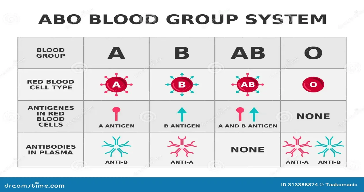
Biosynthesis of blood group
All individuals generate the H antigen or O antigen, which is basically the precursor of any blood group. The H antigen is synthesised by a gene known as fucosyltransferase, which adds fucose to the oligosaccharide sequence (N-acetyl Glucosamine and Galactose). When the N-acetyl galactosamine monosaccharide sugar gets attached to this precursor, it forms the A antigen, and when galactose sugar is synthesized at the place of N- acetyl galactosamine, it forms the B antigen. In the case of the AB blood group, both of these monosaccharides are attached to the oligosaccharide chain. [16][17]
In forensics, blood acts as a biological fluid that can be found on multiple crime scenes, such as homicide, sexual assault, attempted murder, and others. Blood can be found on any of the surfaces, such as the floor, tiles, footwear, soil, weapon that has been used while committing the crime, clothes of the suspect and victim, and at many other places. Blood evidence is examined to link the crime scene, the victim, and the suspect with the help of forensic examination as per Locard’s principle of exchange, which says that whenever two objects come in contact, both of them leave traces on each other, which helps in forensic examination to conclude the commission of the crime.[4]
Material Used
In the present study, some materials were used from the laboratory premises of Regional Forensic Science Laboratory, Chhatrapati Sambhajinagar such as cotton gauze pieces, sample tubes, racks, spoke, washing tubes, commercially available antiserum of ERYCLONE company, cavity slides and slide box, fresh blood cells of group A, B and O. Along with these, Incubator and compound microscope were also used.
Procedure
An experimental study of absorption-elution technique was conducted to evaluate the effect of different numbers of washes and different dilutions of anti-sera on ABO blood grouping. These hypothetical variations were decided after considering the concept of serial dilution. The results of agglutination reactions were then recorded and compared with every hypothetical situation.
Fresh blood samples of A, B and O blood groups were used to prepare blood stains on cotton fabric and were allowed to dry and then used for the actual study. In this study, we worked on two aspects:
I. The evaluation of the effect of different numbers of washing on ABO grouping that were given to the antiserum of a working dilution (Antiserum A 1:8 and Antiserum B 1:4). Here the dilution of saline and antiserum was decided based on the findings of antiserum's titre using serial dilution method, the selected dilutions were kept constant to minimize the dilution related changes, and to study the effect of washings independently. The number of washings chosen were 2, 4, 6, 8 and 10.
Serial Dilution of Anti A and Anti B serum
Commercially available antiserums are highly concentrated and come in bulk and before using them, it is essential to evaluate the working dilution of the antiserums. They are diluted with normal saline. Serial dilution is prepared by first taking 0.1ml of Normal Saline and 0.7ml of the anti serum and was given a gentle mix. In the next nine tubes, 0.4 ml of normal saline was added to each of the tube and then from the very first tube 0.4 ml of content was added to the second one and from the second tube, 0.4ml of content was taken and transferred to the third tube and the same step was continued till last tube.
|
TUBE NUMBER |
TITRE |
|
1 |
1:8 |
|
2 |
1:16 |
|
3 |
1:32 |
|
4 |
1:64 |
|
5 |
1:128 |
|
6 |
1:256 |
|
7 |
1:512 |
|
8 |
1:1024 |
|
9 |
1:2048 |
|
10 |
1:4096 |
Then, each of the dilutions was tested with the known cells by adding one drop of known cells and diluted serum to record the end point of agglutination reaction, and the working dilution was selected accordingly for the experimental conduction.
Also in the present study, the factor of washing cycles was hypothetically initially set to 2 and then was increased by factor of 2 at every other rack and was increased by the same factor to record the agglutination reaction until expected results are observed.
II. The evaluation of the effect of different dilutions of antiserum on ABO grouping with the fixed number of washings (3.5) on ABO blood grouping. The different dilutions were selected around the titrated value to observe the minor changes, if any. The chosen dilutions for Antiserum A were Undiluted, 1:4, 1:6, 1:8, and 1:10. The dilutions for Antiserum B were Undiluted, 1:3, 1:4, 1:5 and 1:6. The effect of variable anti sera dilution with constant washings cycles is being studied independently.
∗1 part of Normal Saline and the rest of the part of antiserum.
∗In this study, there were no changes made for the dilution and washings for antiserum H as it already has least concentration and the main motive here is to study the cross reactivity of A and B group.
Absorption- elution method.
Absorption elution method works on the principle of immunological assay as it involves the reaction between antigen and antibody. It was discovered by Vittorio Siracusa in 1923. It is one of the most reliable serological techniques for ABO blood grouping in forensic science. It is a sensitive technique to determine the blood group of any forensic exhibits.
Absorption- small square pieces of dried blood stains were cut and absorbed with one drop of antiserum and kept at low temperature (4℃) overnight which led to the occurrence of antigen and antibody reaction.
Washing- the absorbed antigen and antibody were given washings as per the with chilled saline so that the un-bound antibodies gets washed away.
Elution- washed samples were incubated at 56℃ of temperature to break the bonds between antigen and antibody that basically leads to elution of antibodies.
Indicator cells- Cell suspension of known blood samples were added and were left undisturbed that allowed the reaction to take place.
Microscopic examination- The cell's suspension was then observed for the clumping or agglutination reaction. The cells that show agglutination read the positive result and the one that shows the free cells gives the negative results.
RESULTS AND DISCUSSION:
The agglutination reactions were observed for different washings and anti sera dilution simultaneously under the microscope at different numbers of washes and different dilutions of antiserum for unstained cotton cloth and stained standard samples of A, B and O. In these result tables, ‘-’ means negative, ‘++++’ means very strong positive, ‘+++’ means strong positive, ‘++’ mildly positive, ‘+’ means weak positive, u/d means undiluted.
The observations are recorded in the form of table and are illustrated below:
∗ Table I to V illustrates the effect of differential washings on ABO grouping.
|
|
ANTI A (1:8) |
ANTI B (1:4) |
ANTI H (u/d) |
WASHES |
REMARKS |
OBSERVATION |
|
UNSTAIN |
- |
- |
- |
2 |
- |
- |
|
Standard A |
++++ |
+++ |
- |
2 |
Interference of B |
AB |
|
Standard B |
- |
++++ |
- |
2 |
No Interference |
B |
|
Standard O |
+ |
+++ |
++++ |
1.5 |
Interference of A and B |
ABO |
Table I: Two washes given to Anti A(1:8) and Anti B (1:4)
|
|
ANTI A (1:8) |
ANTI B (1:4) |
ANTI H (u/d) |
WASHES |
REMARKS |
OBSERVATION |
|
Unstain |
- |
- |
- |
4 |
- |
- |
|
Standard A |
++++ |
++ |
- |
4 |
Interference of B |
AB |
|
Standard B |
- |
++++ |
- |
4 |
No Interference |
B |
|
Standard O |
- |
++ |
++++ |
1.5 |
Interference of B |
BO |
Table II: Four washes given to Anti A(1:8) and Anti B (1:4)
|
|
ANTI A (1:8) |
ANTI B (1:4) |
ANTI H (u/d) |
WASHES |
REMARKS |
OBSERVATION |
|
Unstain |
- |
- |
- |
6 |
- |
- |
|
Standard A |
+++ |
- |
- |
6 |
No Interference |
A |
|
Standard B |
- |
+++ |
- |
6 |
No Interference |
B |
|
Standard O |
- |
- |
+++ |
1.5 |
No Interference |
O |
Table III : Six washes given to Anti A(1:8) and Anti B (1:4)
|
|
ANTI A (1:8) |
ANTI B (1:4) |
ANTI H (u/d) |
WASHES |
REMARKS |
OBSERVATION |
|
Unstain |
- |
- |
- |
8 |
- |
- |
|
Standard A |
++ |
- |
- |
8 |
No Interference |
A |
|
Standard B |
- |
+++ |
- |
8 |
No Interference |
B |
|
Standard O |
- |
- |
++++ |
1.5 |
No Interference |
O |
Table IV: Eight washes given to Anti A(1:8) and Anti B (1:4)
|
|
ANTI A (1:8) |
ANTI B (1:4) |
ANTI H (u/d) |
WASHES |
REMARKS |
OBSERVATION |
|
Unstain |
- |
- |
- |
10 |
- |
- |
|
Standard A |
+ |
- |
- |
10 |
- |
Weak A |
|
Standard B |
- |
+ |
- |
10 |
- |
Weak B |
|
Standard O |
- |
- |
++++ |
1.5 |
No Interference |
O |
Table V: Ten washes given to Anti A(1:8) and Anti B (1:4)
As per the agglutination reactions observed under the microscope, it is observed in table I, when the washing cycles were 2, inter reactivity of Anti B was highly observed with Anti A and Anti H. When the washing cycles were increased to 4, in table II, it was observed that inter reactivity of Anti B was there with Anti A and Anti H but less than 2 washings. On the other hand, at six washing cycles (table III), there was no inter reactivity observed of Anti B with Anti A and Anti H and expected results were read. In table IV and V, it can be seen when the washing cycles were 8 and 10 respectively, interference of Anti B disappeared but along with that the expected results for gradation of agglutination got decreased due to overwashing. It can be said that variable changes in the agglutination reaction were observed with the measurable changes in washing cycles. Unexpected cross reactivity of Anti B was observed with Anti A and Anti H at initial washing cycles, but with the progressive change in the number of washing cycles given to the stains absorbed with tirated Anti-sera dilution, the interference of Anti B gradually improved and gave the expected results at 6 washings.
In the present study with present scenarios and blood samples, it was observed that the washing cycle of ‘six’ appeared to be favorable over four, eight or higher than that.
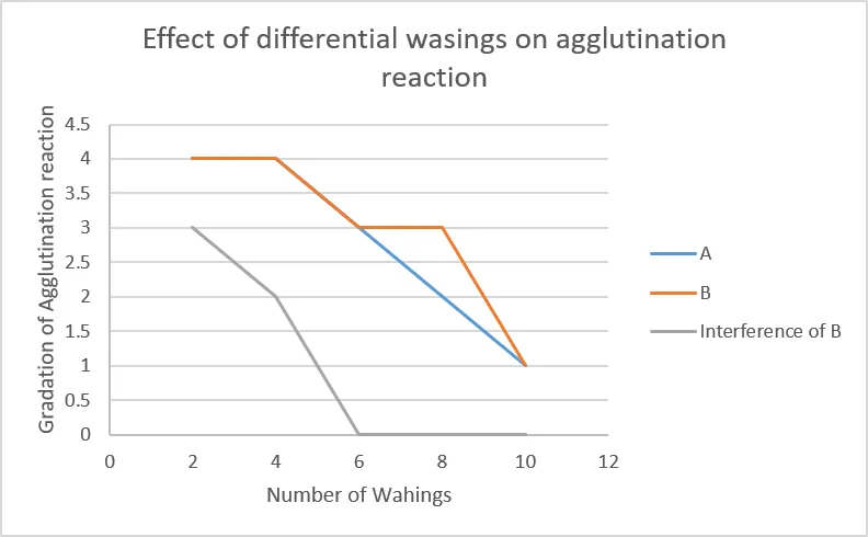
Graph I: Effect of differential washings on agglutination reaction.
From the plotted graph, it can be clearly seen that with the increase in the number of washing cycles, the gradation of agglutination reaction decreases. Now, if we observe this graph, it can easily be interpreted that when the washing cycles were increased the interference of Anti B couldn’t be observed. At six washing cycles the cross reactivity reaction was not there.
∗ Table VI to X illustrates the effect of different anti sera dilution on ABO grouping.
|
|
ANTI A (u/d) |
ANTI B (u/d) |
ANTI H (u/d) |
WASHES |
REMARKS |
OBSERVATION |
|
UNSTAIN |
- |
- |
- |
3.5 |
- |
- |
|
Standard A |
++++ |
+++ |
- |
3.5 |
Interference of B |
AB |
|
Standard B |
- |
++++ |
- |
3.5 |
No interference |
B |
|
Standard O |
- |
+++ |
+++ |
1.5 |
Interference of B |
BO |
Table VI: 3.5 washings given to undiluted serums.
|
|
ANTI A (1:4) |
ANTI B (1:3) |
ANTI H (u/d) |
WASHES |
REMARKS |
OBSERVATION |
|
UNSTAIN |
- |
- |
- |
3.5 |
- |
- |
|
Standard A |
++++ |
+++ |
- |
3.5 |
Interference of B |
AB |
|
Standard B |
- |
++++ |
- |
3.5 |
No Interference |
B |
|
Standard O |
- |
+++ |
++++ |
1.5 |
Interference of B |
BO |
Table VII: 3.5 washings given to Anti A(1:4) and Anti B (1:3)
|
|
ANTI A (1:6) |
ANTI B (1:4) |
ANTI H (u/d) |
WASHES |
REMARKS |
OBSERVATION |
|
Unstain |
- |
- |
- |
3.5 |
- |
- |
|
Standard A |
++++ |
++ |
- |
3.5 |
Interference of B |
AB |
|
Standard B |
- |
++++ |
- |
3.5 |
No Interference |
B |
|
Standard O |
- |
++ |
++++ |
1.5 |
Interference of B |
BO |
Table VIII: 3.5 washings given to Anti A(1:6) and Anti B (1:4)
|
|
ANTI A (1:8) |
ANTI B (1:5) |
ANTI H (u/d) |
WASHES |
REMARKS |
OBSERVATION |
|
Unstain |
- |
- |
- |
3.5 |
- |
- |
|
Standard A |
++++ |
+ |
- |
3.5 |
Interference of B |
AB |
|
Standard B |
- |
++++ |
- |
3.5 |
No Interference |
B |
|
Standard O |
- |
+ |
++++ |
1.5 |
Interference of B |
BO |
Table IX: 3.5 washings given to Anti A(1:8) and Anti B (1:5)
|
|
ANTI A (1:10) |
ANTI B (1:6) |
ANTI H (u/d) |
WASHES |
REMARKS |
OBSERVATION |
|
Unstain |
- |
- |
- |
3.5 |
- |
- |
|
Standard A |
++ |
- |
- |
3.5 |
No Interference |
A |
|
Standard B |
- |
++ |
- |
3.5 |
No Interference |
B |
|
Standard O |
- |
- |
++++ |
1.5 |
No Interference |
O |
Table X: 3.5 washings given to Anti A(1:10) and Anti B (1:6)
In table VI, when the undiluted antisera was added, it was observed that there was significant interference of Anti B with Anti A and Anti H. Secondly, when the dilutions were increased to 1:4 for Anti A and 1:3 for Anti B (table VII), there were no significant changes observed in the interference of Anti B with Anti A and Anti H and the results were the same as of table VI . In table VIII, where the dilution for Anti A is 1:6 and Anti B with the dilution of 1:4, it was observed that inter reactivity of Anti B was there with Anti A and Anti H but it was comparatively less and least interference was observed when the dilution was 1:8 for Anti A and 1:5 for Anti B as per table IX. Lastly, when the dilutions were increased to 1:10 for Anti A and 1:6 for Anti B, as per the observation noted in table number X, it can clearly be seen that there is no interference of Anti B with Anti A and Anti H. Hence, It can be said that with the successive increment in the dilution of antiserums while keeping the number of washings fixed (i.e., 3.5) the interference of group B gradually disappeared and gave the expected result at Anti A 1:8, 1:10 and Anti B 1:4, 1:5. As it can clearly be seen that at Anti A 1:8 and Anti B 1:4, the cross reactivity of Anti B was observed but it is almost negligible and when the Anti A was diluted to 1:10 and Anti B to 1:5, it was observed that the cross reactivity of Anti B did not appear at all but on the other hand the intensity of agglutination reaction also got decreased for the expected results in the present experimental study with the present scenarios.
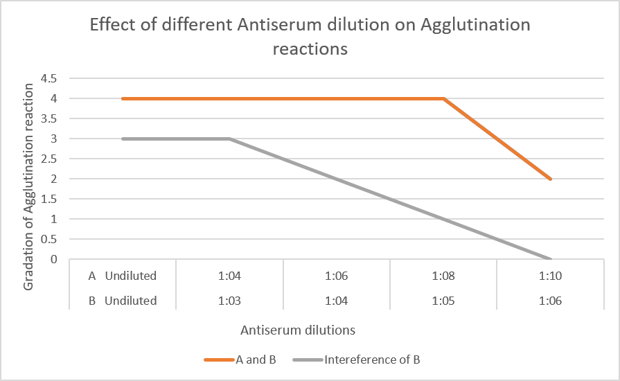
Graph II: Effect of different Antiserum dilution on Agglutination reaction.
From the plotted graph, it can be observed that at lower dilutions, the gradation of agglutination reaction is high and as the dilutions of antiserum were increased the gradation of agglutination reaction decreased. Also, the grey line here shows the interference of Anti B in the findings and it is observed that interference could not be seen at the experimental conditions where dilution of Anti A is 1:8/1:10 and Anti B is 1:4/1:5.
CONCLUSION
In this experimental study, it was predominantly observed that with fewer number of washings and lesser dilution of Antiserums, the inter-reactivity of other group was observed but with the progressive change in washings and dilutions this inter-reactivity started reducing and then at certain experimental condition of washings (six washes) and dilutions (Anti A 1:8/1:10) and (Anti B 1:5/1:6), the inter- reactivity of other group could not be observed. Also, after crossing the experimental conditions, the gradation of agglutination gradually started decreasing and showed weak reactions for the expected results. Also, it was consistently observed that it is ‘Anti B’ which was showing non-specific reactivity with other groups which somehow disappeared with the change in the washing cycles and dilution of Antiserums. In conclusion to this present experimental study, it can be said that carefully monitoring the reaction patterns of anti sera dilutions and washing cycles may serve as a useful laboratory practice for minimizing interference and improving the reliability of ABO grouping.
ACKNOWLEDGEMENT
We show our sincere gratitude to our Director Dr. V. J. Thakare Sir, Deputy Director Shri N. R. Gosavi Sir, our Assistant Director Smt. V. R. Padale Ma’am and Shri S. A. Sable Sir and Assistant Chemical Analyser Dr. H. H. Shah Sir for their supervision while conducting this experimental study and our laboratory attendant Shri R. R. Jaiswal Sir who helped us at every step during this practice.
SCOPE AND INTENDED USE OF STUDY
The present study is solely to evaluate and document the observations obtained under the experimental conditions investigated. The findings are not intended to challenge, replace any existing method or established practice. Rather, the study provides an experimental evaluation of selected procedural parameters and may serve as a basis for further research and validation.
REFERENCES


Rushikesh Laxmanrao Munde*, Sanya Gulati, Evaluation Of Differential Washings And Anti-Serum Dilution Protocols For ABO Blood Group Determination Using Absorption- Elution Technique: An Experimental Study, Int. J. Sci. R. Tech., 2026, 3 (9), 697-706. https://doi.org/10.5281/zenodo.23081093
 10.5281/zenodo.23081093