

We use cookies to ensure our website works properly and to personalise your experience. Cookies policy
Department of [Science Laboratory Technology], [Ladoke Akintola University of Technology University], [Ogbomoso, Oyo State, Nigeria
Tuberculosis (TB) remains a major global health challenge, particularly in low- and middle-income countries, due to increasing drug resistance and oxidative tissue damage. Picralima nitida has traditionally been used to treat infectious diseases, including TB, but its therapeutic potential requires scientific validation. This study evaluated the in vitro antioxidant and antitubercular activities of methanolic leaf and bark extracts of P. nitida. Methanolic extracts of P. nitida leaf and bark were screened for their phytoconstituents. Antioxidant activities were evaluated using standard antioxidant assays, including DPPH, ABTS, Nitric oxide (NO) scavenging activities, Ferric reducing antioxidant power (FRAP) and Lipid peroxidation inhibition. The leaf and bark extracts were screened for anti-TB activity against three different tuberculosis isolates using agar diffusion method in Middlebrook 7H10 agar. Data was analyzed using one-way ANOVA. Phytochemical screening identified alkaloids, flavonoids, phenols, saponins, steroids, cardiac glycosides, tannins and reducing sugars in both methanolic leaf and bark extracts of P. nitida, with alkaloids, phenols and saponins relatively abundant, while antioxidant activities increased with concentration: DPPH from 20.22 ± 4.69% to 44.68 ± 0.27% in the leaf and 31.07 ± 10.42% to 42.06 ± 9.38% in the bark; ABTS from 55.51 ± 2.00% to 68.54 ± 0.76% in the leaf and 33.27 ± 4.42% to 47.85 ± 3.42% in the bark; nitric oxide from 22.97 ± 2.01% to 26.70 ± 8.08% in the leaf and 57.33–64.83% in the bark; and FRAP from 64 mg/g AAE to 72 mg/g AAE in the leaf and 53 mg/g AAE to 74 mg/g AAE in the bark, with lipid peroxidation inhibition increasing from 57.23 ± 6.02% at 10 mg/ml in the leaf to 67.06 ± 9.43% at 30 mg/ml in the leaf and the bark respectively. The extracts also demonstrated concentration-dependent antitubercular activity, with 1.0 mg/ml producing 56–57% inhibition for the leaf and 55–58% for the bark against M. tuberculosis H37Rv and clinical isolates, although rifampicin and isoniazid which are standard drugs showed greater inhibition. Statistical analysis confirmed significant concentration-dependent effects (p < 0.001, ?² = 0.88 - 0.91). This result supports that P. nitida leaf and bark extracts possess significant antioxidant and moderate antitubercular activities, supporting their ethnomedicinal use. The bark extract demonstrated superior antioxidant properties, while both extracts showed comparable antimycobacterial activity. These findings justify further bioassay-guided fractionation and in vivo studies toward developing novel therapeutic agents.
Tuberculosis (TB) is a major infectious disease caused primarily by Mycobacterium tuberculosis (MTB) and transmitted mainly through the inhalation of airborne particles expelled by individuals with active pulmonary TB. The pathogen belongs to the Mycobacterium tuberculosis complex (MTBC), which comprises several closely related species, including M. bovis, M. africanum, M. microti, M. caprae, M. pinnipedii, M. canettii, and M. mungi. Although several members of the complex can cause disease in humans, M. tuberculosis is responsible for the vast majority of TB cases worldwide (Heliyon, 2022).
TB primarily affects the lungs but can also involve other organs, resulting in extrapulmonary disease. Infection may exist as latent or active TB. Latent TB infection is generally asymptomatic, while approximately 5 - 10% of infected individuals may develop active disease during their lifetime. Active TB is commonly characterized by persistent cough, fever, night sweats, weight loss, fatigue, and, in some cases, haemoptysis (Ying et.al., 2022).
Despite decades of advances in diagnosis, treatment, and prevention, TB remains one of the world's leading causes of infectious disease-related morbidity and mortality. The burden is particularly pronounced in low- and middle-income countries, where limited healthcare resources, delayed diagnosis, inadequate treatment coverage, and gaps in disease surveillance continue to hinder effective TB control (WHO, 2023). Nigeria remains among the countries with a high TB burden globally, with substantial gaps in case detection and notification, further highlighting the need for improved diagnostic, preventive, and therapeutic strategies (WHO, 2023).
Although effective first- and second-line antitubercular drugs are available, the continued burden of TB is complicated by challenges such as prolonged treatment duration, adverse drug reactions, poor treatment adherence, and the emergence of drug-resistant M. tuberculosis strains. These challenges have sustained the need for the discovery and development of new, safe, effective, and affordable antitubercular agents. However, investment in TB drug discovery and development has historically remained relatively limited compared with other major diseases, partly because of the high cost and long duration of pharmaceutical research and development (Alsayed et.al., 2023).
In addition to the direct effects of infection, oxidative stress has been implicated in the pathophysiology of several infectious and inflammatory diseases, including TB. Oxidative stress occurs when there is an imbalance between the production of reactive oxygen species and the ability of antioxidant defense systems to neutralize them. Antioxidants are substances capable of inhibiting or reducing oxidative reactions and protecting biological molecules against oxidative damage. Interest in antioxidants initially emerged from their ability to prevent oxidation and deterioration in industrial and food products, but the concept later gained significant biological and medical relevance following the recognition of oxidative processes in living systems (Flieger et.al., 2021).
Natural products, particularly medicinal plants, remain an important source of bioactive compounds with potential therapeutic applications. Medicinal plants contain diverse phytochemicals, including alkaloids, flavonoids, phenolic compounds, tannins, saponins, and terpenoids, many of which have demonstrated antioxidant, antimicrobial, anti-inflammatory, and other pharmacological activities. Consequently, medicinal plants continue to provide valuable candidates for the discovery of novel therapeutic agents against infectious diseases, including TB (Riaz et,al., 2023, Guptal et.al., 2021).
Picralima nitida (family Apocynaceae), commonly known as “Abeere” among the Yoruba people of Southwestern Nigeria, is a medicinal plant widely used in West African traditional medicine for the management of various conditions, including infectious diseases, malaria, pain, fever, and inflammatory disorders (Akbasi et.al., 2021). Previous phytochemical investigations have demonstrated the presence of several biologically active constituents in the plant, particularly alkaloids and other secondary metabolites, which may contribute to its pharmacological activities (Akbasi et.al., 2021). However, despite its longstanding ethnomedicinal use, scientific evidence regarding the antioxidant and antitubercular potential of the leaf and bark extracts of P. nitida remains limited.
Therefore, this study investigates the in vitro antioxidant and antitubercular properties of methanolic leaf and bark extracts of Picralima nitida to provide scientific evidence supporting the potential of P. nitida as a source of natural compounds for the development of novel therapeutic agents against tuberculosis and oxidative stress-related conditions.
MATERIALS AND METHODS
2.1 Reagents
All reagents were of analytical grade and obtained from Sigma Aldrich Chemical, Germany
2.2 Plant Materials
Fresh leaves and bark of Picralima nitida were gathered from Odo Oba, Oyo State, Nigeria, on March 29, 2025. The plant sample was identified and authenticated in the herbarium of Ladoke Akintola University of Technology, Ogbomoso, Oyo State.
2.3 Preparation of plant extract
Healthy leaves and bark of P. nitida were thoroughly washed with clean tap water to remove debris, cut into smaller pieces, and air-dried at room temperature in a well-ventilated, dust-free environment until a constant weight was obtained. The dried samples were separately pulverized using a mechanical grinder and sieved to obtain uniform particle sizes. A total of 4,600g of leaf powder and 3,400g of bark powder were stored separately in airtight containers under desiccated conditions before extraction.
The powdered samples were separately macerated in analytical-grade methanol for 72hr with occasional stirring. The resulting extracts were filtered through double-layered cheesecloth lined with cotton wool and subsequently through Whatman No. 1 filter paper. The filtrates were concentrated under reduced pressure at 40°C using a rotary evaporator (Büchi Rotavapor R-210, Switzerland) and freeze-dried to obtain powdered crude extracts. The extraction yielded 128g and 105g of methanolic leaf and bark extracts respectively, which were stored in sterile amber bottles under refrigeration until further analysis (Larson et.al., 2026).
2.4 Phytochemical Screening
Preliminary phytochemical analysis of Picralima nitida leaf and bark extracts was carried out to qualitatively and quantitatively to determine the presence of secondary metabolites using the method described by Yoon, (2025). The crude methanol extract was subjected to specific chemical tests to detect major classes of phytochemicals, including alkaloids, flavonoids, saponins, cardiac glycosides, tannins, phenolic compounds, and steroids (Yoon et.al., 2025).
2.5 In vitro Antioxidant assay
2.5.1 Determination of DPPH Radical Scavenging Activity
The DPPH radical scavenging activity of the methanolic extracts was determined spectrophotometrically according to the method described by Gulcin (2023). A 0.3mM DPPH solution was prepared by dissolving 0.03g of DPPH in 250ml of methanol. Extract stock solutions (10mg/ml) were prepared by dissolving 0.1g of each extract in 10ml of methanol and subsequently diluted to obtain concentrations ranging from 100 to 500µg/ml.
For the assay, 1ml of 0.3mM DPPH solution was mixed with 1ml of each test solution and incubated in the dark at room temperature for 30min. Absorbance was measured at 517nm, and the percentage DPPH radical scavenging activity was calculated using the following formula:
DPPH scavenging activity = (Ao – As/Ao) x 100
Ao = absorbance without sample
As = absorbance with sample.
2.5.2 Determination of Ferric Reducing Antioxidant Power (FRAP)
The ferric reducing antioxidant power (FRAP) of the extracts was determined spectrophotometrically using the method of Oyiazu, (1986). A 2.5ml extract was mixed with 2.5ml of 200mM sodium phosphate buffer and 2.5ml of 1% potassium ferricyanide and incubated at 50°C for 20 min. The reaction was terminated by adding 2.5ml of 10% trichloroacetic acid, followed by centrifugation at 2,000 × g for 10 min. Thereafter, 5ml of the supernatant was mixed with 5ml of distilled water and 1ml of 0.1% ferric chloride. Absorbance was measured at 700nm, and the reducing power was expressed as ascorbic acid equivalents.
2.5.3 Determination of ABTS Scavenging Ability
The ABTS radical cation (ABTSâº) scavenging activity was determined by reacting equal volumes of 7mM ABTS and 2.45mM potassium persulfate, followed by incubation in the dark at room temperature for 14hrs. The resulting ABTS⺠solution was diluted with ethanol to an absorbance of 0.70 ± 0.02 at 734 nm. For the assay, 1.0ml of the ABTS⺠working solution was mixed with 10µL of the sample, vortexed gently, and incubated at room temperature for 6 min. Absorbance was measured at 734nm against a solvent blank (Oyiazu,, 1986), and the percentage inhibition was calculated as follows:
2.5.4 Determination of Nitric Oxide Radical (NO) Scavenging Assay
Nitric oxide was generated from sodium nitroprusside and measured by Griess reaction (Marcoci et al., 2021). A 1ml of 10mM sodium nitroprusside prepared in phosphate buffer (pH 7.4) was mixed with 1ml of the sample at different concentrations and incubated at 25°C for 150 min under light. Thereafter, 1ml of the reaction mixture was mixed with an equal volume of Griess reagent and allowed to stand at room temperature for 10 min. Absorbance was measured at 540nm, and the percentage inhibition was calculated using the following formula:
2.5.5 Determination of Malondialdehyde (MDA) Concentration
Lipid peroxidation inhibition was assessed by measuring malondialdehyde (MDA) formation using the thiobarbituric acid reactive substances (TBARS) method of (Zeb et, al., 2016). A 0.5ml of 10% egg yolk homogenate was mixed with 0.1ml of the extract at different concentrations and made up to 1.0ml with distilled water. Lipid peroxidation was initiated by adding 0.05ml of 0.07M FeSOâ‚„, followed by incubation at 37°C for 30 min. Thereafter, 1.5ml each of 10% trichloroacetic acid (TCA) and 0.67% thiobarbituric acid (TBA) were added. The mixture was vortexed, heated at 95°C for 15 min, cooled, and centrifuged at 3,000 rpm for 10 min. Absorbance of the supernatant was measured at 532nm, and percentage inhibition of lipid peroxidation was calculated using the following formula:
2.6 Antitubercular Study
Mycobacterial isolates
Three M. tuberculosis isolates from three different patients identified and reference susceptible strain H37Rv were collected from the TB reference laboratory, Department of Medical Microbiology, University College Hospital, Ibadan
Determination of Colony Forming Units (CFU)
The ten- fold dilution of standard 1mg/ml M. tuberculosis suspension was streaked on Middlebrook 7H10 media for determination of CFU in the presence or absence of plant extract. Media inoculation without plant extract served as the control. M. tuberculosis suspension of 1mg/ml is equivalent to MacFarland standard 1 (Smith-Jeficoat et.al., 2022). One loopful (0.6ul) of this suspension was streaked on the media using 3mm bacteriological loop. The plant extracts were incorporated separately on the media at concentrations of 0.2mg/ml; 0.4mg/ml; 0.6mg/ml; 0.8mg/ml and 1mg/ml of extract dissolved into 100 ml of culture medium prior to inspissation. The inoculated culture media containing extracts and the controls were incubated at 370C for eight weeks. Reading of the culture media was taken weekly. Percentage inhibition of each test was calculated by mean reduction in number of colonies on extract containing medium as compared to extract free control medium.
Statistical Analysis
Data are presented as mean ± standard deviation (SD). The data were analysed using one-way analysis of variance (ANOVA), followed by an appropriate post-hoc test, with statistical significance set at P < 0.05.
RESULTS
3.1 Phytochemical Analysis
The phytochemical analysis of the crude methanolic extract of Picralima nitida demonstrated that the plant is a rich source of bioactive secondary metabolites, notably alkaloids, phenols, saponins, cardiac glycosides, flavonoids, and steroids. The relatively high concentrations of these compounds, especially flavonoids, saponins and alkaloids suggest that they may collectively contribute to the antitubercular, antioxidant, and other pharmacological activities observed in subsequent experimental analyses. The abundance of these bioactive compounds supports the plant's pharmacological versatility and potential therapeutic value. These findings are consistent with previous reports that have identified similar phytoconstituents in P. nitida and related species within the Apocynaceae family (Eze et.al., 2022), highlighting its chemical richness and medicinal promise. Overall, these findings provide scientific validation for the ethnomedicinal use of P. nitida in traditional healthcare systems and reinforce its potential as a candidate for further phytopharmacological and drug discovery studies.
Table 1: Qualitative Phytochemical Screening of P. nitida Extracts
| Phytochemicals | Leaf | Bark |
|---|---|---|
| Alkaloids | +++ | +++ |
| Flavonoids | + | + |
| Phenols | +++ | +++ |
| Saponins | +++ | +++ |
| Steroids | + | + |
| Tannin | ++ | ++ |
| Cardiac glycosides | ++ | ++ |
| Terpenoids | ++ | ++ |
+++ Present in very high concentration
++ Present in moderate concentration
+ Present in low concentration
Table 2: Quantitative Phytochemical Screening of P. nitida Extracts
| Phytochemicals | Leaf | Bark |
|---|---|---|
| Alkaloids | 121.35 ± 0.72 | 119.42 ± 1.4 |
| Phenol (mg GAE/g extract) | 92.45 ± 0.84 | 91.38 ± 1.6 |
| Saponin (mg DE/g extract) | 209.79 ± 2.50 | 219 ± 4.3 |
| Flavonoid (mg QE/g extract) | 326.64 ± 1.39 | 289 ± 6.1 |
| Tannin (mg TAE/g extract) | 64.89 ± 1.75 | 67 ± 4.6 |
3.2 In vitro Antioxidant Activity of the leaf and bark of the extracts of P. nitida
The antioxidant potential of the leaf and bark extracts was assessed through a series of six validated in vitro bioassays: DPPH radical scavenging, ferric reducing antioxidant power (FRAP), ABTS radical cation decolorization, Nitric Oxide (NO) radical inhibition, and Malondialdehyde (MDA) lipid peroxidation inhibition.
The results are presented in Figures 1 - 5.
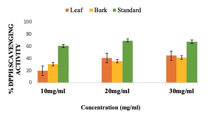
Figure 1: DPPH Radical Scavenging Activity of Leaf and Bark Extracts Compared with Ascorbic Acid Standard
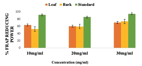
Figure 2: Ferric Reducing Antioxidant Power (FRAP) of Leaf and Bark Extracts Compared with Ascorbic Acid Standard
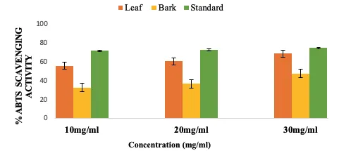
Figure 3: ABTS Radical Scavenging Activity of Leaf and Bark Extracts Compared with Trolox Standard
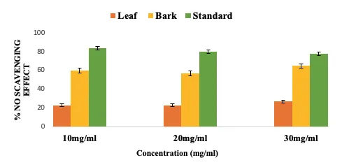
Figure 4: Nitric Oxide Radical Inhibition of Leaf and Bark Extracts Compared with Quercetin Standard
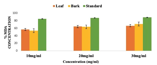
Figure 5: Malondialdehyde (MDA) Inhibitory Activity of Leaf and Bark Extracts Compared with Quercetin Standard
Result of Antioxidant Activities:
The antioxidant activities of the leaf and bark extracts were evaluated using DPPH, ABTS, nitric oxide (NO) scavenging, ferric reducing antioxidant power (FRAP), and lipid peroxidation inhibition assays.
DPPH radical scavenging activity increased with extract concentration in both samples. The leaf extract exhibited activity ranging from 20.22 ± 4.69% at 10 mg/ml to 44.68 ± 0.27% at 30 mg/ml, while the bark extract ranged from 31.07 ± 10.42% to 42.06 ± 9.38%.
Similarly, ABTS radical scavenging activity increased from 55.51 ± 2.00% to 68.54 ± 0.76% in the leaf extract and from 33.27 ± 4.42% to 47.85 ± 3.42% in the bark extract across the tested concentrations.
NO scavenging activity in the leaf extract increased slightly from 22.97 ± 2.01% to 26.70 ± 8.08%, whereas the bark extract exhibited higher activity, ranging from 57.33% to 64.83%.
For FRAP, the leaf extract exhibited reducing capacities ranging from 61.54 to 71.03 mg/g AAE, while the bark extract ranged from 52.53 ± 9.15 to 73.94 ± 10.21 mg/g AAE.
Lipid peroxidation inhibition increased from 57.23 ± 6.02% to 67.06 ± 9.43% in the leaf extract and from 54.03 ± 2.91% to 72.05 ± 5.03% in the bark extract. Overall, the leaf extract exhibited higher DPPH and ABTS activities, whereas the bark extract showed greater NO scavenging, FRAP, and lipid peroxidation inhibition.
This version is better suited to a Results section because it reports the findings without adding interpretation that belongs in the Discussion.
3.3 Antitubercular activity of the leaf and bark of the extracts of P. nitida
The antitubercular activity of the crude leaf and bark extracts of Picralima nitida against Mycobacterium tuberculosis isolates was assessed using mean colony-forming unit (CFU) counts and percentage inhibition, with the activities of standard antitubercular drugs included for comparative evaluation. The findings are presented in Tables 4 and 5, respectively.
Result of antitubercular activities
The colony-forming unit (CFU) counts of methanolic leaf and bark extracts of Picralima nitida and the standard antitubercular drugs against Mycobacterium tuberculosis isolates are presented in Table 4.
Both the leaf and bark extracts demonstrated a concentration-dependent reduction in mycobacterial growth, as evidenced by the progressive decrease in CFU counts with increasing extract concentrations (0.2–1.0 mg/ml). The leaf extract reduced the CFU count of the H37Rv isolates from 124 CFU/ml in the control to 55 CFU/ml at 1.0 mg/ml, while the bark extract reduced the count from 122 to 51 at the same concentration.
A similar trend was observed among the MTB1, MTB2, and MTB3 isolates, indicating that both plant extracts exhibited inhibitory activity against the tested M. tuberculosis strains. However, the reduction in CFU counts was less pronounced than that observed with rifampicin and isoniazid under the experimental conditions.
Rifampicin demonstrated the most substantial reduction in CFU counts, decreasing the H37Rv isolate from 126 in the control to 5 at 1.0 mg/ml. Isoniazid also exhibited inhibitory activity, although its response varied among the tested isolates.
The leaf extract exhibited a concentration-dependent increase in percentage inhibition across all tested isolates. At 1.0 mg/mL, the percentage inhibition ranged from 56% in H37Rv and MTB1 to 59% in MTB3. Similarly, the bark extract showed increasing inhibitory activity with increasing concentration, with percentage inhibition ranging from 52% to 54% among the tested isolates at 1.0 mg/mL, except for the values as presented in the table.
The highest percentage recorded for the leaf extract was 59% against MTB3, while the bark extract achieved its highest recorded inhibition of 54% against MTB2. These findings indicate that the leaf extract exhibited slightly higher percentage inhibition than the bark extract at the maximum tested concentration.
In comparison, rifampicin demonstrated substantially higher inhibitory activity, with percentage inhibition ranging from 89% to 97% at 1.0 mg/ml. Isoniazid also showed considerable inhibitory activity, ranging from 73% to 89% at the same concentration, depending on the isolate (Table 5).
The observed increase in percentage inhibition with increasing extract concentration suggests a concentration-dependent antitubercular effect of the plant extracts. Nevertheless, the lower inhibitory activity compared with the standard drugs indicates that the crude extracts were less potent under the conditions of this assay.
Table 4: CFU Counts of Crude Methanol Leaf and Bark Extracts of Picralima nitida and Standard Drugs Against Mycobacterium tuberculosis Isolates.
| Extract/Drug | Isolate | Control | 0.2 mg/mL | 0.4 mg/mL | 0.6 mg/mL | 0.8 mg/mL | 1.0 mg/mL |
|---|---|---|---|---|---|---|---|
| Leaf Extract | H37Rv | 124 | 101 | 92 | 78 | 60 | 55 |
| MTB1 | 120 | 108 | 92 | 78 | 64 | 54 | |
| MTB2 | 141 | 122 | 102 | 84 | 70 | 59 | |
| MTB3 | 138 | 116 | 98 | 82 | 68 | 56 | |
| Bark Extract | H37Rv | 122 | 98 | 91 | 75 | 58 | 51 |
| MTB1 | 118 | 102 | 88 | 78 | 61 | 52 | |
| MTB2 | 131 | 116 | 95 | 84 | 64 | 53 | |
| MTB3 | 132 | 110 | 93 | 77 | 62 | 55 | |
| Rifampicin | H37Rv | 126 | 58 | 40 | 31 | 15 | 5 |
| MTB1 | 130 | 74 | 52 | 40 | 29 | 14 | |
| MTB2 | 140 | 65 | 50 | 39 | 25 | 12 | |
| MTB3 | 132 | 55 | 40 | 29 | 20 | 4 | |
| Isoniazid | H37Rv | 136 | 68 | 55 | 40 | 25 | 15 |
| MTB1 | 138 | 105 | 80 | 62 | 51 | 38 | |
| MTB2 | 146 | 80 | 71 | 60 | 49 | 31 | |
| MTB3 | 144 | 68 | 51 | 39 | 22 | 16 |
Table 5: Percentage Inhibition (%) of Crude Methanol Leaf and Bark Extracts of Picralima nitida and Standard Drugs Against Mycobacterium tuberculosis Isolates.
| Extract/Drug | Isolate | 0.2 mg/ml | 0.4 mg/ml | 0.6 mg/ml | 0.8 mg/ml | 1.0 mg/ml |
|---|---|---|---|---|---|---|
| Leaf Extract | H37Rv | 19 | 26 | 37 | 52 | 56 |
| MTB1 | 10 | 23 | 35 | 47 | 56 | |
| MTB2 | 14 | 27 | 40 | 50 | 58 | |
| MTB3 | 16 | 28 | 41 | 51 | 59 | |
| Bark Extract | H37Rv | 15 | 25 | 34 | 51 | 53 |
| MTB1 | 9 | 22 | 33 | 45 | 52 | |
| MTB2 | 11 | 26 | 39 | 47 | 54 | |
| MTB3 | 13 | 24 | 38 | 48 | 53 | |
| Rifampicin | H37Rv | 54 | 68 | 75 | 88 | 96 |
| MTB1 | 43 | 60 | 69 | 78 | 89 | |
| MTB2 | 54 | 64 | 72 | 82 | 91 | |
| MTB3 | 58 | 70 | 78 | 85 | 97 | |
| Isoniazid | H37Rv | 50 | 60 | 71 | 82 | 89 |
| MTB1 | 24 | 42 | 55 | 63 | 73 | |
| MTB2 | 45 | 52 | 59 | 66 | 79 | |
| MTB3 | 53 | 65 | 73 | 85 | 89 |
MTB = Mycobacterium tuberculosis Cc = Number of colonies in control medium CFU = Colony Forming Units Ct = Number of colonies in treated medium
DISCUSSION
Antioxidant Activities
The antioxidant activity of the crude methanol leaf and bark extracts of Picralima nitida were evaluated using DPPH, ABTS, nitric oxide (NO) scavenging, ferric reducing antioxidant power (FRAP), and lipid peroxidation inhibition assays. The extracts demonstrated antioxidant activity across the tested concentrations, although variations were observed between the leaf and bark samples depending on the assay employed. These findings suggest that the extracts contain constituents with different antioxidant capacities and mechanisms of action.
The DPPH radical scavenging activity of both extracts increased with increasing concentration. The leaf extract exhibited an increase in activity from 20.22 ± 4.69% to 44.68 ± 0.27% at the concentration of 10mg/ml, while the bark extract showed an increase from 31.07 ± 10.42% to 42.06 ± 9.38% at 30mg/ml concentrations. The observed concentration-dependent response may be attributed to the presence of antioxidant phytochemicals capable of donating hydrogen atoms or electrons to neutralize free radicals. Previous investigations of P. nitida have reported the presence of polyphenols, flavonoids, alkaloids, and other secondary metabolites, which may contribute to its antioxidant properties (Koudou et al., 2013). However, the contribution of individual compounds to the present DPPH activity requires further characterization.
The higher DPPH scavenging activity observed in the leaf extract at the maximum tested concentration may be associated with differences in the composition and abundance of phenolic and flavonoid compounds. Phenolic compounds are recognized for their ability to participate in free radical neutralization through electron donation and hydrogen atom transfer. A previous study on P. nitida reported antioxidant activity in methanol leaf extracts and identified polyphenolic constituents among the phytochemical groups present. This provides a possible phytochemical basis for the activity observed in the present investigation, although the phenolic and flavonoid contents of the current extracts were not provided.
The ABTS assay also revealed an increase in radical scavenging activity with increasing extract concentration. The leaf extract demonstrated higher ABTS inhibition, increasing from 55.51 ± 2.00% to 68.54 ± 0.76%, compared with the bark extract, which increased from 33.27 ± 4.42% to 47.85 ± 3.42%. The higher activity of the leaf extract may reflect differences in the concentration or composition of compounds capable of reacting with the ABTS radical cation. Phenolic compounds and flavonoids are potential contributors to this activity, as they possess chemical groups that can participate in antioxidant reactions. Nevertheless, antioxidant activity measured by ABTS cannot be attributed to a particular compound without chemical identification and correlation studies.
In contrast to the DPPH and ABTS results, the bark extract exhibited substantially higher nitric oxide scavenging activity than the leaf extract. The bark extract showed inhibition ranging from 57.33% to 64.83%, whereas the leaf extract demonstrated lower activity, ranging from 22.97 ± 2.01% to 26.70 ± 8.08%. This difference indicates that the antioxidant response of the extracts varies according to the radical system employed. The higher nitric oxide scavenging activity of the bark extract may be associated with differences in the abundance of alkaloids, phenolic compounds, tannins, or other constituents capable of interacting with reactive nitrogen species. However, this proposed relationship cannot be confirmed without phytochemical profiling and compound-specific investigations.
The ferric reducing antioxidant power (FRAP) assay showed reducing capacities ranging from 61.54 to 71.03 mg/g AAE in the leaf extract and from 52.53 ± 9.15 to 73.94 ± 10.21 mg/g AAE in the bark extract. The relatively high reducing capacity observed in both extracts suggests the presence of electron-donating constituents that may reduce ferric ions under the assay conditions. Phenolic compounds, flavonoids, and other reducing phytochemicals may contribute to this activity. The higher maximum FRAP value recorded for the bark extract may reflect differences in its reducing constituents compared with the leaf extract. However, FRAP measures reducing capacity in a chemical assay and does not directly establish antioxidant effectiveness in biological systems.
Lipid peroxidation inhibition increased with increasing concentration in both extracts. The leaf extract exhibited inhibition ranging from 57.23 ± 6.02% to 67.06 ± 9.43%, while the bark extract showed inhibition ranging from 54.03 ± 2.91% to 72.05 ± 5.03%. The higher maximum inhibition observed in the bark extract suggests a greater capacity under the experimental conditions to inhibit the processes measured by the lipid peroxidation assay. This activity may be related to the presence of phenolic compounds, flavonoids, and other secondary metabolites that can interact with reactive species involved in lipid oxidation. Nevertheless, the precise mechanism and contribution of individual bioactive compounds require further investigation.
The differences in antioxidant activity between the leaf and bark extracts may be explained by variation in their phytochemical composition, concentration of active constituents, and the chemical principles underlying each antioxidant assay. The leaf extract demonstrated higher DPPH and ABTS radical scavenging activities, whereas the bark extract showed greater nitric oxide scavenging, FRAP, and lipid peroxidation inhibition at the reported maximum concentrations. These findings emphasize that antioxidant activity is assay-dependent and that no single test provides a complete assessment of the antioxidant potential of a plant extract. Similar investigations of P. nitida have reported the presence of multiple phytochemical classes, including alkaloids, flavonoids, tannins, and polyphenols, supporting the possibility of a complex contribution from several constituents.
Overall, the crude methanol leaf and bark extracts of P. nitida exhibited measurable antioxidant activity across the different assay systems. The observed activity may be associated with the presence of bioactive secondary metabolites, particularly phenolic compounds, flavonoids, tannins, and alkaloids.
Antitubercular Activities
The methanolic leaf and bark extracts of Picralima nitida demonstrated concentration-dependent inhibitory activity against Mycobacterium tuberculosis isolates, as evidenced by the progressive reduction in colony-forming unit (CFU) counts and corresponding increase in percentage inhibition. At the highest tested concentration of 1.0 mg/mL, the leaf extract exhibited percentage inhibition ranging from 56% to 59%, whereas the bark extract demonstrated inhibition ranging from 52% to 54%. This observation suggests that the extracts possess constituents capable of suppressing mycobacterial growth under experimental conditions. The observed activity may be associated with the presence of bioactive secondary metabolites previously reported in P. nitida, including alkaloids, tannins, polyphenols, steroids, and other phenolic compounds (Saidu et al., 2014). The slightly higher percentage observed with the leaf extract compared with the bark extract may reflect differences in the qualitative and quantitative composition of bioactive constituents in the two plant parts.
Alkaloids are among the important phytochemical constituents identified in P. nitida and may contribute to the antimicrobial properties of the plant. Several indole alkaloids, including akuammine, akuammidine, and akuammicine, have been reported from different parts of the plant. Previous studies have documented antimicrobial activities associated with P. nitida extracts and its alkaloid-containing fractions. However, the specific contribution of these alkaloids to the antitubercular activity observed in the present study remains to be established. Their potential involvement may be related to interactions with cellular processes or membrane-associated functions, although such mechanisms should not be inferred as confirmed for M. tuberculosis without targeted investigations.
The presence of phenolic compounds and tannins may also be relevant to the observed inhibitory activity. These phytochemicals are recognized for their diverse biological properties, including antimicrobial effects, and have been identified in P. nitida. Their activity may involve interactions with microbial proteins, cellular structures, or other biological targets. Nevertheless, the present study did not isolate or characterize individual phenolic compounds from the leaf and bark extracts; therefore, their direct contribution to the inhibition of M. tuberculosis cannot be conclusively established.
Phytochemical investigations of P. nitida have reported variation in the distribution of secondary metabolites among plant organs. Consequently, differences in alkaloid, phenolic, tannin, or other metabolite concentrations could potentially contribute to the variation in antitubercular activity observed in this study. However, this interpretation remains hypothetical in the absence of comparative quantitative phytochemical profiling of the leaf and bark extracts.
In comparison with the plant extracts, rifampicin and isoniazid demonstrated higher percentage inhibition against the tested isolates. At 1.0 mg/mL, rifampicin exhibited inhibition ranging from 89% to 97%, while isoniazid showed inhibition ranging from 73% to 89%. The comparatively lower activity of the crude extracts may be related to the presence of multiple constituents with varying biological activities, differences in the concentration of active compounds, and the limited availability of compounds capable of exerting activity against M. tuberculosis. Furthermore, the standard drugs contain established active pharmaceutical ingredients, whereas crude extracts represent complex mixtures whose activity depends on their chemical composition.
The concentration-dependent increase in inhibition observed for both extracts indicates that increasing extract concentration was associated with reduced mycobacterial growth. This pattern may suggest a dose-related inhibitory response; however, concentration-dependent activity alone does not establish the specific mechanism or potency of the extracts. Further investigations involving quantitative phytochemical analysis, chromatographic separation, compound identification, and evaluation of the activity of isolated compounds are necessary to clarify the relationship between the bioactive constituents of P. nitida and its antitubercular effects.
Overall, the findings provide preliminary evidence of the inhibitory potential of crude methanol leaf and bark extracts of P. nitida against the tested M. tuberculosis isolates. The presence of alkaloids, phenolic compounds, tannins, and other secondary metabolites may contribute to this activity, but their specific roles require experimental confirmation. The results support further phytochemical and pharmacological investigations of P. nitida as a potential source of compounds with antitubercular activity.
REFERENCES



Akintola O. A., Oginni O. C., Kehinde B. D., Evaluation of in Vitro Antioxidant and Antitubercular Activities of Methanolic Leaf and Bark Extracts of Picralima Nitida, Int. J. Sci. R. Tech., 2026, 3 (10), 243-254. https://doi.org/10.5281/zenodo.23142656
 10.5281/zenodo.23142656