

We use cookies to ensure our website works properly and to personalise your experience. Cookies policy
Department of Pharmacy, JES's S.N.D. College of Pharmacy Babhulgoan (Yeola), India
Digital eye strain (DES), also referred to as computer vision syndrome, has emerged as an important ocular and visual health concern associated with prolonged exposure to computers, smartphones, tablets, and other digital displays. It is characterized by a combination of ocular and visual manifestations such as dryness, burning, irritation, blurred vision, eye fatigue, headache, and difficulty in maintaining near focus. Reduced blinking, increased tear evaporation, tear-film instability, sustained accommodation, and environmental and ergonomic factors contribute substantially to these symptoms. In addition, prolonged exposure to high-energy visible blue light has been associated with photooxidative stress, mitochondrial dysfunction, inflammatory signaling, and impairment of antioxidant defense mechanisms, providing a biological rationale for exploring antioxidant and anti-inflammatory interventions The aim of this review is to comprehensively evaluate the scientific rationale, formulation approaches, characterization requirements, and potential therapeutic relevance of biodegradable nutraceutical-loaded hydrogel patches as an emerging strategy for managing digital eye strain.
The human eye is a highly specialized sensory organ with a complex anatomical and physiological organization that enables visual perception and maintenance of ocular homeostasis. Anatomically, the eye can be broadly divided into anterior and posterior segments. The anterior segment comprises the cornea, conjunctiva, aqueous humour, iris, ciliary body, and crystalline lens, whereas the posterior segment includes the sclera, choroid, retina, and retinal pigment epithelium [1]. The ocular surface, particularly the cornea and conjunctiva, represents the primary interface between the eye and the external environment and is continuously exposed to environmental, mechanical, and digital-device-associated stressors.
Rapid technological advancement during the digital era has transformed communication, education, professional activities, entertainment, and access to information. Computers, smartphones, tablets, laptops, e-readers, and other digital display devices have consequently become integral components of everyday life. Although these technologies provide substantial social, educational, and occupational benefits, prolonged and continuous screen exposure has simultaneously generated concerns regarding ocular and visual health. Digital eye strain (DES), also referred to as computer vision syndrome (CVS), is among the most frequently reported consequences of prolonged digital-device use [2].
Digital eye strain is characterized by a collection of recurrent ocular and visual symptoms that develop or worsen during or after digital-screen use. The Tear Film & Ocular Surface Society has described DES in terms of the appearance or exacerbation of ocular symptoms and signs associated with digital-device use. CVS is a closely related terminology that has traditionally been used to describe visual and ocular complaints associated with sustained computer and screen-based activities and has increasingly been recognized as an occupational and public health concern [3,4]. The widespread integration of digital devices into education and professional environments has further increased daily screen exposure, particularly among students, office workers, healthcare professionals, and individuals engaged in technology-intensive occupations [5,6]. The clinical manifestations of DES are diverse and may include ocular dryness, burning, irritation, foreign-body sensation, blurred or fluctuating vision, ocular fatigue, difficulty maintaining near focus, and headache. Prolonged screen use may also be accompanied by extra-ocular symptoms such as neck, shoulder, and back discomfort and generalized fatigue, contributing to the overall functional burden of the condition [5,7]. Continuous screen viewing for periods of approximately two hours has been reported to be sufficient to induce or aggravate symptoms in susceptible individuals [1,8]. However, symptom severity is influenced by several factors, including duration and frequency of screen exposure, viewing distance, font size, ambient illumination, screen characteristics, posture, blink behavior, refractive status, and pre-existing ocular-surface abnormalities.
1.1 Ocular Surface and Tear-Film Homeostasis
The ocular surface is a dynamic functional unit comprising the cornea, conjunctiva, lacrimal glands, meibomian glands, eyelids, and nasolacrimal drainage system. These structures interact through coordinated neural, vascular, immune, endocrine, and epithelial mechanisms to preserve ocular integrity and visual performance [12]. As the principal interface between the eye and the surrounding environment, the ocular surface provides mechanical, chemical, antimicrobial, and immunological protection while maintaining a smooth and hydrated optical surface.
The tear film plays a central role in maintaining ocular-surface homeostasis. It provides lubrication, facilitates nutrient and oxygen exchange, contributes to antimicrobial defense, and maintains a smooth refractive surface essential for high-quality vision. The aqueous component is primarily supplied by the lacrimal glands and contains water, electrolytes, proteins, enzymes, and other protective factors. Corneal and conjunctival epithelial cells contribute mucins that facilitate tear spreading and interaction with the ocular surface, whereas the meibomian glands produce a lipid-rich layer that reduces excessive tear evaporation and contributes to tear-film stability [13]. Prolonged digital-device use can disturb this finely regulated system. Concentrated visual attention during screen viewing is frequently accompanied by a reduction in spontaneous blink rate and incomplete blinking. These changes may increase tear evaporation and compromise tear-film stability. Disturbances in tear-film homeostasis can subsequently contribute to increased tear osmolarity, ocular-surface irritation, epithelial stress, and inflammatory signaling [13]. Therefore, tear-film instability and ocular-surface dysfunction represent important physiological components of the pathophysiology of DES.
1.2 Digital Eye Strain and Ocular-Surface Dysfunction
DES is a multifactorial condition rather than a single structural ocular disease. Its manifestations may arise from the interaction of ocular-surface stress, accommodative and binocular visual demands, environmental conditions, and individual susceptibility. Reduced blinking and prolonged visual fixation can increase evaporative stress on the tear film, whereas sustained accommodation and convergence during near-screen activities may contribute to visual fatigue and difficulty refocusing [15]. Several pre-existing conditions may further increase susceptibility to DES, including dry-eye disease, uncorrected refractive errors, binocular-vision abnormalities, contact-lens use, and inappropriate visual ergonomics [15]. Environmental factors such as low humidity, air-conditioning, airflow directed toward the face, inadequate illumination, and screen glare may further aggravate ocular discomfort. Consequently, effective management of DES requires consideration of both symptomatic ocular-surface support and the underlying environmental and behavioral factors contributing to screen-related discomfort.
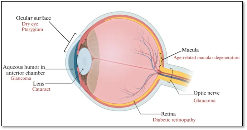
Figure 1: Oxidative Stress and Eye Diseases
1.3 Oxidative Stress and Inflammatory Considerations
In addition to mechanical and tear-film-related mechanisms, oxidative stress and inflammatory processes have received increasing attention in relation to ocular-surface health. The ocular tissues possess endogenous antioxidant systems that protect cellular components from reactive oxygen species and other oxidative challenges. Persistent environmental or metabolic stress may disturb the balance between reactive oxygen species generation and antioxidant defense, potentially affecting epithelial integrity and cellular homeostasis.
Prolonged exposure to digital displays, particularly in the context of intense visual activity and associated environmental stressors, has therefore generated interest in antioxidant-based approaches for supporting ocular health. High-energy visible light, including the blue-light region of the visible spectrum, has been investigated for its potential contribution to photooxidative processes; however, the clinical significance of blue-light exposure in DES remains an area of continuing investigation. Accordingly, antioxidant and anti-inflammatory interventions should be considered as potential supportive strategies rather than definitive treatments for all manifestations of DES.
1.4 Nutraceuticals as Potential Supportive Interventions
In parallel with topical approaches, nutritional strategies have attracted considerable interest for maintaining ocular health. Nutraceuticals are food-derived or naturally occurring bioactive substances that may provide physiological or health-promoting effects beyond basic nutritional requirements. Several nutrients and phytochemicals, including omega-3 polyunsaturated fatty acids, lutein, zeaxanthin, anthocyanins, vitamin A, and other antioxidant compounds, have been investigated for their potential roles in ocular-surface and visual health [14].
The proposed benefits of these bioactive compounds include antioxidant protection, modulation of inflammatory pathways, support of epithelial integrity, and potential improvement of tear-film-related functions. Their application in DES is particularly relevant because oxidative imbalance, inflammation, tear-film instability, and ocular-surface stress may occur concurrently in individuals with prolonged digital-device exposure. Nevertheless, the effectiveness of nutraceutical interventions depends on factors such as bioavailability, stability, dose, absorption, metabolism, and delivery to the intended site of action.
1.5 Hydrogel-Based Delivery Platforms
Hydrogels are three-dimensional hydrophilic polymeric networks capable of absorbing and retaining substantial quantities of water while maintaining structural integrity. Their networks may be formed through covalent cross-linking or reversible physical interactions, including hydrogen bonding, ionic interactions, hydrophobic interactions, and other intermolecular forces [9–11]. The resulting hydrated and flexible structure provides several characteristics desirable for localized delivery systems, including moisture retention, conformability, mechanical softness, and the ability to incorporate a wide range of active compounds.
The physicochemical characteristics of hydrogels make them attractive carriers for pharmaceutical agents, cosmetic ingredients, plant-derived compounds, and nutraceuticals. Bioactive substances incorporated within the polymeric network may be released through diffusion, swelling-controlled transport, polymer relaxation, erosion, or biodegradation. Depending on the formulation and route of application, hydrogels may additionally provide hydration and prolonged contact with the target surface [9–11].
For DES-associated discomfort, hydrogel-based patches may offer a multifunctional platform capable of combining localized delivery of bioactive compounds with hydration, cooling, and prolonged contact with the periorbital region. Their soft and flexible nature may also provide improved patient comfort compared with conventional rigid delivery systems. However, it is important to distinguish periocular hydrogel patches, which are applied to the skin surrounding the eye, from ocular inserts or ocular-surface hydrogels, which are designed for direct contact with ocular tissues. Their formulation requirements, safety considerations, residence mechanisms, and regulatory considerations may differ substantially.
1.6 Biodegradable Polymers and Their Relevance to Ocular Delivery
Biodegradability is an important consideration when designing hydrogel-based systems intended for repeated or prolonged use. Biodegradable polymers can undergo controlled breakdown into smaller products that can subsequently be metabolized, eliminated, or cleared through physiological processes, depending on their chemical composition and site of application. In appropriately designed systems, biodegradation may also contribute to controlled release of the incorporated bioactive compound.
For ocular and periocular applications, the selected polymer should possess appropriate biocompatibility, low irritation potential, adequate mechanical integrity, predictable hydration behavior, and controlled degradation characteristics. The polymer should not produce unacceptable local inflammation, cytotoxicity, or prolonged accumulation at the application site. Furthermore, polymer degradation can influence the release kinetics and local availability of incorporated nutraceuticals. Therefore, polymer selection must consider not only biodegradability but also physicochemical stability, cross-linking characteristics, swelling behavior, mechanical properties, release performance, and biological compatibility [31].
These requirements become particularly important for DES because the condition is commonly associated with repeated and prolonged exposure to digital devices. A patient-friendly delivery platform should therefore ideally provide comfort, ease of application, reproducible bioactive delivery, and an acceptable safety profile during repeated use.
1.7 Rationale for Nutraceutical-Loaded Biodegradable Hydrogel Patches
The combination of nutraceutical bioactives with biodegradable hydrogel technology provides an opportunity to integrate several potentially beneficial functions within a single delivery platform. A suitably designed hydrogel patch may act as a hydrated reservoir for nutraceutical compounds while providing localized and prolonged contact with the periorbital region. Depending on its anatomical site and formulation design, the system may provide antioxidant, anti-inflammatory, moisturizing, soothing, and cooling effects. The incorporation of nutraceuticals into a hydrogel matrix may additionally address some limitations associated with conventional administration, including poor stability, rapid clearance, limited residence time, and inconsistent local availability of bioactive compounds. The polymeric matrix can potentially protect sensitive constituents from environmental degradation and modulate their release according to the physicochemical properties of the active ingredient and the characteristics of the polymer network.
|
Structure |
Structure & Function |
Relevance to DES / CVS (mechanism & symptoms) |
References |
|
Tear film |
A three-layered coating over the cornea (lipid, aqueous, mucin) that lubricates, protects, and provides a smooth optical surface. |
Screen viewing reduces blink rate and increases incomplete blinks, causing greater tear evaporation, tear-film instability, dryness, and irritation core features of DES-associated dry eye. |
[16,29] |
|
Cornea |
Transparent anterior surface providing most of the eye’s refractive power; densely innervated for sensation. |
Exposed corneal nerve endings detect dryness and irritation; tear-film breakup and surface stress from screen use produce burning, foreign-body sensation, and pain typical of DES. |
[18,28] |
|
Conjunctiva |
Thin mucous membrane covering the sclera and inner eyelids; contributes mucin to tears and immune defence. |
Dryness and inflammation from reduced blinking and tear instability cause conjunctival redness, grittiness, and foreign-body sensation seen in DES/CVS. |
[16-26] |
|
Iris and pupil |
Control the amount of light entering the eye via pupil size. |
Glare, excessive brightness, and rapid contrast changes on screens increase visual discomfort and light sensitivity, aggravating DES symptoms. |
[17-29]
|
|
Lens |
Transparent, flexible structure that changes shape (accommodation) to focus near and distant objects. |
Prolonged near work increases accommodative demand; sustained focusing can produce blur, difficulty refocusing, and eyestrain in DES. |
[16-25] |
|
Ciliary muscle |
Smooth muscle that changes lens curvature during accommodation. |
Sustained contraction during near viewing contributes to asthenopia (eye fatigue), aching around the eyes, and focusing fatigue characteristic of CVS. |
[17-26] |
|
Retina |
Light-sensitive tissue (rods and cones) that converts focused light into electrical signals. |
Receives the focused image during screen tasks; while not typically structurally damaged by ordinary screen use, prolonged exposure can contribute to visual fatigue and transient blur. |
[16-29] |
|
Macula and fovea |
Central retinal region responsible for high-acuity, detailed vision. |
Continuously engaged during reading and screen-based near tasks; high demand can exacerbate symptoms like blurred vision and difficulty sustaining focus in DES. |
[17-26] |
|
Optic nerve |
Carries visual signals from retina to brain. |
Transmits visual information; ordinary screen use does not usually cause structural optic nerve damage, though visual fatigue may be perceived centrally. |
[16-29] |
Table 1: DES Ocular Structures, Function and Relevance
2. Core Molecular Pathways of Digital Eye Strain
Digital eye strain (DES) is a multifactorial condition involving interactions between ocular-surface stress, prolonged visual demand, oxidative imbalance, inflammatory responses, and retinal/visual processes. Although reduced blinking, tear-film instability, and sustained accommodation are established contributors to DES, increasing experimental evidence has highlighted several molecular pathways that may provide a mechanistic basis for the cellular consequences associated with prolonged digital-device exposure. In particular, blue-light-associated photooxidative stress, mitochondrial dysfunction, inflammatory signaling, impaired antioxidant defenses, alterations in visual-cycle homeostasis, and the gut–retina axis represent interconnected pathways of potential relevance. However, the strength of evidence differs among these mechanisms; some, particularly blue-light-induced oxidative and mitochondrial injury, are strongly supported by experimental models, whereas their direct contribution to routine human DES remains an evolving area of research [32–34].
2.1 Blue Light–Induced Photooxidative Stress
Digital displays emit visible light containing wavelengths within the short-wavelength region of the visible spectrum. Blue or high-energy visible (HEV) light has attracted particular attention because photons at shorter wavelengths possess relatively greater energy and can interact with endogenous ocular chromophores. Under excessive experimental exposure conditions, these interactions may increase the generation of reactive oxygen species (ROS), including superoxide anion, hydrogen peroxide, and hydroxyl radicals. Excessive ROS can subsequently initiate lipid peroxidation, protein oxidation, nucleic-acid damage, and cellular stress responses [32,33]. Experimental studies have demonstrated that blue-light exposure can increase ROS production and reduce cellular viability in ocular-surface and retinal models, providing a biological rationale for investigating photooxidative stress as one potential component of screen-associated ocular stress.
The retina is particularly susceptible to oxidative injury because of its high metabolic activity, high oxygen consumption, abundance of polyunsaturated fatty acids, and continuous exposure to light. Retinal pigment epithelial (RPE) cells and photoreceptors therefore depend on efficient antioxidant mechanisms to maintain redox homeostasis. Excessive photooxidative stress can disrupt cellular membranes, mitochondrial function, and photoreceptor/RPE homeostasis. Experimental evidence indicates that blue-light exposure can induce oxidative damage and apoptosis in retinal cells, while antioxidant interventions can attenuate some of these effects [32,33]. Nevertheless, the role of blue light in human DES should be interpreted cautiously. Experimental studies often employ controlled light intensities and exposure conditions that may not correspond directly to typical exposure from smartphones, tablets, or computer displays. Furthermore, clinical studies of blue-light filtering interventions have produced mixed findings regarding improvement of computer-related eye discomfort. Therefore, blue-light-induced photooxidative stress should be considered a potential contributing molecular pathway rather than the sole cause of DES [33,34].
2.2 Mitochondrial Dysfunction and Drp1-Mediated Fission
Mitochondria are central regulators of cellular energy production, redox homeostasis, calcium signaling, and programmed cell death. Ocular tissues, particularly retinal neurons and RPE cells, possess high metabolic requirements and consequently depend heavily on mitochondrial integrity. Excessive oxidative stress can impair mitochondrial membrane potential, electron-transport-chain activity, ATP generation, and mitochondrial quality-control mechanisms. Dysfunctional mitochondria can subsequently generate additional ROS, establishing a positive feedback loop between mitochondrial injury and oxidative stress [35,36]. Mitochondrial dynamics are regulated through coordinated processes of fission and fusion. Dynamin-related protein 1 (Drp1) is a major GTPase involved in mitochondrial fission. Under pathological stress conditions, increased Drp1 activity and recruitment to mitochondria can promote excessive mitochondrial fragmentation. Experimental investigation of blue-light exposure in retinal neuronal R28 cells demonstrated increased Drp1 expression, reduced mitofusin-2 (MFN2) expression, enhanced mitochondrial fission, increased ROS generation, disruption of mitochondrial membrane potential, and apoptosis. Importantly, pharmacological inhibition or genetic suppression of Drp1 attenuated mitochondrial fragmentation, ROS production, membrane-potential disruption, and apoptosis, supporting a mechanistic role for Drp1-mediated mitochondrial fission in blue-light-induced retinal cellular injury [35].
Recent experimental evidence has further strengthened this mechanistic relationship. Blue-light exposure has been reported to increase Drp1-dependent mitochondrial fission and subsequently activate an NF-κB/p65–NOX4 signaling axis, leading to increased mitochondrial ROS generation and retinal ganglion-cell injury. Inhibition of Drp1 or NOX4 reduced oxidative stress and protected against cellular damage in experimental models [36]. These findings suggest that mitochondrial dynamics may act as an important molecular bridge connecting photooxidative stress with inflammatory and apoptotic pathways.
2.3 Inflammatory Cascade Activation
Oxidative stress and mitochondrial dysfunction can activate intracellular inflammatory signaling pathways, creating an important connection between cellular stress and ocular inflammation. Reactive oxygen species can stimulate nuclear factor-kappa B (NF-κB), mitogen-activated protein kinase (MAPK), and inflammasome-associated signaling. Activation of these pathways can increase the expression and release of pro-inflammatory mediators such as interleukin (IL)-1β, IL-6, tumor necrosis factor-α (TNF-α), and other inflammatory molecules. Persistent activation of these pathways can impair epithelial integrity, alter cellular homeostasis, and amplify ocular-surface discomfort [37,38].
NF-κB is particularly important because it functions as a central transcriptional regulator of inflammatory gene expression. Oxidative stress, hyperosmolarity, cellular injury, and mitochondrial dysfunction can activate NF-κB signaling. Experimental blue-light studies have additionally demonstrated interaction between mitochondrial fission and NF-κB signaling, with Drp1-dependent mitochondrial dysfunction associated with increased p65 activation and downstream NOX4-mediated ROS production [36]. Thus, oxidative stress, mitochondrial dysfunction, and inflammation may operate as an interconnected molecular network rather than as independent pathways. Inflammasome signaling may provide another level of inflammatory amplification. Cellular damage and mitochondrial dysfunction can generate danger-associated molecular signals capable of activating inflammasome complexes, including the NLRP3 inflammasome. Activation of such pathways can promote maturation of IL-1β and IL-18 and contribute to persistent inflammatory responses. Although inflammasome activation is well established in ocular-surface and retinal inflammatory disorders, its specific contribution to routine DES requires further investigation. Accordingly, inflammatory signaling should be regarded as an important candidate mechanism linking oxidative stress with ocular discomfort and tissue dysfunction rather than as a proven single molecular cause of DES.
2.4 Impaired Antioxidant Defenses
Under physiological conditions, ocular tissues possess sophisticated antioxidant systems that neutralize ROS and protect cellular components from oxidative damage. Important enzymatic defenses include superoxide dismutase (SOD), catalase, glutathione peroxidase (GPx), glutathione reductase, and related antioxidant enzymes, while non-enzymatic defenses include reduced glutathione, vitamins, carotenoids, and other endogenous molecules. Maintenance of an appropriate balance between ROS generation and antioxidant capacity is essential for preserving ocular-surface and retinal homeostasis [39,40].
The nuclear factor erythroid 2-related factor 2 (Nrf2)–antioxidant response element (ARE) pathway represents a major regulator of endogenous cytoprotective defense. Under oxidative conditions, Nrf2 can translocate to the nucleus and activate genes involved in ROS detoxification, glutathione synthesis, and cellular protection, including heme oxygenase-1 (HO-1) and NAD(P)H quinone dehydrogenase 1 (NQO1). Impairment of this pathway can increase cellular susceptibility to oxidative injury [39]. Blue-light-associated oxidative stress may place substantial demands on these antioxidant systems. Experimental studies have shown alterations in antioxidant enzymes following blue-light exposure in ocular cells, including changes in SOD and GPx-related responses [40]. Therefore, prolonged oxidative challenge combined with inadequate antioxidant protection may promote accumulation of ROS and intensify mitochondrial and inflammatory injury. This mechanism provides a particularly relevant rationale for the investigation of nutraceuticals containing antioxidant compounds capable of directly scavenging ROS or enhancing endogenous antioxidant pathways.
2.5 Visual Cycle Disruption
The visual cycle is a highly coordinated biochemical process that regenerates visual chromophore required for phototransduction. It involves continuous interaction between photoreceptors and the RPE, where all-trans-retinal generated following photon absorption is enzymatically converted through a series of reactions toward 11-cis-retinal, which is subsequently supplied back to photoreceptors for regeneration of visual pigments. Efficient functioning of this cycle is essential for sustained visual perception and adaptation to changing light conditions. Prolonged visual stimulation and oxidative stress may place additional metabolic demands on photoreceptors and RPE cells. The visual cycle itself generates reactive aldehyde intermediates, particularly retinaldehyde, which can participate in oxidative reactions when cellular detoxification and clearance mechanisms become overwhelmed. Accumulation of reactive retinal derivatives can contribute to oxidative modification of cellular proteins and lipids and may compromise RPE and photoreceptor homeostasis. Blue-light-associated retinal stress has consequently been investigated in relation to photoreceptor and RPE injury and disturbances in retinal homeostasis [32,41]. However, direct evidence demonstrating that ordinary digital-screen exposure causes clinically significant disruption of the visual cycle in humans with DES remains limited. Therefore, visual-cycle disturbance should be presented as a potential downstream molecular consequence of retinal oxidative stress and prolonged photic stimulation, rather than as an established primary mechanism of DES.
2.6 Gut–Retina Axis Modulation
The gut–retina axis represents an emerging concept linking intestinal microbiota, microbial metabolites, systemic immunity, and retinal homeostasis. The intestinal microbiome can influence host metabolism, immune regulation, epithelial-barrier integrity, and systemic inflammatory status. Alterations in gut microbial composition or intestinal permeability may increase exposure to microbial products and metabolites capable of influencing systemic inflammatory pathways and distant tissues, including the retina [42,43]. Short-chain fatty acids (SCFAs), including acetate, propionate, and butyrate, are among the important microbial metabolites involved in immune and metabolic regulation. Changes in microbial composition can modify the production and availability of these metabolites and may consequently influence inflammatory and oxidative pathways. Evidence from retinal diseases such as age-related macular degeneration and diabetic retinopathy suggests associations between gut-microbiome alterations, systemic inflammation, and retinal pathology [42,43]. The relevance of the gut–retina axis to DES is currently hypothetical and emerging, rather than directly established. Nevertheless, this pathway is particularly interesting for nutraceutical-based approaches because dietary bioactive can influence both antioxidant status and gut microbial composition. Nutraceuticals with antioxidant, anti-inflammatory, prebiotic, or microbiota-modulating properties may therefore provide a broader biological influence extending beyond direct ocular effects. Further studies are required to determine whether modulation of the gut–retina axis can meaningfully influence digital-screen-associated ocular symptoms.
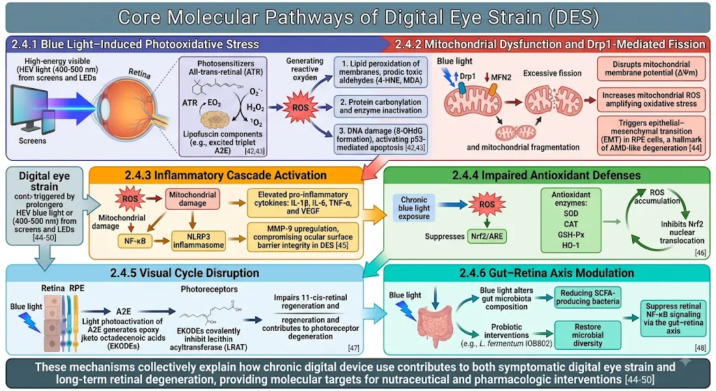
Figure 2: Core Molecular Pathways of Digital Eye Strain (DES)
3. Nutraceuticals Used Against Digital Eye Strain
3.1 Definition of Nutraceuticals
The term nutraceutical was introduced by Stephen De Felice in 1989 and was derived from the words “nutrition” and “pharmaceutical.” Nutraceuticals are broadly described as food-derived or naturally occurring biologically active substances that may provide physiological or health-promoting benefits beyond their basic nutritional functions. This broad category includes nutrients, vitamins, minerals, carotenoids, polyphenols, flavonoids, fatty acids, peptides, probiotics, prebiotics, and other bioactive compounds obtained from food or food-associated sources [44,45]. Because the term does not have a universally harmonized regulatory definition across countries, nutraceuticals are more appropriately considered a broad scientific and functional category rather than a standardized pharmaceutical classification [44,45].
Nutraceuticals have attracted considerable interest in ocular-health research because the eye, particularly the retina and ocular surface, is highly susceptible to oxidative and inflammatory stress. High metabolic activity, continuous exposure to light, and the abundance of polyunsaturated fatty acids make ocular tissues particularly dependent on effective antioxidant defense mechanisms. Nutraceutical compounds possessing antioxidant, anti-inflammatory, photoprotective, membrane-stabilizing, or immunomodulatory properties may therefore contribute to the maintenance of ocular homeostasis [46,47]. The relevance of nutraceuticals to digital eye strain (DES) arises from the multifactorial nature of the condition. DES, also referred to as computer vision syndrome or digital asthenopia, encompasses ocular and visual symptoms such as dryness, irritation, burning, blurred or fluctuating vision, ocular fatigue, headache, and difficulty maintaining near focus following prolonged digital-device use. Although tear-film instability, reduced blinking, accommodative demand, and ergonomic factors represent important contributors, oxidative and inflammatory mechanisms have also received increasing attention [48,49]. Consequently, nutraceuticals capable of modulating oxidative stress and inflammatory responses may provide supportive benefits for individuals experiencing screen-associated ocular discomfort.
For the proposed biodegradable nutraceutical-loaded hydrogel patch, nutraceuticals can be regarded as the bioactive functional components responsible for potentially providing antioxidant, anti-inflammatory, photoprotective, cytoprotective, or ocular-surface-supporting effects. Incorporation into a hydrogel matrix may offer opportunities for localized application and prolonged contact compared with conventional topical administration. However, the suitability of an individual nutraceutical depends on its chemical structure, molecular weight, aqueous solubility, lipophilicity, chemical stability, susceptibility to oxidation, required concentration, biological safety, and compatibility with the selected polymeric network [50,51]. Therefore, nutraceutical selection should be considered simultaneously with polymer selection and formulation design.
3.2 Classification of Nutraceuticals
Nutraceuticals can be classified according to their chemical composition, biological source, physiological function, or method of production. Based on chemical characteristics, major groups include vitamins, minerals, carotenoids, fatty acids, polyphenols, flavonoids, anthocyanins, proteins and peptides, carbohydrates, probiotics, prebiotics, and other naturally occurring bioactive molecules [45,52]. Based on origin, nutraceuticals may be obtained from plant, animal, microbial, marine, or mineral sources. They may also be categorized as traditional nutraceuticals, which are obtained directly from conventional foods or naturally occurring food components, and non-traditional nutraceuticals, which may involve enrichment, fortification, modification, or biotechnological processing [52]. From the perspective of ocular health and DES, particular interest has been directed toward carotenoids, anthocyanins, flavonoids, polyphenols, omega-3 fatty acids, and antioxidant vitamins. These compounds have been investigated because of their ability to modulate oxidative stress, inflammatory responses, cellular signaling, membrane stability, and retinal or ocular-surface function [46,47,53]. Their relevance is further strengthened by the molecular pathways discussed previously, in which excessive reactive oxygen species, mitochondrial dysfunction, inflammatory signaling, and impaired antioxidant defenses may contribute to ocular stress associated with prolonged digital-device use.
Carotenoids such as lutein and zeaxanthin are particularly important because they are concentrated within ocular tissues and can participate in light filtering and antioxidant protection. Anthocyanins and other polyphenolic compounds have attracted attention because of their free-radical-scavenging and anti-inflammatory properties, whereas omega-3 polyunsaturated fatty acids have been investigated for their potential effects on inflammatory pathways and ocular-surface function [46,53]. Vitamins with antioxidant or ocular-supportive roles, particularly vitamins A, C, and E, have also been considered important components of nutritional strategies for maintaining ocular health. For hydrogel-patch development, chemical classification is particularly useful because the physicochemical properties of each nutraceutical strongly influence its incorporation and release from the polymeric matrix. Hydrophilic compounds may readily distribute within the aqueous phase of a hydrogel, whereas poorly water-soluble compounds such as certain carotenoids may require solubilization, emulsification, nanoencapsulation, cyclodextrin complexation, or other formulation approaches. Consequently, nutraceutical classification is not only biologically relevant but also directly influences formulation strategy, loading efficiency, stability, release kinetics and biological performance.
3.3 Nutraceuticals from Herbal Sources for Ocular Hydrogel Patches
Plant-derived nutraceuticals represent an especially attractive group for the development of ocular-health formulations because plants provide structurally diverse carotenoids, flavonoids, anthocyanins, phenolic acids, tannins, terpenoids, and other antioxidant constituents. Many of these compounds exhibit free-radical-scavenging, metal-chelating, anti-inflammatory, membrane-protective, and cytoprotective activities in experimental systems [53,54]. Several herbal-derived compounds have been investigated in relation to ocular oxidative stress and retinal protection. Polyphenolic compounds such as curcumin, resveratrol, quercetin, epigallocatechin gallate, and related phytochemicals have demonstrated antioxidant and anti-inflammatory activities in experimental ocular models. Anthocyanin-rich extracts, particularly those obtained from berries and other deeply pigmented fruits, have also attracted interest because of their potential antioxidant and vascular or retinal protective effects [55,56]. Similarly, carotenoid-rich botanical sources can provide lutein, zeaxanthin, and related pigments that participate in ocular photoprotection and antioxidant defense [46,57].
The potential importance of these compounds to DES is associated with their ability to target pathways implicated in oxidative and inflammatory stress. Polyphenols and flavonoids may directly neutralize reactive oxygen species and may additionally modulate intracellular signaling pathways such as NF-κB, MAPK, and Nrf2. Some phytochemicals have also demonstrated the ability to preserve mitochondrial function and reduce oxidative damage in experimental cellular systems [54–56]. These properties make herbal nutraceuticals attractive candidates for further investigation in localized delivery systems designed to support ocular or periocular health. However, translating evidence from oral nutraceutical supplementation into a hydrogel patch requires careful consideration. Most available clinical evidence for ocular nutraceuticals has been generated following dietary or oral administration, and systemic absorption, metabolism, and tissue distribution may differ substantially from localized topical delivery. Furthermore, several plant-derived compounds exhibit poor aqueous solubility, chemical instability, rapid oxidation, limited permeability, or low bioavailability, which can restrict their therapeutic utilization [50,58]. These limitations highlight the need for appropriate formulation strategies.
Biodegradable hydrogel matrices may provide a promising approach for addressing some of these limitations. The hydrated polymer network can act as a reservoir for selected bioactive
compounds and may protect susceptible constituents from environmental degradation. Depending on the physicochemical properties of the nutraceutical and polymer, controlled release may be achieved through diffusion, swelling, polymer relaxation, erosion, or biodegradation. A hydrogel patch applied to the periorbital region may additionally provide hydration and a cooling or soothing sensation, potentially complementing the biological activity of the incorporated nutraceutical. Nevertheless, the terminology and route of application must be carefully defined. A periocular hydrogel patch applied to the skin surrounding the eye is pharmacologically and anatomically different from an ocular insert or ocular-surface hydrogel intended for direct contact with the cornea or conjunctiva. Consequently, claims regarding ocular bioavailability, corneal penetration, retinal delivery, or direct treatment of ocular tissues should not be made solely on the basis of periorbital application. For the proposed system, the intended site of application and mechanism of action should therefore be clearly established during formulation development and supported by appropriate permeation, retention, safety, and efficacy studies [58,59].
Overall, herbal nutraceuticals provide a diverse reservoir of potentially useful antioxidant and anti-inflammatory compounds for the development of biodegradable hydrogel patches. Their combination with advanced polymeric delivery systems may overcome some limitations associated with conventional administration by improving local retention, protecting unstable bioactives and enabling controlled release. However, the therapeutic potential of these systems for DES remains an emerging research area, and systematic evaluation of formulation performance, ocular/periocular safety, bioactivity, stability, and clinical efficacy is necessary before such products can be considered established interventions [59,60].
|
Herbal source |
Important nutraceutical/ bioactive |
Major pharmacological activity |
Potential relevance to DES |
Potential role in hydrogel patch |
References |
|
Marigold (Tagetes erecta L.) |
Lutein, zeaxanthin |
Antioxidant; blue-light filtering; macular photoprotection |
May counter light-associated oxidative stress and support visual performance |
Candidate antioxidant/photoprotective active |
[59,63] |
|
Bilberry (Vaccinium myrtillus L.) |
Anthocyanins/anthocyanosides |
Antioxidant; possible support of tear secretion |
Particularly relevant to dry-eye component of DES |
Candidate antioxidant and ocular-surface-supporting active |
[64] |
|
Saffron (Crocus sativus L.) |
Crocin, crocetin |
Antioxidant, anti-inflammatory, neuroprotective |
Potential protection against oxidative retinal stress |
Candidate antioxidant/neuroprotective active |
[65,66] |
|
Turmeric (Curcuma longa L.) |
Curcumin/curcuminoids |
Antioxidant, anti-inflammatory |
Potential relevance to inflammatory and oxidative ocular processes |
Candidate anti-inflammatory antioxidant |
[ 67,68] |
|
Green tea (Camellia sinensis L.) |
Catechins, particularly EGCG |
Antioxidant and anti-inflammatory |
Potential protection against oxidative/inflammatory ocular stress |
Candidate polyphenolic antioxidant |
[69,70] |
|
Grape seed extract (Vitis vinifera L.) |
Anthocyanins/polyphenols |
Antioxidant; possible ocular-surface benefits |
Potential relevance to dry-eye symptoms associated with prolonged screen use |
Candidate antioxidant active |
[71] |
|
Chokeberry (Aronia melanocarpa) |
Anthocyanins |
Antioxidant |
Potential support for ocular surface and visual function |
Candidate polyphenolic active |
[ 72] |
|
Honeysuckle (Lonicera caerulea) |
Anthocyanins, iridoids |
Antioxidant and potential ocular-surface support |
May support tear-film-related parameters |
Candidate botanical antioxidant |
[72] |
Table 2: Herbal-Source Nutraceuticals of Potential Interest for a DES Hydrogel Eye Patch
3.3.1 Marigold (Tagetes erecta) – Lutein and Zeaxanthin
Marigold (Tagetes erecta L.) is an important botanical source of the xanthophyll carotenoids lutein and zeaxanthin and is widely utilized for the production of carotenoid-rich nutritional ingredients. Marigold extracts contain predominantly lutein, together with smaller quantities of zeaxanthin and their corresponding esters. Following absorption, these xanthophyll carotenoids are selectively distributed within ocular tissues, particularly the macular region, where they contribute to optical filtering of short-wavelength visible light and provide antioxidant protection against photooxidative stress [63,64].
Lutein and zeaxanthin are of particular interest in the context of prolonged digital-device use because oxidative stress and visually demanding near work may contribute to visual fatigue and ocular discomfort. Their ability to absorb portions of the blue-light spectrum and neutralize reactive oxygen species provides a mechanistic rationale for their investigation as supportive nutritional agents for screen-associated visual stress. A randomized controlled study conducted in individuals with frequent electronic-screen exposure reported that six months of lutein and zeaxanthin supplementation was associated with improvements in selected ophthalmic parameters, including Schirmer test results, photostress recovery, and tear-film breakup time, although not all subjective outcomes differed significantly from placebo [65]. These findings suggest potential benefits of lutein and zeaxanthin for ocular function in individuals with high screen exposure; however, evidence from oral supplementation cannot be directly extrapolated to a topical hydrogel patch.
Marigold-derived lutein and zeaxanthin are therefore attractive candidates for incorporation into nutraceutical delivery systems because of their antioxidant and photoprotective characteristics. Nevertheless, both compounds are highly lipophilic and exhibit poor aqueous solubility. Their incorporation into a hydrophilic hydrogel therefore presents formulation challenges related to solubilization, dispersion, chemical stability, protection against oxidation, and uniform distribution within the polymeric matrix [64,66]. Appropriate approaches such as lipid-based carriers, emulsification, cyclodextrin complexation, nanoencapsulation, or incorporation of suitable solubilizing excipients may be considered to improve their formulation performance.
3.3.2 Bilberry (Vaccinium myrtillus) – Anthocyanins
Bilberry (Vaccinium myrtillus L.) is a recognized botanical source of anthocyanins, including delphinidin-, cyanidin-, malvidin-, petunidin-, and peonidin-derived glycosides. These polyphenolic pigments possess pronounced antioxidant activity and have been investigated for their potential effects on vascular, retinal, and ocular-surface function [67]. Their ability to scavenge reactive oxygen species and modulate oxidative and inflammatory signaling makes bilberry anthocyanins relevant to nutraceutical approaches targeting ocular stress. The potential relevance of bilberry to DES is supported indirectly by evidence obtained from ocular-surface and dry-eye research. A randomized, double-blind, placebo-controlled study of standardized bilberry extract in individuals with dry-eye symptoms reported improvement in tear secretion and systemic antioxidant status following oral supplementation [68]. Since ocular dryness and discomfort constitute important components of DES, such findings provide a rationale for further investigation of bilberry-derived anthocyanins in screen-associated ocular stress.
However, the available evidence predominantly concerns oral administration, and the pharmacokinetic behavior of anthocyanins following topical or periocular application may differ substantially. Furthermore, anthocyanins may be susceptible to degradation depending on pH, temperature, oxygen exposure, light, and formulation environment. Consequently, incorporation into a hydrogel system would require careful optimization of polymer composition, pH, antioxidant protection, packaging, and storage conditions [67,69]. These considerations make bilberry anthocyanins promising but formulation-sensitive candidates for biodegradable nutraceutical hydrogel patches.
3.3.3 Saffron (Crocus sativus) – Crocin and Crocetin
Saffron (Crocus sativus L.) contains several biologically active constituents, among which crocin and crocetin are particularly relevant to ocular-health research. These carotenoid-derived compounds have demonstrated antioxidant, anti-inflammatory, neuroprotective, and cytoprotective properties in experimental studies [70]. Saffron-derived bioactives have been investigated in relation to retinal function and disorders involving oxidative and neurodegenerative mechanisms, including age-related macular degeneration and other retinal conditions [71]. The potential relevance of crocin and crocetin to DES is primarily related to their ability to modulate oxidative stress and inflammatory pathways. Because prolonged visual activity and screen-associated environmental stress may increase oxidative burden, compounds capable of enhancing cellular resistance to oxidative injury may provide supportive benefits. Clinical and experimental studies involving saffron supplementation have reported effects on visual function and retinal parameters, although these findings primarily concern retinal disorders rather than DES itself [70,71]. Accordingly, saffron-derived bioactives should presently be regarded as potential nutraceutical candidates rather than established treatments for digital eye strain. Their incorporation into a biodegradable hydrogel patch may be explored to provide localized delivery of antioxidant compounds; however, appropriate studies are required to establish their stability, loading capacity, release behavior, permeation, safety, and biological activity following topical application.
3.3.4 Turmeric (Curcuma longa) – Curcumin
Curcumin is the principal biologically active polyphenolic constituent of turmeric (Curcuma longa L.). It has been extensively investigated because of its antioxidant, anti-inflammatory, antimicrobial, and cytoprotective activities. Curcumin can interact with several molecular pathways associated with oxidative stress and inflammation, including NF-κB, MAPK, and Nrf2-associated signaling. These characteristics have generated considerable interest in its application to ocular disorders involving oxidative injury and inflammation, including experimental dry-eye and ocular-surface conditions [72,73]. From a DES perspective, curcumin is of interest because oxidative stress and inflammatory signaling have been proposed as components of the biological response associated with prolonged digital-device use. Its antioxidant activity may help neutralize reactive oxygen species, while its anti-inflammatory effects may potentially modulate inflammatory signaling associated with ocular-surface stress. However, direct clinical evidence demonstrating that curcumin specifically prevents or treats DES remains limited, and its potential role should therefore be considered supportive and investigational. A major limitation of curcumin is its extremely low aqueous solubility, chemical instability under certain environmental conditions, and limited bioavailability. These characteristics can restrict its effective utilization in conventional formulations [73,74]. Consequently, advanced delivery systems, including nanoparticles, liposomes, nanoemulsions, polymeric carriers, and hydrogel-based systems, have been investigated to improve its solubility, stability, retention, and controlled release. Incorporation of curcumin into a biodegradable hydrogel could therefore provide an opportunity to overcome some of these limitations while maintaining prolonged contact with the intended application site [74].
3.3.5 Green Tea (Camellia sinensis) – Catechins
Green tea (Camellia sinensis L.) is a rich source of polyphenolic catechins, including epigallocatechin gallate (EGCG), epigallocatechin, epicatechin gallate, and epicatechin. Among these constituents, EGCG has received particular attention because of its potent antioxidant and anti-inflammatory properties. Green-tea catechins can scavenge reactive oxygen species and influence several cellular signaling pathways involved in oxidative stress, inflammation, apoptosis, and cellular protection [75]. The potential relevance of green-tea catechins to DES is associated with the proposed contribution of oxidative stress and inflammatory responses to screen-related ocular discomfort. Experimental ocular research suggests that catechins may exert protective effects against oxidative and inflammatory injury in ocular tissues [75,76]. These properties make green-tea-derived bioactives potential candidates for incorporation into nutraceutical delivery systems designed to support ocular or periocular health. However, direct evidence demonstrating the effectiveness of green-tea catechins in DES, particularly following topical hydrogel administration, remains limited. In addition, catechins can undergo oxidation and degradation during processing and storage, which may influence formulation stability and biological activity. Therefore, hydrogel formulation should consider pH, oxygen exposure, light protection, polymer–polyphenol interactions, and appropriate packaging [76]. Further experimental studies are necessary to determine whether localized delivery of catechins can provide meaningful benefits for digital-screen-associated ocular symptoms.
3.3.6 Grape (Vitis vinifera) – Anthocyanins and Polyphenols
Grape (Vitis vinifera L.) represents an important dietary source of polyphenolic compounds, including anthocyanins, flavonoids, proanthocyanidins, and other phenolic constituents. These compounds exhibit antioxidant and anti-inflammatory properties and have been investigated for their potential role in maintaining ocular and vascular health [77]. Grape-derived polyphenols may neutralize reactive oxygen species and modulate cellular pathways involved in oxidative stress and inflammation. The potential application of grape-derived bioactives to DES is supported primarily by their relevance to ocular-surface oxidative stress and dry-eye-related symptoms. A randomized, double-blind, placebo-controlled study investigating an anthocyanin oligomer preparation derived from grape skin reported beneficial effects in individuals with dry-eye disease [78]. Because ocular dryness, irritation, and discomfort are major components of DES, these findings provide a rationale for further investigation of grape-derived anthocyanins and polyphenols in screen-associated ocular stress.
Nevertheless, the available clinical evidence predominantly involves oral supplementation rather than localized delivery. Consequently, the efficacy and safety of grape-derived bioactives cannot be assumed to be equivalent following incorporation into a periocular hydrogel patch. In addition, the chemical stability of anthocyanins and polyphenols can be influenced by pH, temperature, oxygen, light, and interactions with other formulation components. Therefore, formulation-specific investigations are required to determine loading efficiency, stability, release characteristics, skin compatibility, and biological activity following hydrogel incorporation [77–79]. Overall, the herbal nutraceuticals discussed above represent chemically diverse candidates with complementary mechanisms potentially relevant to DES. Lutein and zeaxanthin provide carotenoid-based photoprotective and antioxidant activity; bilberry anthocyanins offer potent polyphenolic antioxidant activity; crocin and crocetin provide antioxidant and potential retinal-supportive effects; curcumin combines antioxidant and anti-inflammatory activity; green-tea catechins provide polyphenolic redox and inflammatory modulation; and grape-derived anthocyanins and polyphenols offer additional antioxidant and ocular-surface-supportive potential. Their different physicochemical characteristics, however, necessitate individualized formulation strategies when incorporated into biodegradable hydrogel matrices.
4.1 Methods of Formulation of Biodegradable Hydrogel Eye Patches
The selection of an appropriate formulation method for biodegradable nutraceutical-loaded hydrogel patches depends on several factors, including the chemical nature and stability of the nutraceutical, polymer characteristics, desired mechanical properties, degree of biodegradability, intended site of application, swelling behavior, required release profile, and sensitivity of the active constituent to heat, light, oxygen, solvents, and crosslinking conditions. The method must also provide adequate uniformity, reproducibility, structural integrity, and compatibility with the intended route of administration. Commonly investigated approaches for hydrogel-based ocular and periocular delivery systems include solvent casting, freeze–thaw physical crosslinking, ionic gelation, chemical crosslinking, photopolymerization, and advanced additive-manufacturing techniques such as three-dimensional (3D) printing [80–84].
4.1.1 Solvent-Casting Method
Solvent casting is one of the simplest and most widely applicable techniques for preparing thin polymeric films, membranes, and patch-like hydrogel systems. In this approach, the selected biodegradable or biocompatible polymer is dissolved or uniformly dispersed in an appropriate solvent or aqueous medium. The nutraceutical is subsequently incorporated under controlled stirring or homogenization to obtain a uniform formulation. Depending on the intended mechanical characteristics, plasticizers, secondary polymers, humectants, or other functional excipients may be incorporated. The resulting homogeneous dispersion is transferred onto a suitable mould or flat casting surface and subjected to controlled drying to remove the solvent and form a thin polymeric film. The dried film can subsequently be peeled from the casting surface and cut into patches of predetermined dimensions [81,82].
For nutraceutical-loaded systems, solvent casting offers several advantages, including relatively simple processing, low equipment requirements, flexibility in controlling polymer concentration, and the possibility of incorporating thermolabile bioactive compounds under mild processing conditions. Film thickness, polymer concentration, plasticizer concentration, drying conditions, and casting volume can be adjusted to modify flexibility, swelling, mechanical strength, and release characteristics [81,83]. The method is therefore particularly attractive for preliminary development of biodegradable periocular hydrogel patches. However, several formulation challenges must be considered. Inadequate mixing may produce nonuniform nutraceutical distribution, whereas inappropriate drying conditions may result in shrinkage, cracking, excessive brittleness, or residual solvent. Poorly soluble nutraceuticals may undergo precipitation or crystallization during solvent evaporation, leading to dose nonuniformity and altered release behavior. In addition, volatile or oxidation-sensitive phytochemicals may be lost or degraded during processing. For a formulation intended for application around the eye, the selection of solvents and excipients must therefore prioritize biocompatibility, and residual solvent levels should be appropriately controlled [80,82,84].
4.1.2 Freeze–Thaw Crosslinking
Freeze–thaw processing is a physical crosslinking technique particularly associated with poly (vinyl alcohol) (PVA)-based hydrogel systems. During the freezing stage, water molecules form ice crystals and the polymer chains become concentrated within the unfrozen regions. Subsequent thawing promotes formation of crystalline domains and intermolecular interactions that act as physical crosslinking points within the polymer network. Repeated freeze–thaw cycles generally increase the extent of network formation and can modify the mechanical strength, swelling characteristics, porosity, and diffusion-controlled release properties of the resulting hydrogel [82,85].
An important advantage of freeze–thaw crosslinking is that it can reduce or eliminate the need for potentially cytotoxic chemical crosslinking agents. This feature is particularly attractive for systems intended for prolonged contact with sensitive biological tissues. The number and duration of freeze–thaw cycles, polymer concentration, molecular weight, freezing temperature, and thawing conditions can be optimized to control the structural characteristics of the hydrogel [85,86]. For nutraceutical-loaded systems, however, the stability of the active compound must be considered carefully. Repeated exposure to low temperatures and subsequent thawing may influence the physical state, aggregation, oxidation, or activity of sensitive bioactive compounds. Accordingly, the nutraceutical may be incorporated before network formation or loaded after formation of the polymeric matrix, depending on its physicochemical stability and desired release characteristics [82,86]. Freeze–thaw processing may therefore be particularly useful for developing physically crosslinked PVA-containing biodegradable or bioresorbable composite hydrogel systems, provided that the overall polymer composition satisfies the intended safety and degradation requirements.
4.1.3 Ionic Gelation
Ionic gelation is a mild crosslinking approach that utilizes electrostatic interactions between oppositely charged polymers or between polymers and multivalent ions. The method is particularly useful for natural polymers such as alginate and chitosan. Alginate, an anionic polysaccharide, can undergo ionic crosslinking in the presence of divalent cations such as calcium ions, producing an interconnected three-dimensional network. Conversely, positively charged chitosan can interact electrostatically with negatively charged crosslinking agents such as sodium tripolyphosphate (TPP) or with anionic polymers including alginate and hyaluronic acid [80,87]. The mild aqueous processing conditions associated with ionic gelation are advantageous for incorporating thermolabile and oxidation-sensitive nutraceuticals. Because extensive organic solvents and high temperatures can often be avoided, the technique is potentially suitable for incorporating plant-derived compounds such as anthocyanins, flavonoids, polyphenols, and other sensitive bioactives [80,87]. Chitosan–alginate and chitosan–hyaluronic acid combinations may also provide opportunities to modify mucoadhesion, hydration, swelling, and release properties.
For biodegradable nutraceutical-loaded hydrogel patches, the concentration of polymer and ionic crosslinker must be carefully optimized. Excessive crosslinking can produce a dense network with reduced swelling and slower nutraceutical diffusion, whereas insufficient crosslinking may result in inadequate mechanical strength, excessive swelling, and rapid erosion [87,88]. The ionic environment may also influence the stability and ionization state of incorporated nutraceuticals. Consequently, optimization of polymer ratio, crosslinker concentration, crosslinking time, pH, and ionic strength is essential.
4.1.4 Chemical Crosslinking
Chemical crosslinking involves the formation of covalent bonds between polymer chains, producing a relatively stable three-dimensional hydrogel network. Depending on the polymeric components, several chemical strategies can be employed, including aldehyde-mediated crosslinking, carbodiimide-mediated coupling, click reactions, Michael addition, and other covalent crosslinking approaches [84,89]. Chemical crosslinking can provide greater structural stability and allows precise modification of network density, swelling behaviour, degradation rate, and release kinetics. Photo cross linkable polymers such as gelatin methacrylate (GelMA) represent another important class of chemically crosslinked hydrogel materials. GelMA contains methacrylated functional groups that can undergo polymerization in the presence of an appropriate photoinitiator following exposure to light. Such systems have attracted interest for biomedical applications because their network properties can be adjusted by controlling polymer concentration, degree of functionalization, photoinitiator concentration, and irradiation conditions [89,90].
Despite these advantages, chemical crosslinking requires particular attention in ocular and periocular applications. Unreacted monomers, crosslinking agents, photoinitiators, reaction by-products, or residual solvents may potentially produce irritation, cytotoxicity, or inflammatory responses. Therefore, appropriate purification, washing, residual-material analysis, physicochemical characterization, and biological safety evaluation are necessary before considering a chemically crosslinked hydrogel for ocular-related application [84,89]. For nutraceutical-loaded systems, the chemical compatibility of the active constituent with the crosslinking chemistry must also be established because reactive functional groups or processing conditions may alter the stability or biological activity of sensitive phytochemicals.
4.1.5 Photopolymerization
Photopolymerization enables rapid formation of hydrogel networks through light-induced polymerization of photocrosslinkable functional groups. In a typical approach, a photocrosslinkable polymer is combined with a suitable photoinitiator and exposed to a controlled wavelength and intensity of light. The resulting polymerization generates a three-dimensional hydrogel network, and the degree of crosslinking can be regulated through polymer concentration, exposure time, light intensity, and photoinitiator concentration [89,90]. An important advantage of photopolymerization is the ability to produce hydrogel structures with defined geometry and relatively precise spatial control. This characteristic is valuable for customized ocular inserts, microstructured systems, and other geometrically controlled delivery platforms. Photopolymerization may also allow rapid fabrication with minimal mechanical manipulation of the formulation [90]. Nevertheless, the method requires careful evaluation for nutraceutical delivery. Exposure to ultraviolet or high-energy visible radiation may degrade photosensitive compounds, while some photoinitiators and unreacted components may raise concerns regarding biological compatibility. Therefore, visible-light-compatible photoinitiators, optimized irradiation conditions, and appropriate purification strategies may be required. The final formulation must also be evaluated for residual photoinitiator, cytotoxicity, degradation behavior, and active-ingredient stability [89,90].
4.1.6 3D Printing/Advanced Fabrication
Three-dimensional printing represents an advanced fabrication approach capable of producing hydrogel-based structures with precise control over geometry, thickness, internal architecture, porosity, and spatial distribution of formulation components. In extrusion-based bioprinting and related approaches, a polymeric hydrogel or bioink is deposited layer-by-layer according to a predefined digital design. Crosslinking may occur simultaneously or after printing through ionic, chemical, enzymatic, or photochemical mechanisms [90–92]. For ocular drug-delivery applications, 3D printing offers the possibility of producing customized inserts and patient-specific structures with controlled dimensions and potentially tailored release characteristics. The technique may also permit incorporation of multiple materials or active ingredients within different regions of the same structure. Recent investigations into 3D-printed biodegradable ocular and conjunctival delivery systems demonstrate the growing potential of additive manufacturing for personalized ocular drug delivery [90,93]. However, successful 3D printing requires careful control of formulation rheology, viscosity, shear response, extrusion pressure, printing temperature, layer adhesion, crosslinking kinetics, and structural fidelity. These requirements become particularly important when incorporating nutraceuticals because many plant-derived compounds are sensitive to temperature, oxidation, light, and mechanical processing. The printing process must therefore be selected to preserve the chemical integrity and biological activity of the active compound [91,93].
4.2 Evaluation Parameters for the Developed Hydrogel Patch
Following formulation, the patch should undergo systematic evaluation. Important physicochemical tests include appearance, thickness, weight variation, surface pH where applicable, moisture content, swelling index, gel fraction, mechanical strength, tensile properties, folding endurance, and morphology. For nutraceutical-loaded systems, drug/nutraceutical content, entrapment efficiency, content uniformity, chemical stability, and antioxidant activity should also be determined. [94] In-vitro release studies should be conducted using a suitable release medium, with sampling at predetermined intervals. Mathematical models such as zero-order, first-order, Higuchi, and Korsmeyer–Peppas models can be used to investigate the release mechanism. The model should be selected according to the formulation and data characteristics rather than merely choosing the model with the highest correlation coefficient. [74,84] For an ocular formulation, additional studies should include sterility, microbial limits, cytotoxicity, ocular irritation, hemocompatibility where relevant, mucoadhesion, corneal/conjunctival compatibility, and ex-vivo permeation. If the formulation is designed as an external periocular patch, skin irritation, sensitization, transepidermal water loss, skin permeation, and local tolerability become important evaluation parameters. [75,84,90]
CONCLUSION
Digital eye strain (DES) is a multifactorial condition associated with prolonged digital-device use and characterized by dryness, irritation, visual fatigue, blurred vision, headache, and related discomfort. Reduced blinking, tear-film instability, prolonged near fixation, oxidative stress, mitochondrial dysfunction, and inflammatory pathways may collectively contribute to its development. Nutraceuticals such as lutein, zeaxanthin, bilberry anthocyanins, crocin, crocetin, curcumin, green-tea catechins, and grape polyphenols show potential because of their antioxidant, anti-inflammatory, and photoprotective properties. However, most available evidence is based on oral supplementation or experimental ocular models, and their effectiveness when delivered through hydrogel patches remains to be established. Biodegradable hydrogels offer a promising platform for localized delivery because of their hydration, flexibility, biocompatibility, swelling, and controlled-release properties. Nevertheless, formulation development must address nutraceutical stability, loading efficiency, mechanical strength, degradation, release behaviour, irritation, and safety. Overall, nutraceutical-loaded biodegradable hydrogel patches represent a promising research-stage approach for supporting ocular comfort in DES, but comprehensive in-vitro, ex-vivo, in-vivo, and clinical studies are required to establish their safety, efficacy, and therapeutic potential.
REFERENCES

Mrunal Ghuge*, Vikram Saruk, Ramdas Darade, Manoj Garad, Sandhya Kungar, Gayatri Landge, Formulation And Characterization Biodegradable Nutraceutical-Loaded Hydrogel Patches For Combating Digital Eye Strain: A Comprehensive Review, Int. J. Sci. R. Tech., 2026, 3 (8), 1014-1036. https://doi.org/10.5281/zenodo.22122487
 10.5281/zenodo.22122487