

We use cookies to ensure our website works properly and to personalise your experience. Cookies policy
Department of Pharmaceutics, JKKMIHSCP, T.N. Palayam, Tamil Nadu, India
Telmisartan is a medication used to treat high blood pressure, and it belongs to a class of drugs called angiotensin II receptor blockers. However, it has low solubility in water, which can lead to inconsistent absorption when taken by mouth. The study was designed to create and test telmisartan-loaded transferosomes that were added to a Carbopol gel for delivering the drug through the skin. Pre-formulation studies, including the physical appearance, solubility, melting point, and UV spectral analysis, along with drug-excipient compatibility testing using FTIR, were carried out. Five formulations (F1-F5) were made using the reverse phase evaporation method with soya lecithin and either Span 60, Span 80, or Tween 80 as edge activators. They were tested for zeta potential, particle size, entrapment efficiency, surface shape, and percentage yield. The vesicles were added to Carbopol 934P gel, and the gel was tested for pH, how easily it spreads, the amount of drug it contains, its thickness, and how much drug is released in a lab setup using Franz diffusion cells. Formulation F4 had the best overall results: it had a particle size of 158.7 nm, a zeta potential of -31.6 mV, an entrapment efficiency of 89.5%, and a percentage yield of 91.2%. The SEM image showed mostly round-shaped bubbles with smooth outer surfaces. The pH of the F4 gel was 5.91, its spreadability was 8.02 g.cm/s, the drug content was 97.98%, and the viscosity was 2369 cps. It released the slowest and most steady way (55.8% over 6 hours), and the release followed the Korsmeyer-Peppas model with n = 0.74, which shows anomalous (non-Fickian) transport. The use of telmisartan-loaded transferosomal gel is a promising method for achieving sustained and controlled transdermal delivery.
Hypertension is when your systolic blood pressure is 140 mmHg or higher and/or your diastolic blood pressure is 90 mmHg or higher. High blood pressure is a major cause of stroke, heart attack, heart failure, and chronic kidney disease. However, the rate at which people around the world keep their blood pressure under control is still not high enough.1 Many medicines used to lower blood pressure work less well when their effects are wearing off compared to when they are strongest. That's why it's important to use medicines that keep blood pressure under control throughout the entire time between doses.2
Telmisartan, a drug that blocks the non-peptide angiotensin II type 1 (AT1) receptor, has a terminal elimination half-life of roughly 24 h. Studies have demonstrated that telmisartan can effectively control the last 6 h of the dosing interval, both alone and with hydrochlorothiazide.2,3 Telmisartan also acts as a partial agonist of PPARγ, which has anti-inflammatory, antioxidant, and neuroprotective effects,4,5 but its solubility is highly dependent on pH, which hinders oral dissolution and oral bioavailability.6
Vesicular carriers have been studied for delivering telmisartan, such as proniosomal tablets, nanovesicular transfersomes, and transdermal methods.7-9 Transdermal delivery helps avoid the liver's first-pass effect and leads to more stable levels of the drug in the blood. However, the stratum corneum, which is just 10 to 15 micrometers thick, is the main obstacle that limits how well the drug can pass through the skin.9,10 Transferosomes are special types of vesicles made from phospholipids and an edge activator, which is a type of surfactant that helps them change shape. Their elastic membranes enable them to pass through pores that are smaller than their own size and allow them to move across the skin following the transdermal hydration gradient.10-12 They can transport drugs with varying levels of solubility, shield them from breakdown, and function as a storage site for gradual release.12,13 Transferosomes have been used in gels for different drugs.14,15
The current study focused on making telmisartan-loaded transferosomes using the reverse phase evaporation technique, adding them into a Carbopol 934P gel, and checking the characteristics of the vesicles and the gel for physical and chemical features as well as how the drug is released in a lab setting.
MATERIALS AND METHODS
MATERIALS
The ingredients used in the preparation were telmisartan, soya lecithin, Span 60, Span 80, Tween 80, Carbopol 934P, chloroform, methanol, glycerin and distilled water. The equipment used consisted of a rotary flask evaporator, an analytical balance, a probe sonicator, a cooling centrifuge, a pH meter, a Brookfield viscometer, a Franz diffusion cell, and a UV-visible spectrophotometer model UV 1800 from Shimadzu, Japan.
Preformulation studies
The color, smell, and appearance of the drug were observed. Solubility was tested in water, methanol, ethanol, and an alkaline solution. The melting point was determined by using the open capillary method. For λmax, 10 mg of the drug was dissolved in 5 mL of methanol, then diluted to 100 mL using phosphate buffer at pH 6.8. The solution was scanned between 200 and 700 nm. Standard solutions were prepared with concentrations between 10 and 50 micrograms per milliliter to make a calibration curve. Drug compatibility with other ingredients was tested using FTIR on a KBr pellet over the wavelength range of 4000 to 400 cm⻹.
Preparation of telmisartan-loaded transferosomes
Transferosomes were made using the reverse phase evaporation technique. The drug (10 mg), soya lecithin, and surfactant were dissolved in a mixture of chloroform and methanol (in a ratio of 2:1) and left for 24 hours until a thin film was formed. The film was mixed with a phosphate buffer solution at a pH of 6.8 and heated to 55 °C. It was then treated with a probe sonicator operating at 20 kHz for 2 minutes. Table 1 presents the compositions of F1-F5.
Table 1: Composition of Telmisartan-Loaded Transferosomes
| Formulation | Drug (mg) | Lecithin (mg) | Span 60 (mg) | Span 80 (mg) | Tween 80 (mg) | Chloroform:methanol |
|---|---|---|---|---|---|---|
| F1 | 10 | 100 | 50 | - | - | 2:1 |
| F2 | 10 | 200 | 50 | - | - | 2:1 |
| F3 | 10 | 400 | - | 50 | - | 2:1 |
| F4 | 10 | 600 | - | 50 | 50 | 2:1 |
| F5 | 10 | 800 | - | - | 50 | 2:1 |
Evaluation of transferosomes
Zeta potential was measured to show how stable the vesicles are. The size of the particles was measured using dynamic light scattering. Entrapment efficiency was measured by spinning the mixture at 14,000 to 18,000 revolutions per minute for 45 to 60 minutes at a temperature of 4 °C. After spinning, the clear liquid on top was checked using a spectrophotometer to find out how much drug was not trapped. Then, the entrapment efficiency percentage was calculated by taking the difference between the total drug and the free drug, dividing that by the total drug, and multiplying by 100. The surface was looked at using a scanning electron microscope. The percentage yield was calculated as (practical yield/theoretical yield) × 100.
Preparation of transferosomal gel
Carbopol 934P, which is 0.5 grams, was mixed into water with gentle stirring and left to swell for 24 hours. Then, 2 mL of glycerin was added to make the mixture into a gel. Gel bases with higher amounts of Carbopol, ranging from 0.5% to 3.0%, were made (Table 2). Transferosomes (1 gram) were mixed with 10 milliliters of ethanol and spun at 6000 revolutions per minute for 20 minutes to separate the drug that wasn't enclosed; the settled part was then slowly blended into the gel base using mechanical mixing at 25 revolutions per minute for 10 minutes.
Table 2: Carbopol 934P Concentration in the Gel Bases
| Formulation | F1 | F2 | F3 | F4 | F5 |
|---|---|---|---|---|---|
| Carbopol 934P (%) | 0.5 | 1.0 | 2.0 | 2.5 | 3.0 |
Evaluation of the gel
A pH meter was used to check the pH level of the gel. To check how easily the gel spread, 1 gram of it was put between two glass slides. Then, weights were added every 60 seconds. The spreadability value (S) was calculated using the formula S = m × l/t. In this formula, m is the weight on the top slide, l is how far the gel spread, and t is the time it took. The amount of drug was found by first mixing the sample with a buffer solution that has a pH of 6.8, then breaking the vesicles apart using methanol, and finally checking the clear liquid on top. Viscosity was measured using a Brookfield viscometer model DV-II, fitted with a T-bar spindle and a helipath stand.
In vitro drug release and release kinetics
A Franz diffusion cell with a dialysis membrane was used to examine the in vitro release. The gel was placed in the donor area, and the receptor area had phosphate buffer that was maintained at 37 ± 0.5 °C with constant stirring. The sink conditions were maintained stable by taking samples at 0.5, 1, 2, 4, and 6 hours, replacing each sample with the same volume of fresh medium, and then measuring them using a spectrophotometer at a wavelength of 296 nm. The data were analyzed using four different models: zero-order (Qt = Q0 + K0t), first-order (log (100 - Q) = log 100 - k1t/2.303), Higuchi (Q = KH√t), and Korsmeyer-Peppas (Mt/M∞ = Kp·tâ¿).
RESULTS AND DISCUSSION
Preformulation studies
Telmisartan was a white to slightly yellowish, odorless crystalline solid with the molecular formula C33H30N4O2 and a molecular weight of 514.62 g/mol, which matches what has been described before. It was practically insoluble in water, soluble in methanol, slightly soluble in ethanol, and more soluble in alkaline medium. This poor aqueous solubility lends credence to the use of a vesicular carrier.6 The melting point observed was 267-271 °C, compared to the reported range of 261-263 °C, but differences depend on the method and conditions used.16
The analytical wavelength was selected from the UV spectrum of telmisartan in methanol, which showed a λmax at 296 nm. A linear calibration curve with y = 0.0138x - 0.0295 and R² = 0.9998 was observed for 10-50 μg/mL (absorbance 0.109-0.665).
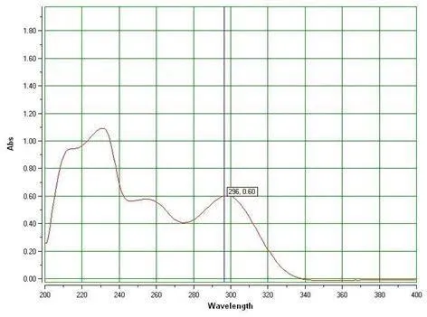
Figure 1 shows the UV spectrum of telmisartan dissolved in methanol, with a maximum absorption wavelength at 296 nm.
The drug and the drug-excipient mixture were tested for compatibility by obtaining FTIR spectra over 4000-400 cm⻹. If the characteristic bands of telmisartan are still visible in the mixture spectrum, it suggests there is no major interaction between the drug and the excipients. [Authors: identify the key bands and verify this conclusion based on your spectra.]
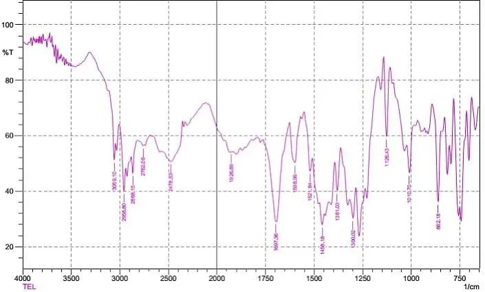
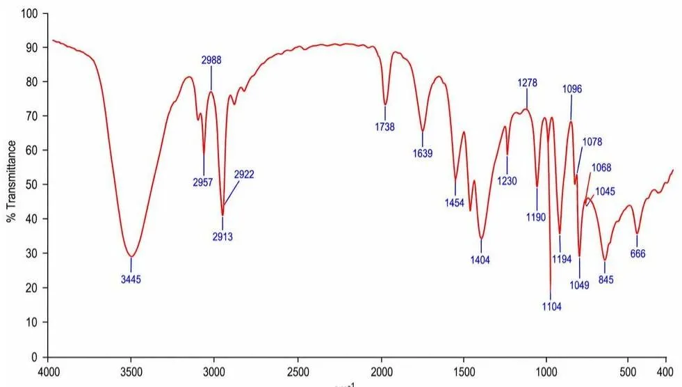
FTIR spectroscopy of telmisartan (top) and its drug-excipient combination (bottom) is shown in Figure 2.
Evaluation of transferosomes
The results of the characterizations are summarized in Table 3. The zeta potential fluctuated between -18.6 and -31.6 mV, and F4 demonstrated the highest magnitude, indicating the most effective electrostatic stabilisation. The particle size decreased from 218.4 nm (F1) to 158.7 nm (F4) and then increased slightly to 169.5 nm (F5). Vesicles in the nanometre range are in agreement with those observed in telmisartan and other transfersomal systems.8,17 Entrapment efficiency was raised from 72.4% (F1) to 89.5% (F4), and percentage yield from 78.4% to 91.2%, with F5 being slightly lower than F4 for both. The improvements in F1-F4 may be attributed to the higher lipid content and the balance between lipids and surfactants, which creates a superior vesicular environment for a lipophilic drug like telmisartan.12,17
Table 3: Characterization of Telmisartan-Loaded Transferosomes and Gels
| Formulation | Zeta potential (mV) | Particle size (nm) | Entrapment efficiency (%) | Yield (%) | Gel pH | Spreadability (g.cm/s) |
|---|---|---|---|---|---|---|
| F1 | -18.6 | 218.4 ± 4.8 | 72.4 ± 1.3 | 78.4 ± 1.2 | 5.62 | 6.82 |
| F2 | -22.4 | 195.6 ± 3.9 | 78.6 ± 1.1 | 82.7 ± 1.0 | 5.71 | 7.14 |
| F3 | -27.8 | 176.3 ± 3.5 | 83.7 ± 1.2 | 86.3 ± 1.1 | 5.83 | 7.63 |
| F4 | -31.6 | 158.7 ± 2.9 | 89.5 ± 0.9 | 91.2 ± 0.8 | 5.91 | 8.02 |
| F5 | -29.2 | 169.5 ± 3.2 | 86.8 ± 1.0 | 88.6 ± 0.9 | 5.78 | 7.46 |
The optimized formulation demonstrated mainly spherical vesicles with smooth, well-defined surfaces and no significant clumping, indicating good physical stability.
[Insert Figure 3: SEM images of optimized telmisartan-loaded transferosomes (F4)]
SEM images of the optimized telmisartan-loaded transferosomes (F4) are shown in Figure 3.
Evaluation of transferosomal gel
The gel had a pH range of 5.62 to 5.91, which is suitable for use on the skin. Its spreadability improved from 6.82 to 8.02 g.cm/s from sample F1 to F4 (Table 3). The drug content varied from 95.65% to 97.98%, with the highest concentration found in formulation F4 at 97.98%, indicating that the formulation retains the drug well. The optimized formulation had a viscosity of 2369 cps. These findings are similar to what other studies have shown about transfersomal gels.14,15
Table 4 shows the results of the in vitro release test. All the gels slowly gave out their contents over the course of 6 hours. F1 had the highest release of 68.7%, while F4 had the lowest release of 55.8%, resulting in F4 having the longest release period. This might happen because the vesicles have more trapped substances and include more lipids and surfactants, which help the drug stay inside longer and prevent it from leaving easily; the vesicles may also function like a storage system.12,13
Table 4: In Vitro Cumulative Drug Release (%) of Telmisartan Transferosomal Gel
| Time (h) | F1 | F2 | F3 | F4 | F5 |
|---|---|---|---|---|---|
| 0.5 | 18.4 | 15.8 | 14.2 | 11.6 | 16.5 |
| 1 | 27.6 | 24.3 | 22.1 | 19.4 | 25.8 |
| 2 | 39.8 | 35.7 | 32.6 | 28.7 | 37.2 |
| 4 | 55.3 | 51.2 | 47.8 | 43.6 | 53.4 |
| 6 | 68.7 | 63.9 | 59.4 | 55.8 | 66.1 |
The data from the release were analyzed using kinetic models, as presented in Table 5. The values of R² for F4 were 0.947 (zero-order), 0.998 (first-order), 0.963 (Higuchi), and 0.996 (Korsmeyer-Peppas). The release of F4 was best described by the first-order and Korsmeyer-Peppas models. The release exponent n ranged from 0.67 to 0.74 across different formulations. A value of 0.74 for F4, which was between 0.5 and 1.0, indicates anomalous, or non-Fickian, transport. This suggests a mix of drug diffusion and matrix swelling or relaxation.18
Table 5: Release Kinetic Parameters (R²) of Telmisartan Transferosomal Gel
| Formulation | Zero-order | First-order | Higuchi | Korsmeyer-Peppas | n |
|---|---|---|---|---|---|
| F1 | 0.974 | 0.999 | 0.999 | 0.998 | 0.67 |
| F2 | 0.977 | 0.998 | 0.9997 | 0.998 | 0.69 |
| F3 | 0.985 | 0.999 | 0.9999 | 0.999 | 0.71 |
| F4 | 0.947 | 0.998 | 0.963 | 0.996 | 0.74 |
| F5 | 0.932 | 0.997 | 0.9996 | 0.998 | 0.70 |
CONCLUSION
Telmisartan-loaded transferosomes were prepared using the reverse phase evaporation technique and then incorporated into a Carbopol gel. The formulation with the highest efficiency was F4, with a particle size of 158.7 ± 2.9 nm, a zeta potential of -31.6 mV, entrapment efficiency of 89.5 ± 0.9%, yield of 91.2 ± 0.8%, drug content of 97.98%, skin-friendly pH of 5.91, and good spreadability. It had the slowest and most consistent effect over 6 hours, showing unusual (non-Fickian) behavior in how it worked. The F4 transferosomal gel is a promising approach for delivering telmisartan through the skin over an extended period with controlled release. We need to run some tests to check how the drug is absorbed by the skin, if it irritates the skin, how it travels through the body, and how stable it is, to make sure this idea is actually possible.
ACKNOWLEDGMENT
The authors thank the Department of Pharmaceutics, JKKMIHSCP, T.N. Palayam, for providing the facilities needed to carry out this work.
REFERENCES

V. Suriya Prasanth, P. Sathish, P. Sripriya, B. Krishnamoorthy, P. Perumal, Formulation and Evaluation of Telmisartan-Loaded Transferosomal Gel for Enhanced Transdermal Delivery, Int. J. Sci. R. Tech., 2026, 3 (11), 172-177. https://doi.org/10.5281/zenodo.23941670
 10.5281/zenodo.23941670