

We use cookies to ensure our website works properly and to personalise your experience. Cookies policy
Dept of MLT, Haldia Institute of Health Sciences, Haldia.
Hemolytic illness, an immune-mediated disease that can be fatal, is brought on by maternal alloantibodies against fetal red blood cell antigens inherited from the father. Fetal anemia, hyperbilirubinemia, hydrops fetalis, and, in extreme circumstances, perinatal mortality these placental-crossing maternal immunoglobulin G antibodies. The incidence of RhD linked HDFN has decreased significantly, however alloimmunization against other blood group antigens such Kell, Rh c, Rh E, Duffy, Kidd, and ABO antigens still causes clinically significant disease. Thus, HDFN remains a significant concern in the fields of immunohematology, transfusion medicine, obstetrics, and neonatology. This review aims to present the current knowledge of the immunohematological mechanism of maternal alloimmunization and fetal hemolysis, especially focusing on clinically important blood group systems and antibody characteristics. Recent prenatal diagnostic developments, There have been notable advancements in early diagnosis and risk stratification of affected pregnancies because of advancements in technology like MCA-PSV Doppler ultrasound, molecular immunohematology, high throughput genotyping, and cell-free fetal DNA for non-invasive fetal blood type genotyping. Newer management techniques, including serial maternal antibody monitoring, intrauterine transfusion, optimized timing of delivery, neonatal phototherapy, exchange transfusion and evidence-based transfusion support, have dramatically improved the survival of fetuses and minimized disease-related complications. In addition, new technologies and innovations such as those based on artificial intelligence that can predict risk, digital immunohematology, automated compatibility testing, FcRn inhibitors, complement targeted therapies, and personalized immunotherapeutic strategies will help further define the diagnosis and management of HDFN. Nevertheless, the world faces many obstacles to achieving these advances, including the lack of access to molecular diagnostics, availability of appropriate blood products, and unequal distribution of health care services. Further multidisciplinary teamwork, technological innovations, and standardized clinical approaches will play a key role in furthering the goals of precision medicine and improving maternal and neonatal outcomes in the management of HDFN.
A potentially fatal immune-mediated condition known as hemolytic causes harm to the developing baby and the mother when a woman's production and transmission of immunoglobulin G (IgG) antibodies destroys fetal erythrocytes, which are blood globulins. This is what occurs when a woman's red blood cell (RBC) immune system attacks fetal RBC antigens that are on the father's RBCs but not on her own [1]. Fetal anemia, hydrops fetalis, hyperbilirubinemia, and neonatal or fetal death can result from progressive hemolysis if it is not addressed. Optimal maternal and newborn outcomes require coordinated, interdisciplinary therapy due to the clinical significance of HDFN as a major complication in transfusion medicine, obstetrics, neonatology, and immunohematology. Over the previous century, the idea of HDFN has evolved considerably. Fetal anemia and neonatal jaundice had no clear explanation until the Rh blood type system was established, and perinatal mortality was high [2]. A significant number of cases of HDFN were found to be caused by mother-produced anti-D antibodies in 1942; the Rh blood type system was established in 1940. Countries that implemented screening and prophylaxis programs during pregnancy saw a marked decline a sharp decline in RhD alloimmunization following the use of anti-D immunoglobulin prophylaxis in the late 1960s and an increase in severe Rh-mediated HDFN. These improvements have not eliminated HDFN as a clinical problem, however, as non-D antigens like Kell, Rh c, Rh E, Duffy, and Kidd blood group antigens continue to cause alloimmunization and ABO incompatibility remains a problem [3]. The prevalence of HDFN varies significantly across the globe and is related to access to advanced fetal medicine services, access to Rh immunoprophylaxis, routine antibody testing, and prenatal care. A comprehensive antenatal screening, combined with the routine use of anti-D immunoglobulin, has caused a marked decline in the occurrence of serious RhD-associated disease in high income countries. By contrast, there are still preventable cases of maternal alloimmunization in many LMICs, where the availability of prenatal testing is low, the blood bank infrastructure is insufficient, and the diagnosis is delayed and prophylactic protocols are not always practiced [4]. Thus, in resource limited areas, HDFN continues to be an important cause of fetal anaemia, neonatal hyperbilirubinemia, hydrops fetalis and perinatal death. The incidence of HDFN has declined significantly in the last few decades, especially in RhD incompatibility, and significant disease remains an occurrence in the world today. The most prevalent cause of neonatal immune hemolysis today is relative to Rh incompatibility, ABO incompatibility is usually less severe. On the other hand, if the blood group antigens involved are clinically important, such as RhD, Kell, Rh c or other blood group antigens, fetal anemia may ensue and require special fetal monitoring and intrauterine transfusion. Despite the significant progress in prenatal diagnosis and fetal therapy, severe HDFN remains associated with high morbidity, such as hydrops fetalis, prematurity, kernicterus, long-term neurological sequelae, hearing loss, and developmental disability if diagnosis and/or treatment are delayed. Immunohematological mechanisms involved in the pathogenesis of HDFN include antigen-antibody interaction, immune-mediated destruction of fetal erythrocytes as well as the placental transmission of maternal IgG antibodies to the developing foetus [5]. Thus, the importance of immunohematology cannot be overstated, ranging from antenatal antibody screening, identification of clinically significant alloantibodies, to fetal antigen genotyping, compatibility testing, choice of blood components, and transfusion support. Recent advances in molecular immunohematology have contributed to the increase in accuracy of risk evaluation and patient-centric patient care: approaches for digital antibody detection, high-throughput blood group genotyping employing mitochondrial DNA, non-invasive fetal blood group genotyping, and cell-free fetal DNA and automated immunohematology platforms. Improvements in the diagnosis of the fetus have been made in the laboratory, as well as in the methods of fetal monitoring and therapeutic interventions. Prenatal measurement of the peak systolic velocity of the middle cerebral artery Doppler ultrasonography has replaced more intrusive methods of fetal hemoglobin testing and image-guided intrauterine transfusions has significantly improved fetal outcomes in more severe fetal anemia. New technologies, such as next generation sequencing, artificial intelligence (AI)-based risk prediction models, precision medicine and new immunomodulatory therapies, will further improve prenatal diagnosis and clinical decision making [6]. Considering the dynamic nature of immunohematology and fetal medicine, a new synthesis of the available evidence is justified. With recent developments in diagnostic technologies, molecular testing, prenatal surveillance, transfusion practices and targeted therapeutic management, the range of HDFN management has grown beyond traditional practices. The purpose of this comprehensive review is to compile all of the existing information in the immunopathogenesis of HDFN, review recent advances in immunohematological investigations and in the prenatal diagnosis of a fetus, review the current clinical management of affected pregnancies and neonates, and outline new innovations, current challenges, and future directions that could affect maternal and fetal outcomes.
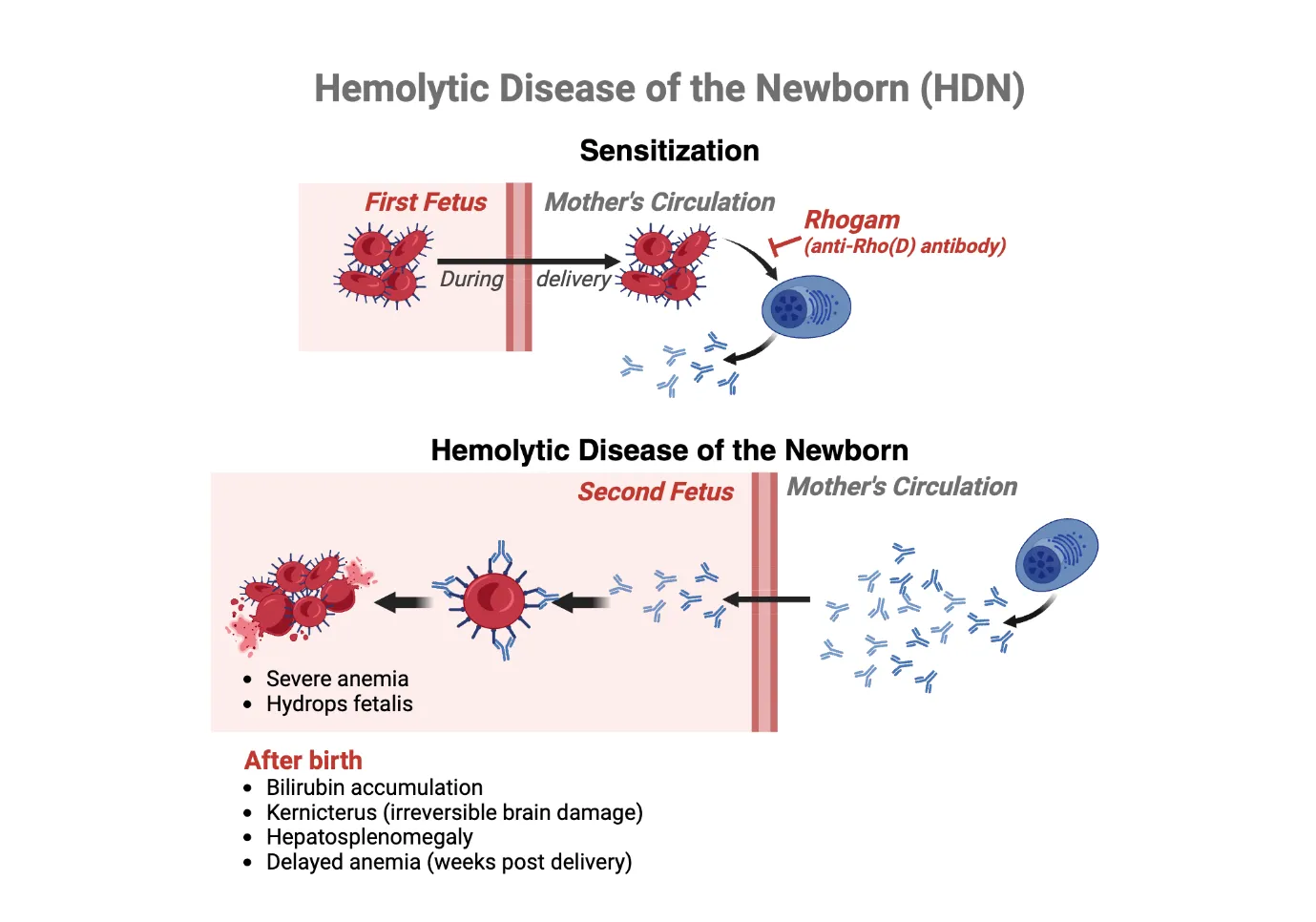
Figure 1: Overview of the Pathogenesis of HDFN
2. Immunohematological Basis of Hemolytic Disease of the Fetus and Newborn
2.1 Maternal Alloimmunization
The basic immunological event that leads Maternal Fetal damage can be caused by alloimmunization and neonatal hemolytic illness. Fetal erythrocytes carry an antigen that is absent from maternal blood, which is the reason why it happens when a pregnant woman with a deficiency in one RBC antigen is exposed to them. Fetomaternal hemorrhage (FMH) happens when red blood cells (RBCs) from the foetus the mother's circulation after a traumatic event during pregnancy, a difficult birth, a miscarriage, in cases of external cephalic version, ectopic pregnancies, abdominal trauma, or invasive prenatal procedures such an amniocentesis or chorionic villus sampling. Fetal blood, even in small amounts, can be enough to sensitize a susceptible mother. Following contact with antigens found on foetal red blood cells are processed by maternal antigen presentation cells, which then pass the antigen on to CD4+ T-helper cells, which activate B lymphocytes that are specific to the antigen [7]. Typically, the present pregnancy is unaffected by the naive B cells that produce low-affinity IgM antibodies as part of the main immune response. Activated B cells undergo a transformation to become memory B cells that last a long time and produce IgG antibodies. Going forward, in the event that the unborn child tests positive for an antigen, this causes a secondary immune response with a stronger affinity, which starts fast and escalates. Fetal anemia and associated complications include antibodies that cross the placenta easily originate in the mother and trigger the immune system to eliminate fetal red blood cells.
2.2 Blood Group Systems Implicated in HDFN
Over three hundred RBC antigens have been discovered, but only a few are of clinical importance in HDFN. Serious illnesses are still mostly caused by the System for Rh blood groups. Antibodies against the most immunogenic Rh antigen, D, are not rare, but antibodies against the Rh c, C, E, and e antigens can cause clinically severe fetal anemia. The incidence of RhD isoimmunization has dropped dramatically, even though anti-D immunoglobulin prophylaxis has been widely used. However, other Rh isoimmunizations can still provide a substantial clinical concern. Neonatal immunological hemolysis is typically caused by the ABO blood group system. When pregnant women of group O bear babies of group A or B, the most prevalent hemoglobin type is ABO-HDFN. During pregnancy, the mother's natural anti-A and anti-B antibodies can cause mild to moderate hemolysis. Severe fetal anemia is rare, as the antigen expression of the fetus is rather small and soluble ABO substance neutralizes the circulating maternal antibody. Because anti-K antibody destruction of foetal erythrocytes also limits the development of foetal bone marrow erythroid progenitor cells, resulting to severe anemia with relatively minor hemolysis, the Kell blood group system is one of the more prominent non-Rh blood group systems. Anemia caused by antibodies targeting the Duffy and Kidd antigens is uncommon but can result in moderate to severe HDFN. Also, antibodies that target MNS blood group antigens (anti-M, anti-S, and anti-s) are essential in clinical practice because they cause newborn hemolysis. Anti-Diᵃ is rarely seen in certain ethnic groups, and can rarely be a factor in HDFN [8].
2.3 Antibody Characteristics
Maternal alloantibodies are pathogenicity dependent on the immunoglobulin class, subclass, antigen specificity and the capacity to cross the placenta. HDFN-causing antibodies are mostly IgG, mostly IgG1 and IgG3, with a high affinity to Fc gamma receptors of placental cells and fetal macrophages. These subclasses are able to pass through the placenta efficiently via the neonatal Fc receptor which starts to appear in the second trimester and becomes more prominent as gestation advances. After crossing the placenta, maternal IgG binds with a specific antigen found on fetal erythrocytes. Complement activation plays a minor role in most instances of HDFN, but some antibodies can activate the classical pathway, thus increasing the immune-mediated hemolysis. The antibody concentration (titer), antigen density on foetal red blood cells (RBCs), IgG subclass distribution, immunogenicity of the antigen, and gestational age of the mother's antibody exposure all play a role in the clinical severity of HDFN. Thus, antibody identification and titration are important elements of fetal risk assessment [9].
2.4 Mechanism of Fetal Hemolysis
The underlying defect of HDFN is the destruction of fetal erythrocytes by antibodies. After transfer to the fetus, maternal IgG antibodies attach to their target antigen molecules on fetal RBC membranes forming antigen–antibody complex. These antibody coated erythrocytes are then removed by phagocytosis by the Fc receptor bearing macrophages in the fetal spleen and liver by extravascular hemolysis. Continued hemolysis leads to a gradual decline in fetal hemoglobin level, which leads to a gradual decrease in fetal hemoglobin level leading to chronic anemia and hypoxia of tissues. So as a result, the production of erythropoietin rises, which induces non-medullary hemopoiesis in various tissues of the developing foetus, including the liver and spleen. In order to maintain an adequate oxygen supply, the heart rate rises with the progression of anemia, resulting in heart failure with high output. When the plasma oncotic pressure drops because of liver failure or congestive heart failure, it causes hydrops fetalis, which manifests as ascites, pleural and pericardial effusions, and widespread fetal edema. Unless prompt diagnosis and treatment are provided, hydrops fetalis can result in intrauterine fetal death or serious neonatal issues. The knowledge of these immunohematological mechanisms is the foundation on which the modern prenatal screening, laboratory diagnostics, fetal monitoring and specific treatment in pregnancies complicated by maternal alloimmunization are built [10].
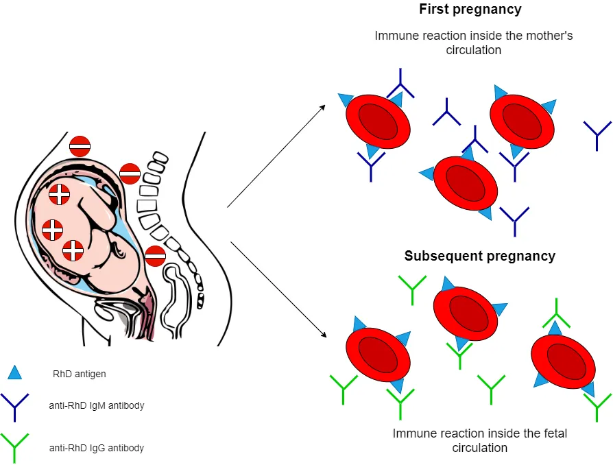
Figure 2: Mechanism of Maternal Alloimmunization Leading to Fetal Hemolysis
3. Risk Factors and Epidemiology
Although significant decreases in severe HDFN cases have occurred after the introduction of anti-D immunoglobulin prophylaxis, HDFN is still a clinically important cause of fetal anemia, hyperbilirubinemia in the newborn and perinatal morbidity in the world. The epidemiology of HDFN is highly different from one population to another and is affected by the pattern of maternal sensitization, distribution of the blood group antigens, availability of prenatal screening, and immunoprophylaxis programmes. However, severe RhD associated HDFN has dramatically reduced in high-income countries as a result of the routine antenatal screening for antibody and universal Rh prophylaxis. Alloimmunization for non-D antigens, including ABO incompatibility, Kell, Rh c, Rh E, Duffy, Kidd and MNS, however, continue to lead to the development of HDFN [11]. There are a number of maternal factors that are associated with an increased risk for alloimmunization and subsequent HDFN. Fetal erythrocytes can sensitize the mother during delivery, miscarriage, abortion or any invasive obstetric procedure. Previous pregnancy is a significant risk factor. Women who have received previous blood transfusion may also be sensitized by previous transfusion to foreign RBC antigens, which can lead to formation of alloantibodies, especially if the antigens have not been matched. If anti-D antibodies are not given correctly, The likelihood of acquiring anti-D antibodies is higher among mothers who do not produce RH but have fetuses that do. An increased risk of maternal sensitization can be observed in several pregnancies due to the increased cumulative exposure to fetal RBC antigens. A number of fetal variables contribute to the disease's severity as well. For HDFN to occur, the antigen must have been passed on to the child from the father. In addition, the density of antigens on fetal erythrocytes affects the amount of antibody binding and hemolysis, and the density of some antigens, such as RhD and Kell, affects the immunogenicity of the antigen and may cause significant fetal anemia [12]. The factors relating to healthcare are important contributors to the persistence of HDFN, particularly in resource-limited environments. Anti-D isoimmunization of mothers is a significant preventable cause of maternal anti-D alloimmunization. As with these examples, limited prenatal screening and poor access to antibody identification, fetal monitoring and specialized transfusion services can delay diagnosis and treatment and put the fetus at risk of developing hydrops fetalis, preterm labor and stillbirth. Hence, adequate antenatal care and access to universal immunohematological testing is needed to minimize HDFN burden worldwide.
Table 1: Risk Factors Associated with HDFN
| Risk factor category | Specific factor | Clinical impact |
|---|---|---|
| Maternal | Previous pregnancy | Increases risk of sensitization through fetomaternal hemorrhage |
| Maternal | Previous blood transfusion | Promotes alloantibody formation against foreign RBC antigens [13] |
| Maternal | Rh-negative mother with Rh-positive fetus | Major risk factor for anti-D alloimmunization |
| Maternal | Multiple pregnancies | Increases cumulative exposure to fetal RBC antigens [14] |
| Fetal | Paternal antigen inheritance | Necessary for maternal-fetal antigen incompatibility |
| Fetal | High antigen density on fetal RBCs | Enhances antibody binding and hemolysis [15] |
| Healthcare | Lack of Rh prophylaxis | Prevents anti-D alloimmunization reduction |
| Healthcare | Limited prenatal screening | Delays diagnosis and increases risk of severe HDFN [16] |
4. Clinical Manifestations
The degree of fetal hemolysis, severity of maternal alloimmunization, the specificity and concentration of maternal antibodies, and the course of hemolytic disease in the fetus and infant are the factors that define the clinical signs. The onset of clinical symptoms might happen during pregnancy, during delivery, or as a consequence in infants who were delivered alive.
Prenatal Manifestations
The most significant and first prenatal symptom of HDFN, the gradual immune-mediated lysis of foetal red blood cells, is foetal anemia. Mild anemia often goes unnoticed, but severe anemia leads to tissue hypoxia and an increase in cardiac output to make up for it. Fetal liver and spleen extramedullary hematopoiesis, sometimes known as hepatosplenomegaly, is a common complication of chronic hemolysis. A potentially fatal syndrome known as hydrops fetalis can develop when anemia worsens over time. Low plasma oncotic pressure and high-output heart failure are the hallmarks of this disease. Ascites, a thick placenta, effusion into the pleura or pericardium, and extensive fetal edema are further signs. There is a considerable risk of intrauterine fetal death associated with hydrops fetalis if prompt assistance is not provided. Severe anemia or hydrops during pregnancy might impede the fetal swallowing abilities, increasing the risk of polyhydramnios and other complications during pregnancy and delivery [17].
Neonatal Manifestations
After delivery, affected neonates often have jaundice, which occurs in antibody-coated erythrocytes are more quickly destroyed in the first twenty-four hours following delivery. Hemolysis causes hyperbilirubinemia because bilirubin continues to be produced, leading to a rapid increase in bilirubin if not treated properly which can cause the the passage of unconjugated bilirubin through the still-developing blood-brain barrier, leading to kernicterus, a severe kind of neurological impairment caused by bilirubin. Neonates can also have moderate to severe anemia along with pallor, tachycardia, poor feeding, lethargy, hepatosplenomegaly and evidence of circulatory failure. Acute, high-output cardiac failure can be caused by severe anemia, respiratory distress and acidosis and shock in severe cases, requiring intensive neonatal care, exchange transfusion, or transfusion of red blood cells [18].
Long-Term Complications
With improvements in prenatal diagnosis and management of the newborn, the rate of survival has greatly increased, but survivors of severe HDFN still have lifelong complications. Infants with prolonged fetal hypoxia or bilirubin neurotoxicity have been found to have impaired neurodevelopment, such as cognitive impairment, motor dysfunction, and cerebral palsy. The other recognized effect of bilirubin-induced neurological injury is "sensorineural hearing loss. In addition, children with the disease can have learning disabilities, speech and language problems and developmental delay, so long-term developmental surveillance, early intervention and multidisciplinary follow-up should be undertaken to maximize neurological and functional outcomes [19].
5. Laboratory Diagnosis and Prenatal Assessment
Prenatal assessment and diagnosis in the laboratory is critical to the detection of pregnancies that pose a high risk of hemolytic disease in the developing baby, assessment of the severity of the disease and appropriate therapy. Immunohematology, molecular diagnosis and fetal imaging advances have greatly enhanced early detection and clinical management of maternal alloimmunization.
5.1 Maternal Testing
Rh blood group compatibility and ABO blood group typing are the first prenatal laboratory tests. These tests are typically performed at the first antenatal visit and are used to find out if the pregnant woman is Rh negative. Immunoassays for alloantibodies such as "anti-D, anti-c, anti-E, anti-K, anti-Fy, and anti-Jk" are routinely conducted using the universal indirect antiglobulin test (IAT). If antibody screening returns a positive result, the specificity and significance of the antibodies can be determined using standardized immunohematological methods, such as antibody identification panels. Antibody titration is performed on an as-needed basis during pregnancy when the presence of clinically relevant antibodies has been confirmed. The risk of fetal anemia increases with increasing antibody titres; nevertheless, the specific link between antibody types varies. Because they inhibit foetal erythropoiesis, anti-K antibodies, for example, can induce severe foetal anemia even in the presence of low antibody levels. Monitoring maternal antibody, therefore, remains an integral part of the process of prenatal risk assessment and dictates the level of intensive fetal monitoring [20].
5.2 Paternal Testing
Paternal testing is useful for information on fetal antigen inheritance and the potential for maternal/fetal incompatibility. The purpose of red blood cell antigen typing is to determine whether the biological father possesses the same antigen that the maternal alloantibody reacts to fetuses whose fathers are antigen-negative will not have a chance of inherit the antigen and the risk of HDFN will be ruled out for that specific antibody. If the father is antigen positive, then zygosity testing is recommended to ascertain if he possesses a single copy of the relevant antigen or not. In contrast to heterozygous fathers, who have an average 50% chance of transferring an antigen to their offspring, homozygous fathers always pass the antigen on to the fetus. Individual prenatal management has been made easier and paternal zygosity has become more accurate due to improvement in molecular genotyping techniques [21].
5.3 Fetal Genotyping
A non-invasive method for prenatal genotyping by utilizing fetal DNA that is free of maternal plasma cells has been possible over the last few years due to recent advances in molecular diagnostics. This method reliably determines the fetal RhD status and a few other clinically important blood group antigens, while avoiding risks to the fetus. A large number of needless invasive tests have been eliminated as a result of cell-free fetal DNA sequencing becoming standard practice in many specialist prenatal care institutions. Traditional polymerase chain reaction (PCR) testing might be employed to ascertain the maternal blood type via means of DNA recovered from amniocentesis or chorionic villus samples. New technologies like next-generation sequencing have made it possible to genotype fetal blood groups has become more accurate and comprehensive, and many blood group genes may now be analyzed concurrently with great analytical sensitivity and specificity. These molecular techniques help to support precision medicine and to accurately detect fetuses at true risk of HDFN [22].
5.4 Fetal Monitoring
Doppler ultrasonography of the middle cerebral artery's peak systolic velocity was the non-invasive diagnostic gold standard for moderate to severe foetal anemia until recently. Anemia lowers blood viscosity, which leads to elevated MCA-PSV levels. If the MCA-PSV is 1.5 times or more the median, additional testing for foetal anemia or intrauterine transfusion may be necessary. Fetal growth, placental thickness, amniotic fluid volume, hepatosplenomegaly, and heart function can all be measured with regular ultrasound exams in conjunction with Doppler. To further assess the baby's well-being, additional Doppler tests can measure blood flow in the umbilical artery and veins [23].
5.5 Neonatal Laboratory Diagnosis
Laboratory evaluation is performed after delivery to confirm the sickness's intensity and its diagnosis. A crucial laboratory test for the diagnosis of immune-mediated hemolysis is the direct antiglobulin test, which detects antibodies and/or complement attached to neonatal erythrocytes. An indirect antiglobulin test (IAT) of maternal or neonatal serum can detect circulating alloantibodies which are not yet bound to erythrocytes. Complete blood count (CBC) assesses hemoglobin concentration and hematocrit, indicating the severity of the anemia, and an elevated reticulocyte count is an indicator of compensatory erythropoiesis. Total serum bilirubin is an important measurement to make in order to determine the severity of hemolysis and the need for or plan for exchange transfusion or phototherapy. Further evidence of active hemolysis is the rise in lactate dehydrochlorase (LDH) levels. The peripheral blood smear usually shows anisopoikilocytosis, polychromasia, nucleated red blood cells, and spherocytes, which are other indications of accelerated destruction of the erythrocytes [24].
5.6 Emerging Diagnostic Technologies
The diagnosis of HDFN is changing as a result of rapid development of laboratory medicine. To measure the amount of DNA from the mother's blood, digital polymerase chain reaction is an extremely sensitive method, which will enhance the accuracy of non-invasive prenatal testing especially in early pregnancy. The potential use of artificial intelligence (AI) based algorithms is also being explored for using maternal antibody titers, fetal genotyping, Doppler data, and clinical parameters to detect the pregnancies at highest risk of severe HDFN. Furthermore, blood group antigen typing using microarray technology allows for the simultaneous detection of multiple clinically relevant blood group antigens and variants that enables fast donor-recipient matching and individualised transfusion approaches. Together, these new technologies will help to further amplify the accuracy of diagnosis, minimize the need for invasive procedures, and improve maternal and neonatal outcomes in HDFN [25].
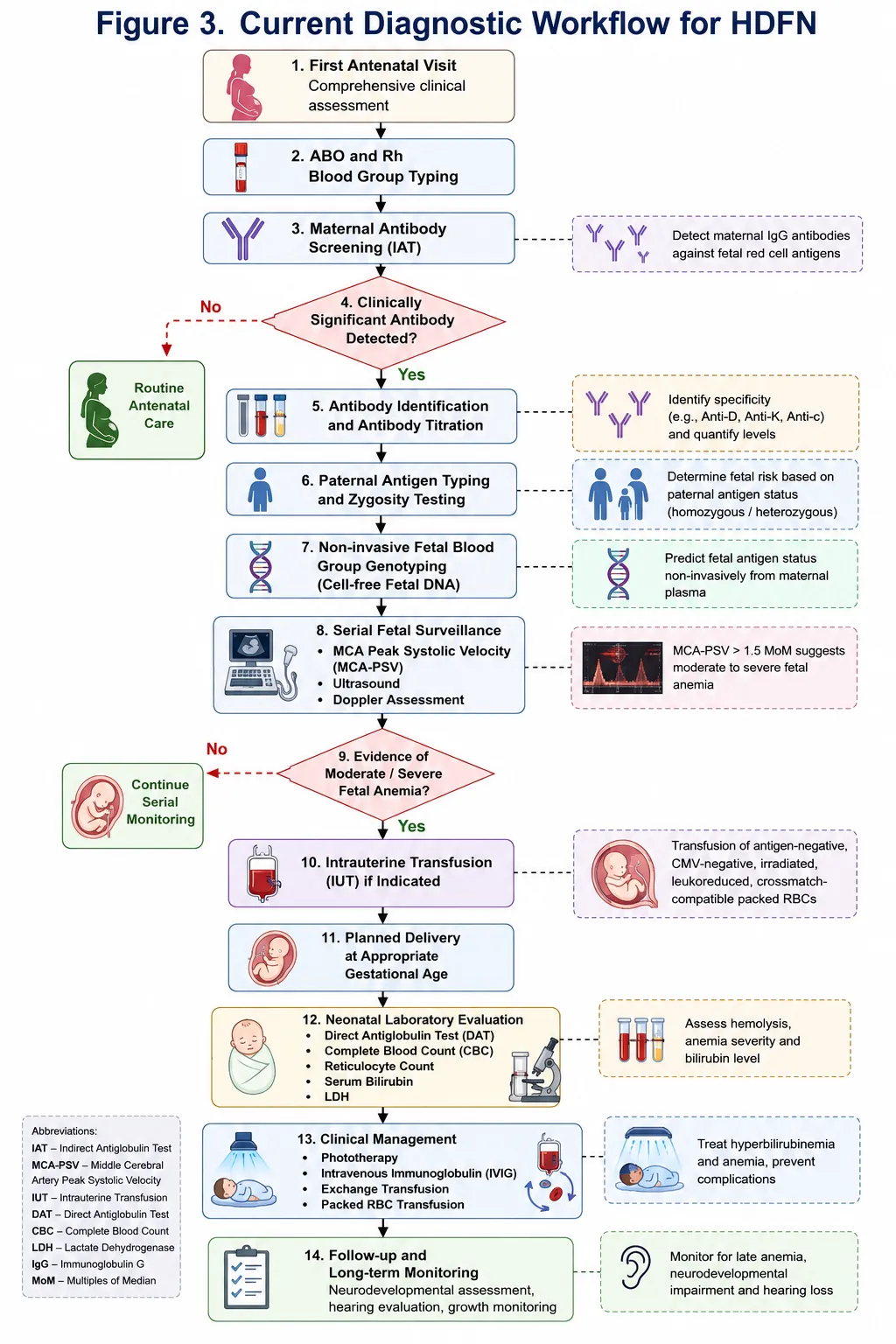
Figure 3: Current Diagnostic Workflow for HDFN
Table 2: Laboratory investigations in HDFN and their clinical significance.
| Investigation | Sample | Purpose | Clinical significance |
|---|---|---|---|
| ABO/Rh typing | Maternal blood | Determine maternal blood group | Initial risk assessment |
| Antibody screening (IAT) | Maternal serum | Detect clinically significant alloantibodies | Identify pregnancies at risk [26] |
| Antibody identification | Maternal serum | Determine antibody specificity | Guides monitoring and management |
| Antibody titration | Maternal serum | Quantify antibody levels | Assess risk of fetal anemia [27] |
| Paternal antigen typing | Paternal blood | Determine fetal antigen inheritance risk | Predict likelihood of HDFN |
| Cell-free fetal DNA testing | Maternal plasma | Non-invasive fetal blood group genotyping | Avoid unnecessary invasive testing [28] |
| MCA-PSV Doppler | Fetus | Detect fetal anemia | Guides intrauterine transfusion |
| Ultrasound | Fetus | Assess growth and hydrops | Monitor disease progression [29] |
| Direct antiglobulin test (DAT) | Neonatal cord/peripheral blood | Detect antibody-coated RBCs | Confirm immune hemolysis |
| Complete blood count (CBC) | Neonatal blood | Evaluate anemia | Determine disease severity [30] |
| Serum bilirubin | Neonatal serum | Assess hemolysis | Guide phototherapy/exchange transfusion |
| Peripheral blood smear | Neonatal blood | Morphological evaluation | Support diagnosis of hemolysis [31] |
6. Current Clinical Management
A multidisciplinary team consisting of obstetricians, neonatologists, experts in fetal medicine, transfusion medicine, and blood banking should handle cases of hemolytic illness in the developing baby. Current management practices include antenatal surveillance, prompt intrauterine intervention, proper timing of delivery, adequate neonatal management and careful blood bank assistance.
6.1 Antenatal Management
Serial maternal antibody monitoring is the first step in the management of pregnancies affected by maternal alloimmunisation. Clinically significant alloantibodies should be periodically titrated in pregnant women to assess the risk for hemolysis. In anti-K-mediated HDFN, antibody titers are less predictive of disease severity, as anti-K antibody does not cause only hemolysis, but also suppresses fetal erythropoiesis. As such, antibody levels should be used in conjunction with clinical and fetal evaluation [32]. Fetal surveillance is necessary once a critical level of antibody level is obtained or once a previous pregnancy has been affected. Fetal growth is monitored by regular ultrasonographic examination, which also assesses the amniotic fluid, placental thickness, hepatosplenomegaly and early features of hydrops fetalis. Fetal monitoring allows the detection of disease progression early on and allows for prompt treatment before irreversible damage occurs. Systolic blood pressure at its highest point within the brain's cortex Doppler ultrasonography is the most reliable method for assessing the foetus without causing any harm. Because fetal anemia lowers blood viscosity, the brain's blood flow velocity is increased. An MCA-PSV greater than 1.5 times the median is the main indication for additional testing and/or intrauterine transfusion. When compared to invasive foetal blood sample, the diagnostic performance and procedure-related hazards of MCA Doppler monitoring are much better [33].
6.2 Intrauterine Transfusion
This is the most often used diagnosis and treatment of severe foetal anemia before viability, specifically through intrauterine transfusion. Intrauterine transfusion has greatly improved foetal survival in pregnancies affected by severe HDFN and is the most common treatment for severe foetal anemia before foetal development (before viability). The main use for IUT is when there is medium to severe fetal anemia confirmed or strongly suspected by the elevated MCA-PSV or fetal blood sampling when clinically indicated. Other features include clinically significant maternal alloantibodies and a rapidly progressive anemia and hydrops fetalis. The success and safety of IUT relies on the proper selection of blood. Transfused RBCs must be group O, Rh compatible, rejecting the associated maternal antigen, compatible with maternal serum in terms of crossmatch, CMV negative, irradiated, leukoreduced, HbS negative and relatively fresh (less than 5-7 days old). The hematocrit of the packed red blood cells should be high (75-85% hematocrit) to maximise the amount of oxygen that can be carried by the transfusion, whilst minimising the quantity of cells being transfused Transfusion of suitable packed red blood cells into the fetal umbilical vein, usually near the placental cord insertion, is a common procedure performed with continuous ultrasound monitoring. Transfusions can be administered to certain individuals through the intrahepatic vein. Until the baby is ready to be born, these infusions can be required every two to four weeks. In high-volume fetal medicine centers, fetal survival has reached 90% or higher and the rate of procedure-related problems has dropped because to increased technology in ultrasound guidance, transfusion method, and fetal monitoring [34].
6.3 Timing of Delivery
The timing of delivery is a balance of the risk of prematurity and continued fetal hemolysis. For pregnancies with mild disease with low maternal antibody levels and reassuring fetal surveillance, delivery is often scheduled at 37-38 weeks of gestation. For moderate disease, the timing of delivery is based on fetal monitoring, prior IUT and fetal condition. When the lungs are considered mature and the neonatal care facilities are available, delivery is typically suggested at 36 to 37 weeks. If the risks of continuing intrauterine treatment are felt to outweigh the benefits of neonatal intensive care, may be warranted, particularly in cases of recurrent fetal anemia, hydrops fetalis or inability to keep the fetus stable with repeated intrauterine transfusion. Multidisciplinary consultation with obstetricians, fetal medicine specialists, and neonatologists should be done individually to make decisions [35].
6.4 Neonatal Management
Post-natal management aims to minimize neurotoxicity due to bilirubin and to correct anaemia. The treatment of neonatal hyperbilirubinemia is first-line intensive phototherapy. By exposing unconjugated bilirubin to blue light, phototherapy can convert it into photoisomers that are soluble in water which can be excreted without the need for hepatic conjugation, leading to a decrease in serum bilirubin. For selected infants with moderate to severe immune-mediated hemolysis, intravenous immunoglobulin can be given to decrease the destruction of antibody-coated erythrocytes via Fc receptors and the necessity of exchange transfusion. Its use is recommended in select cases at this time, but is controversial as a routine, and should be done according to institutional guidelines. Exchange transfusion is the definitive therapy when bilirubin levels are approaching neurotoxic levels with intense phototherapy or when there is severe anemia. This procedure eliminates maternal antibodies, bilirubin and replaces maternal erythrocytes with compatible donor erythrocytes at the same time. Packed red blood cell (PRBC) transfusion may then be needed to alleviate anemia which may be ongoing or delayed due to continued hemolysis or suppression of erythropoiesis. Supportive care measures are complete hydration, nutritional support, maintenance of normothermia, correction of electrolyte disturbances, ongoing cardio-respiratory monitoring, serial bilirubin and hemoglobin determinations and neurological monitoring for early detection of bilirubin-induced neurological dysfunction [36].
6.5 Blood Bank Considerations
Support for safe transfusion is an essential part of the management of HDFN. Fetal and neonatal transfusion recipients should receive antigen negative blood to avoid further immune mediated hemolysis. Pregnant women and babies born prematurely are particularly at danger of contracting the coronavirus, so it's best to use blood components that have tested negative for the virus. It is recommended to irradiate blood products used in fetal transfusions and leukoreduce blood products used in neonatal transfusions in order to prevent such as alloimmunization, febrile non-hemolytic transfusion reactions, graft-versus-host disease, and CMV transmission, respectively. This close relationship between clinicians and the transfusion services is therefore very critical to provide the necessary compatible blood products in time and to maximise the clinical outcome in pregnancies affected by HDFN [37].
7. Recent Advances in HDFN Management
The treatment of foetal and neonatal hemolytic diseases has undergone a dramatic change over the last few years with the advent of molecular diagnostics, fetal medicine, transfusion science, and computational technologies. Modern techniques focus on early detection of risk, individualized obstetric treatment, non-invasive testing and targeted treatment. All of this has led to better diagnosis, fewer complications during the procedure, and better outcomes for the mother and baby.
Molecular Immunohematology
Molecular immunohematology has revolutionized the clinical use of prenatal diagnosis of HDFN. The advantages of using a high throughput blood group genotyping platform for simultaneous determination of multiple genes of the red blood cell antigens circumvent the limitations of conventional serological testing, especially in recently transfused patients or those who have complex antibody profiles. Molecular typing is particularly useful for identification of rare blood group variants and compatible donor units for intrauterine and neonatal transfusion. A major advancement has been the development of the possibility of predicting fetal blood group non invasively from cell-free fetal DNA present in the maternal plasma. This technology is able to accurately identify fetal RhD, and more recently, other clinically important blood group antigens like RhC/c, RhE/e and Kell, without placing the baby at risk with invasive procedures. In antigen-negative fetuses, therefore, fetal monitoring and anti-D prophylaxis may be avoided, allowing a safer and more personalized prenatal care [38].
Precision Medicine
Precision medicine is a burgeoning paradigm impacting the care of the HDFN, specifically by utilizing the characteristics of maternal antibody, fetal genotype, history, Doppler monitoring, and laboratory biomarkers to personalize care. Now, in addition to antibody titers, a number of other clinical factors are being used to carry out personalized fetal monitoring, which allows for early identification of pregnancies at highest risk for severe fetal anemia. In addition, recent developments in molecular blood group typing have enabled the choice of antigen-matched blood transfusion products based on the maternal alloantibody profile and fetal antigen status, further individualizing transfusion. Such individualization reduces the chances of further alloimmunisation, enhances transfusion safety and optimises foetal and neonatal outcomes [39].
Novel Therapeutics
Apart from diagnosis, there are several new novel treatment modalities that are being explored. Antibodies blocking specific immune pathways have been found to be potentially useful for the modulation of maternal alloimmune reactions and thus decreasing antibody-mediated fetal hemolysis. The use of neonatal Fc receptor (FcRn) inhibitors is also an exciting therapeutic strategy, as they directly inhibit the transport of antibodies of pathogenic maternal origin across the placenta, lowering the exposure of antibodies in the foetus during pregnancy. The effects of experimental complement inhibitors that reduce complement-mediated destruction of red blood cells are also under study in certain types of immune hemolysis. In addition, there are several rapidly developing immunomodulatory therapies that are being preclinically and clinically evaluated, such as targeted B-cell modulation and cytokine-directed therapies. These treatments are still largely experimental, but can be useful for pregnancies with severe alloimmunisation that don't respond well to conventional treatment [40].
Artificial Intelligence
Artificial Intelligence (AI) is a vital tool in the process of prenatal medicine and transfusion practice. The machine learning algorithms could be used to combine maternal antibody levels, fetal genotype testing data, ultrasound and Doppler data to create risk prediction models that alert clinicians to pregnancies that need more intensive monitoring or intervention. Automated antibody interpretation using AI-based image analysis and laboratory software is also being developed, which further accelerates the identification of antibodies and helps minimize the inter-observer variability in the process. Further, clinical decision-support systems integrate laboratory and clinical information to support clinical decision making around the timing of surveillance, intrauterine transfusion and delivery planning, incentivising evidence-based clinical decision making [41].
Future Blood Bank Technologies
Improving transfusion safety and workflow efficiency is expected to increasingly depend on digital immunohematology, which involves molecular diagnostics, electronic laboratory information systems and artificial intelligence. Automated interpretation of serological and molecular results could help decrease the number of diagnostic errors and aid in rapid identification of the compatible blood products. Such technologies may serve to reduce transfusion support time, better manage rare antigen negative blood units and reduce turnaround time for pregnancies affected by HDFN. These technologies have the potential to significantly impact precision transfusion medicine and continue to help to enhance maternal and neonatal outcomes as they become more widely used.
Table 3: Recent Advances in Diagnosis and Management of HDFN
| Innovation | Technology | Clinical Benefit |
|---|---|---|
| High-throughput blood group genotyping | Molecular immunohematology | Accurate detection of blood group antigens and rare variants |
| Cell-free fetal DNA (cffDNA) testing | Non-invasive fetal genotyping | Early determination of fetal RhD and other blood group antigens without invasive procedures [42] |
| Next-generation sequencing (NGS) | Comprehensive blood group gene analysis | Improved precision in fetal and parental genotyping |
| Precision medicine | Individualized risk assessment | Personalized fetal surveillance and treatment planning [43] |
| Monoclonal antibody therapies | Immune modulation | Potential reduction of maternal alloimmune response |
| FcRn inhibitors | Block placental IgG transport | Decreased fetal exposure to pathogenic maternal antibodies [44] |
| Complement inhibitors | Target complement activation | Potential reduction in immune-mediated hemolysis |
| AI-based risk prediction | Machine learning algorithms | Early identification of high-risk pregnancies |
| Automated antibody interpretation | Artificial intelligence | Faster and more accurate immunohematology testing [45] |
| Digital immunohematology | Integrated laboratory platforms | Improved workflow, traceability, and patient safety |
| Automated compatibility testing | Robotic crossmatching and molecular typing | Rapid identification of compatible blood products and enhanced transfusion safety [46] |
8. Challenges and Future Perspectives
Although there have been great strides in prenatal diagnosis and fetal therapy, A big clinical and healthcare concern, especially in countries with low or medium income levels, is foetal and neonatal hemolytic disorder. One big drawback is that fetal blood group genotyping, particularly using non-invasive cell free fetal DNA, is not yet possible. While these molecular methods are now common in many high-resource laboratories, they are not being widely adopted due to the lack of laboratory facilities, equipment costs and technical expertise. The high cost of treatment also remains a considerable barrier. Recurrent monitoring of maternal antibodies, serial Doppler ultrasound studies, molecular testing, intrauterine transfusion, prolonged NICU stay and exchange transfusion all are costly to healthcare systems and families and are essential to the management of HDFN. Moreover, the lack of compatible antigen-negative blood units, especially for women who have multiple alloantibodies or rare blood group phenotypes is a continuing dilemma for transfusion services. The challenges are far more severe in low- and middle-income countries where poor performance of the antenatal screening programs, late diagnosis, poor blood bank infrastructure and limited access to fetal medicine specialists result in increased rates of preventable fetal morbidity and mortality. The future of HDFN is likely to move towards more specific, personal and technology-led healthcare. Early identification of alloimmunized pregnancies and the reduction of preventable complications may be possible, if available, with standardized follow-up protocols for all women who are screened for antibodies during their pregnancy. The use of artificial intelligence (AI) in prenatal care could potentially make the process of risk stratification automated, offer personalized fetal monitoring, and aid in the evidence-based clinical decision-making process based on laboratory, imaging, and clinical data. New developments in gene-editing technology such as CRISPR-based methods may offer new solutions for the correction of inherited blood group disorders or modulation of antigen expression someday, but such uses are still experimental and subject to careful consideration of their ethical implications. Further, the creation of customized immunotherapy, such as FcRn inhibitors, customized monoclonal antibodies and other immunomodulators, represents promising opportunities to prevent maternal alloantibody-mediated fetal injury. For the further enhancement of the maternal and neonatal outcomes globally, continued multidisciplinary research, international cooperation, as well as access to innovative diagnostic and therapeutic technologies, would be crucial.
CONCLUSION
In spite of recent remarkable advances in obstetric care, transfusion medicine and neonatal care, A significant immune-mediated condition that continues to impact clinical practice is hemolytic disease of the fetus and infant. Routine prenatal screening for maternal alloimmunization and prompt laboratory evaluation and fetal monitoring are essential elements in the management of maternal alloimmunization to prevent severe fetal anemia and its complications. Immunohematology remains a pivotal role in all facets of disease management from antibody detection and identification, to blood group genotyping, compatibility and providing safe transfusion support. Molecular diagnostics have been greatly enhanced in recent years, with non-invasive fetal blood group genotyping, high throughput molecular typing and advanced Doppler fetal assessment allowing a significant improvement in the accuracy of fetal diagnosis and minimising the need for invasive fetal testing. The effective treatment of HDFN requires a multidisciplinary team effort including obstetricians, neonatologists, transfusion medicine doctors, laboratory immunohematologists, fetal medicine specialists, and blood bank employees. This multi-disciplinary approach will result in timely diagnosis, clinical management based on individual needs and optimal maternal and neonatal outcomes. In the future, further studies are needed to confirm novel immunotherapeutic approaches, to bring novel molecular diagnostics into resource-limited areas, to incorporate artificial intelligence in prenatal risk stratification and to engage precision medicine to ensure personalized care for patients. With ongoing technological advancement, global cooperation and a standardized clinical and treatment approach, further advancement of HDFN prevention, diagnosis and treatment will further limit the impact of HDFN on the lives of mothers and infants around the world.
REFERENCES

Md Masud Rana Shaikh, Samrat Mondal, Antara Acharjya, Hemolytic Disease of the Fetus and Newborn: Recent Advances in Immunohematology, Diagnosis and Clinical Management – A Comprehensive Review, Int. J. Sci. R. Tech., 2026, 3 (10), 786-800. https://doi.org/10.5281/zenodo.23333524
 10.5281/zenodo.23333524