

We use cookies to ensure our website works properly and to personalise your experience. Cookies policy
University College Of Physiotherapy, Baba Farid University Of Health And Sciences, Punjab
Background - Bell’s palsy is a common cause of lower motor neuron facial paralysis leading to functional and psychosocial impairments. Conventional physiotherapy primarily targets muscle function, whereas Nerve Mobilization aims to improve nerve mechanics and physiology. Objectives- To evaluate the effectiveness of Nerve Mobilization compared to conventional physiotherapy in patients with Bell’s palsy. Methods- 30 participants with unilateral Bell’s palsy were randomly allocated into two groups. Group A received Nerve Mobilization of the facial nerve and Coventional Physiogtherapy while Group B received conventional physiotherapy, including facial exercises, facial massage, and electrical stimulation and placebo Nerve Mobilization of the facial nerve. The outcome measure was the Facial Clinimetric Evaluation Scale, assessed at the end of 3rd week and 6th week. Results: Both groups demonstrated significant improvements over the intervention period. However, Group A showed significantly greater enhancement in functional abilities and quality of life compared with Group B at both the 3rd and 6th week assessments (p < 0.05). Conclusion: Nerve Mobilization is more effective than conventional physiotherapy in improving facial function and quality of life in patients with Bell’s palsy.
Bell’s palsy is characterized by the sudden onset of unilateral facial paralysis, which may be partial or complete, following a lower motor neuron pattern.1 The etiology of Bell’s palsy remains uncertain, although it is often associated with viral infections such as herpes simplex virus type 1 and varicella-zoster virus, suggesting an immune-mediated inflammatory response affecting the facial nerve. Clinically, it presents with a sudden onset of facial weakness, numbness, and sometimes pain around the ear, resulting in marked facial asymmetry and functional impairment.2
The annual incidence of Bell's palsy is 15–30 cases per 100,000 people. Bell's palsy has been shown to recover rapidly, with 85% of cases recovering within 3 weeks.3 Facial weakness in Bell’s palsy typically develops rapidly, often reaching its peak within 48 hours, and is usually unilateral, ranging from partial to complete paralysis. Although most patients recover fully, up to 30% may develop long-term complications such as persistent facial weakness, synkinesis, contractures, or asymmetry despite appropriate treatment. Therefore, the likelihood of recovery and the risk of residual deficits remain significant concerns for affected individuals.4 The signs and symptoms of BP appear suddenly within a few hours, resulting in unilateral facial weakness, dropped eyebrow, deviation of the angle of the mouth, and the disappearance of the nasolabial fold. Most of the patients exhibit vestibular symptoms and numbness before exhibiting full-fledged BP.5
The treatment of Bell palsy focuses on maximizing recovery and minimizing associated complications.6 The primary objectives of treatment during the acute phase of Bell’s palsy are to enhance the rate of facial nerve recovery and to prevent corneal complications.7Nerve Mobilization , a technique that applies gentle stretching and mobilization of neural structures, aims to improve nerve gliding and reduce nerve entrapment, thereby facilitating recovery of nerve function. Nerve Mobilization is advocated for the treatment of neurodynamic dysfunction.8 Different techniques that aim to mobilize the peripheral nervous tissue or its surrounding structures have gained considerable attention among therapists and researchers, collectively known as NMs.9 Nerve Mobilization facilitates movement between neural structures and their surroundings (interface) through manual techniques or exercise.8
Therefore, this study aims to evaluate the effects of facial nerve mobilization in Bell’s palsy and to serve as a preliminary reference for future large-scale randomized controlled trials.
Material And Methods
Study design – This study was a randomized controlled trial (RCT) conducted to evaluate the effectiveness of the intervention among the selected participants.
Study settings – the study duration was from September 2025 to March 2026. The study was conducted at the OPD of the University College of Physiotherapy, Faridkot, as well as the IPD and OPD of the Department of Neurology at Guru Gobind Singh Medical College and Hospital, Faridkot. All institutional ethical guidelines were strictly adhered to throughout the study. Before its commencement, ethical approval was obtained from the Institutional Ethics Committee, University College of Physiotherapy, Baba Farid University of Health Sciences, Faridkot (Ref. No. BFUHS/2K25p-TH/22502).
Participants recruitment - After diagnosis by a neurologist, patients aged 18–60 years were referred to the outpatient physiotherapy department with Bell’s palsy. They were invited to participate in the study, and written informed consent was obtained from those who agreed. Participants were then screened according to the inclusion criteria, which required individuals to have moderate to severe acute Bell’s palsy. Only patients classified as Grade IV on the Modified House–Brackmann Scale were included. Both male and female patients with unilateral facial involvement were considered eligible.
Patients were excluded if they were non-cooperative, presented with open wounds or ulcers on the face, had a history of recent surgery in the dental, ear, nose, or throat regions, or had any other neurological disorder.
Sampling method - Participants were recruited using consecutive sampling, whereby all patients presenting to the outpatient department who met the inclusion criteria were enrolled until the required sample size was reached. After enrolment, participants were randomly allocated into two groups in a 1:1 ratio using computer-generated random numbers. Allocation concealment was maintained using sequentially numbered opaque sealed envelopes (SNOSE) containing the group assignments. The envelopes were opened only after participant enrolment.
Outcome variable - The Facial Cilinimetric Evaluation Scale is a validated quality-of-life questionnaire used to assess facial impairment and disability following facial paralysis. It consists of 15 items rated on a 5-point Likert scale, covering six domains: social function, facial movement, facial comfort, oral function, eye comfort, and lacrimal control. Scores are converted to a 0–100 scale, where higher scores indicate better function and quality of life. The scale demonstrates high internal consistency and excellent test–retest reliability, making it a reliable tool for patient-reported outcomes.10
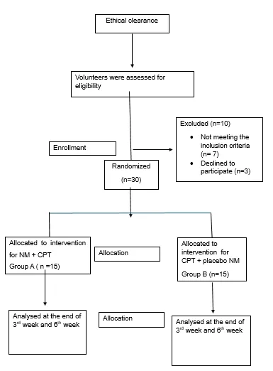
fig. 1.1 CONSORT flow diagram
Intervention - Participants were randomly assigned to two groups using a simple random sampling method: Group A (n = 15) and Group B (n = 15). Group A received facial nerve Nerve Mobilization and conventional physiotherapy, whereas Group B received placebo Nerve Mobilization along with conventional physiotherapy. Both groups underwent a 6-week treatment program consisting of three sessions per week, totaling 18 sessions. Evaluations were carried out at the end of 3rd week, and at 6th week. The outcome measure for each participant was recorded at all time points using individual data collection forms.
GROUP A
Nerve Mobilization
The total duration of Nerve Mobilization was approximately 15 minutes, including rest intervals. The patient was positioned supine with the head slightly turned toward the side being treated.11 The therapist, wearing sterile gloves and using talcum powder to reduce friction, sat at the head end of the treatment table. One hand stabilized the patient’s head, while the other performed facial nerve mobilization.3 The index finger was placed anterior to the external acoustic meatus and directed anteriorly and medially to contact the stylomastoid foramen. The index and middle fingers of the opposite hand were then positioned along the anterior border of the mastoid process to apply posterior-medial mobilization. Circular movements and horizontal traction were performed for 25 repetitions in three sets, with 5-second rests between repetitions and 30-second rests between sets.11
Conventional Physiotherapy
The total duration of the conventional physiotherapy was 45 minutes, including facial massage, facial exercises, and electrical stimulation.
Facial massage was administered in the supine position for 10 minutes and included stroking, circular massage, effleurage, kneading, picking-up, wringing, and hacking techniques to promote circulation and relaxation.3 Facial exercises consisted of ten facial expression movements, including eyebrow raising, nostril flaring, eye closure, frowning, smiling, lip pursing, sucking, crying, and laughing expressions. Each exercise was performed 10 times under supervision and repeated 3–4 times daily at home in front of a mirror to avoid fatigue.3
Electrical stimulation was administered to restore facial muscle tone. Stimulation intensity was determined by eliciting a minimal visible contraction on the unaffected side. The passive electrode was placed over the C7 region, while the active electrode was applied to facial muscle motor points, including the frontalis, orbicularis oculi, zygomaticus, buccinator, nasalis, orbicularis oris, levator muscles, and mentalis. Each motor point received two sets of 30 contractions, while the facial nerve trunk received one set of 60 contractions using faradic current.12
Group B (n=15) – Same as in group A
Conventional Physiotherapy
Conventional physiotherapy is the same as given in group A.
Placebo Neural Mobilisation
The placebo facial nerve mobilisation followed all the procedures of real facial nerve mobilisation without applying horizontal and circular auricular traction.3
Statistical analysis
Data was systematically organized and analyzed using descriptive statistics, including mean and standard deviation. Intra-group comparisons within Group A and Group B for the Facial Clinimetric Evaluation Scale scores were performed using the paired t-test. Inter-group comparisons between Group A and Group B were conducted using the unpaired t-test. The level of statistical significance was set at p < 0.05. Normality of data was assessed using the Kolmogorov–Smirnov and Shapiro–Wilk tests. Given the small sample size (n = 15), greater emphasis was placed on the Shapiro–Wilk test. In Group A, FaCE scores at baseline (0 week), 3 weeks, and 6 weeks were normally distributed (p > 0.05). In Group B, FaCE scores at baseline showed deviation from normality in the Kolmogorov–Smirnov test (p = 0.010), but the Shapiro–Wilk test indicated normal distribution (p = 0.127). At 3 and 6 weeks, FaCE scores were normally distributed (p > 0.05).
Results
Of 40 patients screened for eligibility, 30 patients were included. No participants were lost in follow-up. Assessment was performed by the Facial Clinimetric Evaluation Scale.
The unpaired t-test was also used to compare Facial Clinimetric Evaluation Scale (FaCE) scores between the groups. At baseline, the mean scores were 43.267±4.620 (Group A) and 44.067±5.020 (Group B), with a non-significant t-value (0.454), indicating no difference between groups. At the 3rd week, Group A (66.267±5.812) showed greater improvement than Group B (54.40±4.049), with a mean difference of 11.867 and a significant t-value of 6.488. At the 6th week, the difference increased to 23.333, with Group A scoring 86.933±6.713 and Group B 63.60±3.459, and a t-value of 11.966, confirming a statistically significant difference between the groups.
Top of Form
Bottom of Form
Overall, the findings indicate that although both groups were comparable at baseline, Group A demonstrated significantly greater improvement than Group B over time across all outcome measures, including the Sunnybrook Facial Grading System and the Facial Clinimetric Evaluation Scale. These results suggest that the intervention applied to Group A was more effective in enhancing facial function, reducing synkinesis, and improving quality of life compared to Group B.
|
Unpaired t-test |
Comparison |
|||||
|
Facial Clinimetric Evaluation Scale |
||||||
|
0th day |
3rd week |
6th week |
||||
|
Group-A |
Group-B |
Group-A |
Group-B |
Group-A |
Group-B |
|
|
Mean |
43.267 |
44.067 |
66.267 |
54.40 |
86.933 |
63.60 |
|
S.D. |
4.620 |
5.020 |
5.812 |
4.049 |
6.713 |
3.459 |
|
N |
15 |
15 |
15 |
15 |
15 |
15 |
|
M.D. |
8.0 |
11.867 |
23.333 |
|||
|
df |
28 |
28 |
28 |
|||
|
Unpaired t-test |
0.454 |
6.488 |
11.966 |
|||
|
P value |
0.653 |
<0.001* |
<0.001* |
|||
|
Result |
Non-Significant |
Significant |
Significant |
|||
p>0.05 Non-Significant (NS)
p<0.05 Significant (S)
Table 1.1: Independent t-test for comparison at 0th day, 3rd week, and 6th week score measurements of FaCE between Group A and Group B.
Discussion
The findings of this randomized controlled trial indicate that a 6-week program of facial Nerve Mobilization , when combined with conventional physiotherapy, resulted in greater improvements in facial movement and symmetry in individuals with acute idiopathic facial paralysis compared to conventional physiotherapy alone.
These results are in agreement with those reported by Raed Alharbi et al. (2023)3, who conducted a randomized controlled trial to evaluate the addition of facial nerve Nerve Mobilization to standard conventional physiotherapy in patients with acute Bell’s palsy. Their study demonstrated significantly greater improvements in facial symmetry and voluntary facial movements in the intervention group. Enhanced facial grading scores observed after three weeks suggested that Nerve Mobilization facilitates nerve recovery and improves functional outcomes, thereby supporting the present findings.
Additional support is provided by a case report by Kashoo, Alqahtani, and Ahmad,5 which documented marked improvements in facial symmetry and voluntary movements following facial nerve Nerve Mobilization . These effects were attributed to increased nerve mobility, reduced intraneural pressure, and improved axoplasmic flow. However, as the evidence was based on a single case, its generalizability is limited and should be interpreted with caution. Nonetheless, the report highlights the potential clinical relevance of Nerve Mobilization and underscores the need for further controlled studies.
In contrast, Shahid Ahmed (Heera) et al.2 reported no statistically significant difference when comparing Nerve Mobilization with proprioceptive neuromuscular facilitation techniques. Their findings suggested that Nerve Mobilization was not superior in improving facial function. Such discrepancies indicate that the effectiveness of Nerve Mobilization may depend on variables such as the stage of the condition, treatment protocol, dosage, and duration of intervention.
From a mechanistic perspective, Nerve Mobilization enhances both the mechanical and physiological properties of the nerve, thereby complementing conventional physiotherapy in Bell’s palsy. It promotes normal nerve gliding, reduces adhesions and intraneural pressure, and improves vascular supply, facilitating the healing process. Additionally, it enhances axoplasmic flow, which supports nerve regeneration and conduction. In contrast, conventional physiotherapy primarily focuses on muscle re-education, strengthening, and prevention of complications such as contractures and synkinesis, without directly addressing nerve mobility.
Therefore, the combined application of Nerve Mobilization and conventional physiotherapy appears to yield superior outcomes, as it simultaneously targets nerve recovery and muscle function, resulting in more comprehensive and effective rehabilitation.
Limitations
The findings of this study should be interpreted with caution due to several limitations. Recovery may have been influenced by the natural course of Bell’s palsy and the combined physiotherapy interventions rather than nerve mobilization alone. Additionally, there is no standardized, scientifically validated protocol for facial nerve mobilization, and the small sample size limits the generalizability and statistical power of the results. Further large-scale randomized controlled trials are needed to establish the specific effectiveness of nerve mobilization in Bell’s palsy rehabilitation.
CONCLUSION
The addition of facial nerve mobilization to conventional intervention was found to be more effective in improving functional abilities and quality of life in patients with Bell’s palsy. These enhanced outcomes may be attributed to the role of Nerve Mobilization in improving nerve mobility, facilitating neural conduction, and promoting better neuromuscular function. Furthermore, this combined approach may help minimize long-term complications such as residual weakness.
REFERENCES

Muskan*, Lalit Arora, Reena Arora, Amarjeet Singh, Shiny George Gill, Impact Of Facial Nerve Mobilization On Functional Abilities And Quality Of Life In Bell’s Palsy, Int. J. Sci. R. Tech., 2026, 3 (7), 601-607. https://doi.org/10.5281/zenodo.21426726
 10.5281/zenodo.21426726