

We use cookies to ensure our website works properly and to personalise your experience. Cookies policy
Department of Pharmaceutics, JKKMIHSCP, T.N. Palayam, Tamil Nadu, India
Breast cancer care needs affordable agents that act on several targets, and the leaf of cowpea (Vigna unguiculata (L.) Walp.), an edible legume, has rarely been tested against human tumour cells. Leaves were extracted sequentially with hexane, petroleum ether, ethanol and water, and the ethanolic extract (11.62% w/w) was studied further. It contained phenols, flavonoids and saponins, with a total flavonoid content of 11.07 mg quercetin equivalents/g and a total antioxidant capacity of 312.50 µg ascorbic acid equivalents/mg, but weak DPPH scavenging (IC50 678.2 µg/mL). LC-MS tentatively annotated 21 features. The extract reduced MCF-7 cell viability in a concentration-dependent manner (96.1% to 34.6% at 6.25-100 µg/mL after 24 h; IC50 59.52 µg/mL by interpolation and 46.59 µg/mL by logistic regression), raised intracellular oxidant load and, at 100 µg/mL, produced apoptotic nuclear morphology on acridine orange/ethidium bromide staining. Docking of five annotated constituents against AKT3 and ERK5 gave binding energies of -5.1 to -9.2 kcal/mol, with demethoxycurcumin and acacetin ranking highest. The extract (0.01% w/w) was formulated into Carbopol 934/HPMC K4M gels. The optimised gel (F4) had pH 6.86, viscosity 26,380 cP, spreadability 24.7 g.cm/s and drug content 99.1%, released 77.4% in 8 h by a Higuchi-type mechanism, and showed no incompatibility by FTIR. These in vitro and in silico findings do not establish selectivity, in vivo efficacy or skin permeation.
Breast cancer is the most frequently diagnosed malignancy in women and a leading cause of cancer death worldwide.1 India has a high mortality-to-incidence ratio because many patients present at a locally advanced or metastatic stage, so progress depends on affordable and well-tolerated systemic agents as well as on earlier detection.1 About 70% of tumours are hormone-receptor positive, and the MCF-7 line used here is an oestrogen- and progesterone-receptor-positive model of that subtype.2 Current cytotoxic therapy has a narrow therapeutic window and is limited by toxicity and acquired resistance.2
Plant secondary metabolites are attractive for this problem because polyphenolic and terpenoid scaffolds tend to engage several targets with modest affinity, and a majority of approved small-molecule anticancer drugs are natural products or derived from them.3,4 Cowpea (Vigna unguiculata (L.) Walp.) is a drought-tolerant legume eaten as seed, pod and young leaf in the tropics, and its chemistry includes flavonoids, phenolic acids, saponins and bioactive peptides.5 Cowpea polyphenol extracts inhibit mammary cancer cell proliferation, with activity following the phenolic content of the plant part examined.6 Most of this work, however, has used seed, seed coat or whole grain; the leaf, where inducible defence metabolites accumulate, has been studied mainly for nutrition and antioxidant capacity.5,7 Where cytotoxicity has been reported, it has often rested on a viability assay alone, and few studies link the chemistry of the extract to a possible molecular target.
A crude extract is also not a dosage form. A topical gel allows a measured quantity of a poorly soluble, labile polyphenol-rich extract to be applied to a defined area and gives control over its release, and it is a reasonable first dosage form for locally accessible disease such as chest-wall recurrence.
This study therefore aimed to (i) prepare sequential extracts of V. unguiculata leaf and characterise the selected ethanolic extract by phytochemical screening, flavonoid estimation, LC-MS profiling and two antioxidant assays; (ii) evaluate its cytotoxicity against MCF-7 cells, with intracellular oxidant load and apoptotic morphology as orthogonal readouts; (iii) dock the principal annotated constituents against AKT3 and ERK5 to generate testable hypotheses; and (iv) formulate the extract as a topical gel and evaluate it.
MATERIALS AND METHODS
Plant material and sequential extraction
Healthy, fully expanded leaves of V. unguiculata were collected at the vegetative to pre-flowering stage, washed, shade-dried to constant weight, powdered (No. 40 sieve) and stored in airtight amber containers. The material was assigned the internal code VUC. Fifty grams of powder was extracted in a Soxhlet apparatus with 500 mL each of hexane, petroleum ether (60-80 °C), ethanol and distilled water in turn, the marc being air-dried between solvents. Extracts were concentrated under reduced pressure at 40-45 °C, dried to constant weight and stored at 4 °C. They are coded VUHE, VUPE, VUEE (ethanolic) and VUAE (aqueous); the ethanolic extract used for the gel is referred to as VUC-E. Percentage yield was calculated as (weight of dried extract/weight of powder) × 100.
Phytochemical screening and total flavonoid content
Each extract was screened by standard tests for phenols, tannins, flavonoids, saponins, terpenoids, alkaloids, glycosides, quinones, fatty acids and steroids, and graded as absent, trace, moderate or abundant. Total flavonoid content of VUEE was measured by the aluminium chloride method at 510 nm against a quercetin curve (1-80 µg/mL) and expressed as mg quercetin equivalents (QE) per gram of extract.8
LC-MS profiling
The methanolic solution of VUEE was filtered (0.22 µm) and separated on a reversed-phase C18 column with a water/acetonitrile gradient (0.1% formic acid in both) over about 40 min. Electrospray ionisation was run in positive and negative modes. Features were annotated by accurate mass and molecular formula against public databases and are reported as tentative (Metabolomics Standards Initiative Level 2/3).9 Each positive-mode annotation was also checked against the exact mass of an adduct of its proposed formula.
Antioxidant assays
Total antioxidant capacity was measured by the phosphomolybdenum method (695 nm; extract 1 mg/mL; ascorbic acid 200-1000 µg/mL; 95 °C, 90 min) and expressed as µg ascorbic acid equivalents (AAE) per mg of extract.10 DPPH scavenging was measured in 96-well plates (extract 0-1000 µg/mL; ascorbic acid 0-50 µg/mL; 30 min in the dark; 517 nm; quadruplicate), and the IC50 was obtained by non-linear regression.11
Cell culture and MTT assay
MCF-7 cells (NCCS, Pune) were grown in DMEM with 10% foetal bovine serum and antibiotics at 37 °C in 5% CO2. VUEE was dissolved in 0.1% DMSO, filter-sterilised and diluted to 6.25, 12.5, 25, 50 and 100 µg/mL. Cells (5 × 10³ per well) were seeded in 96-well plates, allowed to attach for 24 h and exposed to the extract for 24 h (three wells per concentration). After 4 h with MTT, formazan was dissolved in DMSO and read at 540 nm.12 Viability was calculated as (mean OD treated/mean OD control) × 100. The IC50 was obtained by interpolation and independently by four-parameter logistic regression.
Intracellular oxidant load and apoptotic morphology
For oxidant load, cells were treated with 50 or 100 µg/mL extract for 24 h, loaded with 10 µM DCFH-DA for 15 min in the dark and imaged under blue excitation (450-480 nm).13 For apoptotic morphology, cells treated with 25, 50 or 100 µg/mL for 24 h were stained with acridine orange/ethidium bromide (100 µg/mL each) for 5-10 min and imaged. Viable cells show uniform green nuclei, early apoptotic cells green nuclei with condensed chromatin, late apoptotic cells orange-red nuclei with condensed or fragmented chromatin, and necrotic cells uniformly orange-red nuclei of normal organisation.
Molecular docking
Five annotated constituents (acacetin, demethoxycurcumin, 4-methoxycinnamic acid, allocryptopine and quinine) were docked against the pleckstrin-homology domain of AKT3 (PDB 2X18) and the kinase domain of ERK5/MAPK7 (PDB 6HKN).14 Ligands were retrieved as 3D structures, energy-minimised (MMFF94) and charged (Gasteiger). Receptors were prepared by removing water and non-essential heteroatoms and adding polar hydrogens. Docking used AutoDock Vina with the grid centred on the co-crystallised ligand site, and the best pose per pair was analysed.15 4-Methoxycinnamic acid was used as the simplest representative of the methoxycinnamic acid series annotated in the extract.
Formulation of the topical gel
Four gels (F1-F4) containing VUC-E at 0.01% w/w (100 µg/g) were prepared. The extract loading, glycerin (5%) and propylene glycol (5%) were constant; Carbopol 934 was raised from 0.50 to 1.00% w/w across F1-F3 and HPMC K4M from 1.00 to 1.50% w/w in F4 (Table 1). Carbopol was hydrated in water for 12 h under stirring (500 rpm), HPMC K4M was dispersed in water at 80 °C, cooled and hydrated for 4 h, and the extract was dissolved in the glycerin/propylene glycol phase by brief sonication. The phases were combined, made up to 100 g, neutralised with triethanolamine to pH 6.7-7.0 and left for 24 h to release entrapped air.
Table 1: Composition of the VUC-E Topical Gels (% W/W)
| Ingredient | F1 | F2 | F3 | F4 |
|---|---|---|---|---|
| VUC-E | 0.01 | 0.01 | 0.01 | 0.01 |
| Carbopol 934 | 0.50 | 0.75 | 1.00 | 1.00 |
| HPMC K4M | 1.00 | 1.00 | 1.00 | 1.50 |
| Glycerin | 5.00 | 5.00 | 5.00 | 5.00 |
| Propylene glycol | 5.00 | 5.00 | 5.00 | 5.00 |
| Triethanolamine | q.s. to pH 6.7-7.0 | q.s. to pH 6.7-7.0 | q.s. to pH 6.7-7.0 | q.s. to pH 6.7-7.0 |
| Purified water | q.s. 100 g | q.s. 100 g | q.s. 100 g | q.s. 100 g |
Evaluation of the gels
Appearance, homogeneity and grittiness were assessed visually and by finger test. The pH was measured on a 1% aqueous dispersion. Viscosity was measured on a Brookfield viscometer (spindle 6, 10 rpm, 25 ± 1 °C). Spreadability was measured by the parallel-plate method (S = M × L/T; 80 g load, 7.5 cm) and extrudability as the mass extruded from a collapsible tube (5 mm orifice, 500 g load, 30 s). Drug content and in vitro release were determined spectrophotometrically against a VUC-E calibration curve with phosphate buffer pH 7.4 as receptor medium, and the release data were fitted to kinetic models. FTIR spectra (ATR, 4000-400 cm-1) were recorded for VUC-E, Carbopol 934, HPMC K4M, their 1:1:1 physical mixture and the F4 gel. The F4 gel was stored for 90 days at 25 ± 2 °C/60 ± 5% RH and at 40 ± 2 °C/75 ± 5% RH and examined at 0, 30, 60 and 90 days. Gel determinations were made in triplicate and expressed as mean ± SD.
Statistical analysis
Raw MTT optical densities were compared with the untreated control by one-way ANOVA followed by Dunnett's test (p < 0.05 significant). Calibration curves were fitted by least-squares regression. The three wells per concentration are technical replicates from a single plate.
RESULTS AND DISCUSSION
Extractive yield and phytochemical screening
Ethanol gave the highest yield (11.62% w/w; 5.81 g from 50 g), followed by water (8.08%), petroleum ether (4.86%) and hexane (0.34%). The extracts were sticky and ranged from green (VUHE) through black (VUPE) to dark brown (VUEE) and brown (VUAE). Screening followed the polarity gradient (Table 2): fatty acids and steroids were found mainly in the non-polar extracts, whereas phenols, flavonoids and saponins were found chiefly in the ethanolic and aqueous extracts. Because the deeply coloured extracts interfere with colour end-points, these results are only a preliminary map. The ethanolic extract was chosen for all later work because it combined the highest yield, abundant phenol and flavonoid signals and moderate saponin content, with less interference in colorimetric and fluorescence readouts than the aqueous extract.
Table 2: Yield and Qualitative Phytochemical Constituents of the Sequential Extracts of V. unguiculata Leaf
| Parameter | VUHE | VUPE | VUEE | VUAE |
|---|---|---|---|---|
| Solvent | Hexane | Pet. ether | Ethanol | Water |
| Yield (% w/w) | 0.34 | 4.86 | 11.62 | 8.08 |
| Phenols | - | - | +++ | + |
| Tannins | + | - | + | - |
| Flavonoids | + | - | +++ | ++ |
| Saponins | - | - | ++ | ++ |
| Terpenoids | + | - | + | + |
| Alkaloids | - | + | + | - |
| Glycosides | - | - | + | - |
| Quinones | + | - | + | - |
| Fatty acids | + | + | + | - |
| Steroids | ++ | + | + | + |
(-) absent, (+) trace, (++) moderate, (+++) abundant.
The total flavonoid content of VUEE was 11.07 mg QE/g (quercetin curve y = 0.00565x + 0.01902, R2 = 0.9996).This is a modest figure that agrees with descriptions of V. unguiculata leaf as relatively poor in flavonoid diversity.7 The aluminium chloride method under-reports flavanones, flavan-3-ols and isoflavones, so the value is best read as a lower bound for a defined subset of flavonoids.8
LC-MS profile
LC-MS annotated 21 features (11 in positive and 10 in negative mode) in six classes: flavonoids and flavonoid glycosides, cinnamic and benzoic acid derivatives, alkaloids, saturated and polyunsaturated fatty acids, glycerophospholipids and soyasapogenol-type triterpenoid saponins. The saponin and flavonoid classes agree with the qualitative screening. Negative-mode annotations included kotanin, dihydrohesperetin-7-O-neohesperidoside, soyasapogenol B derivatives, demethoxycurcumin, behenic acid, γ-linolenic acid, conjugated linoleic acid, an oxylipin and phospholipids. The exact-mass check of the positive-mode annotations (Table 3) showed that five matched [M+H]+ of their proposed formula (glycerophosphorylcholine, dihydroxybenzoic acid, quinine, allocryptopine and monocrotaline N-oxide), and three (3,5-dimethoxycinnamic acid, arachidic acid and L-threonine) matched [M-H]- exactly and are therefore correct assignments filed under the wrong polarity. The feature annotated as acacetin (m/z 591.54) does not correspond to the aglycone and is better explained as the deprotonated acacetin 7-O-rutinoside (theoretical 591.17). The feature annotated as retrorsine (m/z 722.40; match scores 0.37 and 0.41) was not accounted for by any common adduct and is treated as unassigned.
Table 3: Mass-consistency Check of Positive-Mode LC-MS Annotations of VUEE
| Annotation | Formula | Match score | Observed m/z | Assessment |
|---|---|---|---|---|
| Glycerophosphorylcholine | C8H20NO6P | 0.9813 | 258.00 | Consistent with [M+H]+ |
| Dihydroxybenzoic acid | C7H6O4 | 0.8330 | 155.04 | Consistent with [M+H]+ |
| Quinine | C20H24N2O2 | 0.7147 | 325.1911 | Exact [M+H]+ |
| Acacetin | C16H12O5 | 0.7536 | 591.54 | Not the aglycone ion |
| 3,5-Dimethoxycinnamic acid | C11H12O4 | 0.8465 | 207.0663 | Exact [M-H]- |
| Retrorsine | C18H25NO6 | 0.3745 | 722.3957 | Unassigned |
| Arachidic acid | C20H40O2 | 0.5246 | 311.2956 | Exact [M-H]- |
| Allocryptopine | C21H23NO5 | 0.3590 | 370.00 | Consistent with [M+H]+ |
| L-Threonine | C4H9NO3 | 0.7551 | 118.051 | Exact [M-H]- |
| Monocrotaline N-oxide | C16H23NO7 | 0.6945 | 342.1547 | Exact [M+H]+ |
Three annotations (quinine, allocryptopine and demethoxycurcumin) belong to plant families other than the Fabaceae and carried low to moderate match scores (0.7147 and 0.3590 for the first two). Misannotation without fragmentation confirmation, carry-over or contamination, and endophytic origin (kotanin is of fungal origin) are all possible explanations; no solvent or procedural blank was available. These annotations were retained only because they were carried into the docking study. All identifications are tentative and no authentic standards were run.9
Antioxidant activity
The extract had a total antioxidant capacity of 312.50 µg AAE/mg (ascorbic acid curve y = 0.00197x + 1.9141, R2 = 0.9990), but scavenged DPPH weakly: its IC50 was 678.2 µg/mL against 31.25 µg/mL for ascorbic acid, about 21.7-fold higher. The two assays measure different chemistry: the phosphomolybdenum assay is run in hot acid and captures any reductant, including hydrolysed glycosides and lipophilic species, whereas DPPH responds mainly to free phenolic hydroxyl groups at room temperature.10,11 The LC-MS profile, dominated by methoxylated and glycosylated polyphenols (acacetin and its rutinoside, dimethoxycinnamic acid, a flavanone glycoside), is consistent with high reducing capacity but modest hydrogen donation. A crude extract also carries a large mass of inert material, so its IC50 cannot be compared directly with that of a pure standard.
Cytotoxicity against MCF-7 cells
The extract reduced MCF-7 viability in a monotonic, concentration-dependent manner from 96.12 ± 1.27% at 6.25 µg/mL to 34.63 ± 0.99% at 100 µg/mL (Table 4, Figure 1). One-way ANOVA on raw optical densities was significant (F = 1810.5, p < 0.001), and every concentration differed from the control by Dunnett's test. The IC50 was 59.52 µg/mL by interpolation and 46.59 µg/mL (95% CI 36.18-57.00; R2 = 0.9965) by four-parameter logistic regression. Interpolation between 50 and 100 µg/mL overestimates the IC50 because the curve is concave in that interval, so the IC50 is best stated as 47-60 µg/mL. Both values lie below the 100 µg/mL cut-off used in National Cancer Institute plant-screening criteria and above the 20 µg/mL cut-off for strong activity, so the extract is moderately active.16,17
Table 4: Effect of VUEE on MCF-7 Viability After 24 H (MTT Assay; Mean ± SD of Three Wells)
| Concentration (µg/mL) | Viability (%) | Dunnett p value |
|---|---|---|
| Control | 100.00 ± 0.00 | - |
| 6.25 | 96.12 ± 1.27 | 0.0027 (**) |
| 12.5 | 86.22 ± 1.30 | < 0.001 (***) |
| 25 | 74.47 ± 1.12 | < 0.001 (***) |
| 50 | 53.62 ± 1.05 | < 0.001 (***) |
| 100 | 34.63 ± 0.99 | < 0.001 (***) |
Figure 1: Concentration-response of MCF-7 Cells to VUEE at 24 H: Four-Parameter Logistic Fit with IC50 by Interpolation (Left) and Percentage Viability (Right). Mean ± SD of Three Wells; * p < 0.01, ** P < 0.001 Versus Control (Dunnett's Test).
The MTT assay measures reductive capacity, not cell number, and a coloured, reducing extract can reduce tetrazolium directly; a cell-free control was not run, although such interference would make cytotoxicity appear lower rather than higher.12 Phase-contrast microscopy showed progressive loss of monolayer density, rounding and detachment. No non-tumorigenic comparator line was tested, so a selectivity index could not be calculated and the result should be read as cytotoxicity against a breast cancer line, not selective anticancer activity.
Intracellular oxidant load and mode of cell death
DCFH-DA fluorescence was very low in control cells and increased at 50 and 100 µg/mL, being highest at 100 µg/mL where cells were also rounded (Figure 2). The increase was concentration dependent and occurred over the range that brackets the IC50. Polyphenols can act as antioxidants in a cell-free assay yet generate oxidants in culture medium, and the effect depends on concentration and cell type, so a weak DPPH result and raised oxidant load are not contradictory.18,19 Part of the effect could come from hydrogen peroxide formed by polyphenol autoxidation in the medium, which was not excluded by a catalase control.20 The probe is also non-specific, so the finding is an increase in oxidant load, not in any named species.13 The assessment was qualitative and not quantified by image analysis.
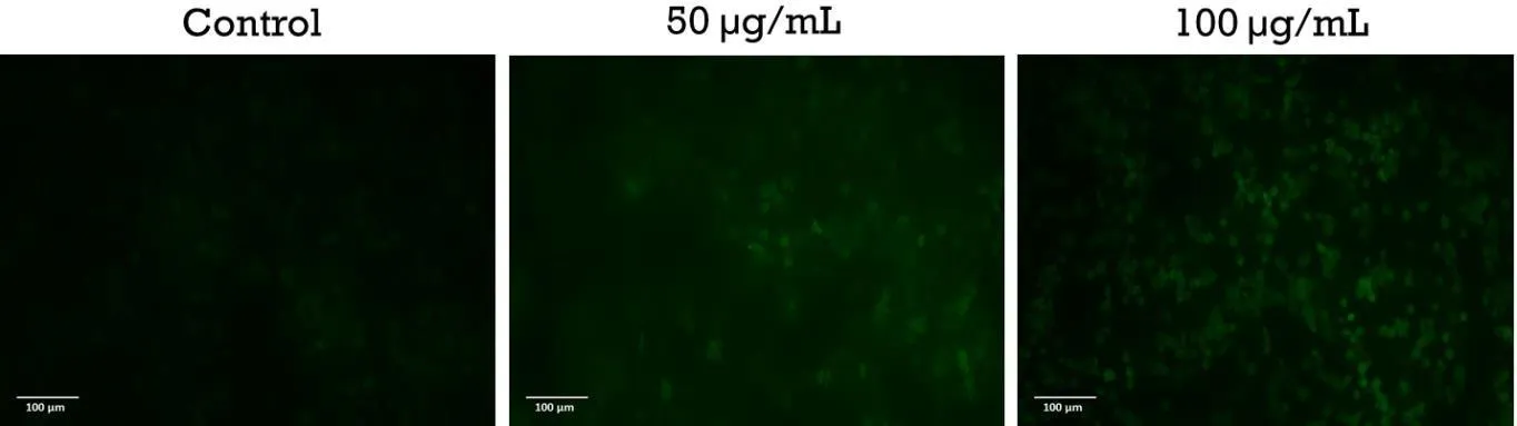
Figure 2: Dcfh-Da Fluorescence of MCF-7 Cells After 24 H: Untreated Control (Left), 50 Μg/ml (Centre) and 100 Μg/ml VUEE (Right).
On acridine orange/ethidium bromide staining, nuclei were uniformly green in control cells and at 25 and 50 µg/mL, whereas at 100 µg/mL light orange and red nuclei with condensed chromatin appeared in quantity and cell number fell (Figure 3). Colour alone does not separate late apoptosis from necrosis, but the chromatin condensation places these cells in the late-apoptotic category, and no population of uniformly orange-red nuclei of normal organisation was described at any concentration. Apoptotic morphology was absent at 50 µg/mL although MTT viability was 53.62% there; this may reflect a cytostatic or metabolic effect, a later onset of apoptosis than the 24 h time point, or an unscored minor apoptotic fraction. Cells were not scored by category, so no apoptotic index is available. MCF-7 cells lack functional caspase-3, so confirmation should target caspase-7 or caspase-9 and not caspase-3.21 The data are consistent with apoptosis but do not prove an oxidative mechanism, which would need rescue with N-acetylcysteine or measurement of mitochondrial membrane potential.
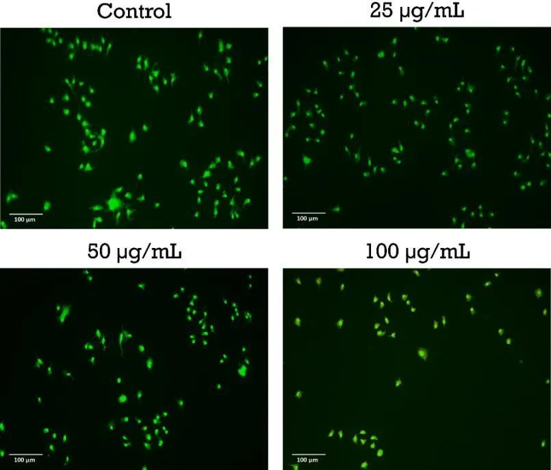
Figure 3: Acridine Orange/Ethidium Bromide Staining of MCF-7 Cells After 24 H with VUEE (Control, 25, 50 and 100 Μg/ml).
Molecular docking
Binding energies ranged from -5.1 to -7.4 kcal/mol at AKT3 and from -6.4 to -9.2 kcal/mol at ERK5 (Table 5, Figure 4). Four of five ligands scored better at ERK5 (mean -7.72 versus -6.38 kcal/mol at AKT3), consistent with a deep, largely hydrophobic ERK5 ATP pocket compared with the shallower, polar phosphoinositide-binding surface of the AKT3 pleckstrin-homology domain.22,23 Demethoxycurcumin scored best overall (-9.2 kcal/mol at ERK5; eleven hydrophobic contacts and hydrogen bonds to THR99, MET140 and ASP200), and acacetin was the most consistent ligand (-7.4 and -8.7 kcal/mol; hydrogen bonds to LYS14, GLU17, THR86 and LYS20 at AKT3, and to MET140 and ASP200 at ERK5). MET140 and ASP200 were contacted by three of five ligands at ERK5 and may anchor that site.
Table 5: Docking Binding Energies (kcal/mol) of Annotated Constituents
| Ligand | AKT3 (2X18) | ERK5 (6HKN) |
|---|---|---|
| 4-Methoxycinnamic acid | -5.1 | -6.4 |
| Acacetin | -7.4 | -8.7 |
| Allocryptopine | -6.6 | -6.5 |
| Demethoxycurcumin | -6.4 | -9.2 |
| Quinine | -6.4 | -7.8 |
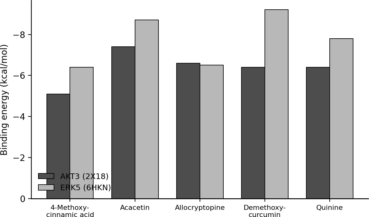
Figure 4: Predicted Binding Energies of Five Annotated Constituents Against AKT3 (2X18) and ERK5 (6HKN); More Negative Values Indicate More Favourable Predicted Binding.
These results are hypotheses, not evidence of target engagement. Redocking validation was not performed, scores within 1-2 kcal/mol are within the noise of the scoring function, and LC-MS was qualitative, so the abundance of each ligand in the extract is unknown. The two highest-scoring ligands differ in reliability: demethoxycurcumin is a Zingiberaceae metabolite and its annotation is doubtful, whereas methoxylated flavones are common in legumes. Acacetin is therefore the better candidate for follow-up, and it has been reported to induce apoptosis in MCF-7 cells as a pure compound.24,25 If the species present is acacetin 7-O-rutinoside, the docked aglycone represents the deglycosylated form, since the glycoside violates the Lipinski criteria and would not fit the pockets.26 A flavonoid content of about 1.1% of the extract mass also makes it unlikely that acacetin alone explains cytotoxicity at about 60 µg/mL, so the activity is probably distributed across several constituents.
Evaluation of the topical gels
All four batches formed pale green, translucent gels that were homogeneous, free of grittiness and free of phase separation after 24 h (Figure 5). The pH of 6.71-6.86 lies within the 5.0-7.0 range generally accepted for topical semisolids. Viscosity rose from 8,250 ± 205 cP (F1) to 26,380 ± 336 cP (F4) and spreadability fell from 29.4 ± 0.8 to 24.7 ± 0.5 g.cm/s (Table 6), the two being strongly negatively correlated (r = -0.975). Weight for weight, Carbopol 934 thickened the gel about twice as efficiently as HPMC K4M. Extrudability fell from 132 ± 4 g (F1) to 108 ± 3 g (F3) and rose slightly to 112 ± 3 g in F4, a difference of the same order as the standard deviations; the greater shear-thinning of the HPMC-containing F4 may explain it. Drug content was 96.8 ± 1.4% to 99.1 ± 0.8% of the label claim. F4 (1.00% Carbopol 934, 1.50% HPMC K4M) was selected as the optimised batch for its highest viscosity, best homogeneity and highest drug content, while its spreadability remained above the acceptance limit of 20 g.cm/s.
Table 6: Physicochemical Characteristics of the VUC-E Gels (Mean ± SD, N = 3)
| Batch | Carbopol 934 / HPMC K4M (%) | pH | Viscosity (cP) | Spreadability (g.cm/s) | Extrudability (g) |
|---|---|---|---|---|---|
| F1 | 0.50 / 1.00 | 6.71 ± 0.03 | 8,250 ± 205 | 29.4 ± 0.8 | 132 ± 4 |
| F2 | 0.75 / 1.00 | 6.76 ± 0.02 | 14,680 ± 271 | 27.1 ± 0.7 | 119 ± 3 |
| F3 | 1.00 / 1.00 | 6.84 ± 0.03 | 20,450 ± 306 | 25.3 ± 0.6 | 108 ± 3 |
| F4 | 1.00 / 1.50 | 6.86 ± 0.01 | 26,380 ± 336 | 24.7 ± 0.5 | 112 ± 3 |
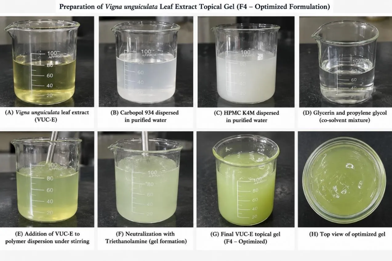
Figure 5: Preparation of the VUC-E Topical Gel: (A) Extract, (B) Carbopol 934 and (C) HPMC K4M Dispersions, (D) Glycerin and Propylene Glycol, (E) Addition of Extract, (F) Neutralisation with Triethanolamine, (G) Final F4 Gel and (H) Top View of the Optimised Gel.
The F4 gel released 77.4 ± 1.0% of its drug content over 8 h, with a release half-time of 4.1 h. Release followed the Higuchi model (R2 = 0.9981) with a Korsmeyer-Peppas exponent n = 0.78, which indicates anomalous, diffusion-dominated transport.
In the FTIR spectra (Figure 6), VUC-E showed a broad O-H stretch (3378 cm-1), aliphatic C-H stretches (2924, 2854 cm-1), a carbonyl band (1712 cm-1), aromatic ring stretches (1608, 1516 cm-1) and C-O stretches (1265, 1045 cm-1). All were retained within 3 cm-1 in the physical mixture with no new band. In the F4 gel the Carbopol carbonyl band was weakened and carboxylate bands appeared at 1552 and 1408 cm-1, the expected result of neutralisation with triethanolamine, while the extract bands at 1606 and 1050 cm-1 persisted. No chemical interaction between the extract and the excipients is indicated.
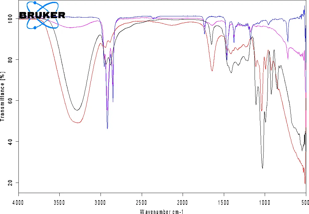
Figure 6: Overlaid FTIR Spectra (4000-400 cm-1) of VUC-E, Carbopol 934, HPMC K4M, Their Physical Mixture and the F4 Gel.
Stability results are in Table 7. At 25 °C/60% RH the F4 gel was essentially unchanged after 90 days (pH -0.04, viscosity -1.3%, drug content -1.8%). At 40 °C/75% RH, pH fell from 6.86 to 6.70, viscosity by 7.1%, spreadability rose from 24.7 to 26.6 g.cm/s and drug content fell by 7.4% to 91.7%, without phase separation. These changes are consistent with slow degradation of phenolic constituents to acidic products, which lowers the pH, partly re-protonates Carbopol and thins the gel. A cool, light-protected store and, in further development, an antioxidant and a preservative would be needed.
Table 7: Stability of the F4 Gel over 90 Days (Mean ± SD, N = 3)
| Condition | Day | pH | Viscosity (cP) | Spreadability (g.cm/s) | Drug content (%) |
|---|---|---|---|---|---|
| 25 °C/60% RH | 0 | 6.86 ± 0.01 | 26,380 ± 336 | 24.7 ± 0.5 | 99.1 ± 0.8 |
| 30 | 6.85 ± 0.02 | 26,310 ± 350 | 24.8 ± 0.4 | 98.6 ± 0.9 | |
| 60 | 6.84 ± 0.02 | 26,190 ± 378 | 24.9 ± 0.5 | 98.0 ± 1.0 | |
| 90 | 6.82 ± 0.03 | 26,040 ± 412 | 25.1 ± 0.6 | 97.3 ± 1.1 | |
| 40 °C/75% RH | 30 | 6.81 ± 0.03 | 25,870 ± 391 | 25.3 ± 0.5 | 96.8 ± 1.0 |
| 60 | 6.76 ± 0.03 | 25,240 ± 424 | 25.9 ± 0.6 | 94.5 ± 1.2 | |
| 90 | 6.70 ± 0.04 | 24,510 ± 455 | 26.6 ± 0.7 | 91.7 ± 1.4 |
The gel was characterised for its physicochemical properties, release and short-term stability only. Skin permeation, dermal deposition and irritation studies were not performed, and the finished gel was not itself tested against MCF-7 cells, so the work describes a delivery system developed and characterised and not a topical treatment demonstrated.
CONCLUSION
The ethanolic leaf extract of V. unguiculata (VUC-E) was rich in phenolic, flavonoid and saponin constituents, had high total reducing capacity but weak DPPH scavenging, and reduced MCF-7 viability in a concentration-dependent manner (IC50 about 47-60 µg/mL) with increased intracellular oxidant load and apoptotic nuclear morphology at 100 µg/mL. Docking suggested that acacetin and demethoxycurcumin may interact with ERK5, and that acacetin is the better candidate for follow-up. A Carbopol 934/HPMC K4M gel containing 0.01% w/w extract (F4) had acceptable pH, viscosity, spreadability, extrudability and drug content, showed no extract-excipient incompatibility by FTIR, and was stable for 90 days at 25 °C but lost 7.4% of its content at 40 °C. The study is in vitro and in silico, used a single cell line without a non-tumorigenic comparator, and relied on tentative LC-MS annotations and unvalidated docking. It therefore does not establish V. unguiculata leaf extract as a treatment for breast cancer. Bioassay-guided fractionation, selectivity testing, mechanistic confirmation and skin permeation and safety studies are needed.
ACKNOWLEDGMENT
The authors thank the Department of Pharmaceutics, JKKMIHSCP, T.N. Palayam, for providing the facilities needed to carry out this work.
REFERENCES

M. Priya, K. Raja, C. V. Navin Rhakul, B. Krishnamoorthy, P. Perumal, Phytochemical Profiling, in Vitro Anticancer Activity, Molecular Docking and Topical Gel Formulation of Vigna Unguiculata (L.) Walp. Leaf Extract, Int. J. Sci. R. Tech., 2026, 3 (11), 153-162. https://doi.org/10.5281/zenodo.23915869
 10.5281/zenodo.23915869