

We use cookies to ensure our website works properly and to personalise your experience. Cookies policy
Department of Pharmacology, KMCH College of Pharmacy, Coimbatore- 641048
Background and Purpose: Glucocorticoid-induced osteoporosis (GIOP) is a leading cause of long-term glucocorticoid therapy, and is associated with decreased bone formation, increased bone resorption, and increased fracture risk. The review highlights the molecular mechanisms, pharmacological properties and experimental evidence demonstrating the therapeutic potential of zoledronic acid in GIOP. Experimental Approach: In vitro and in vivo studies of the mechanisms and pharmacological effects of zoledronic acid in bone loss due to glucocorticoids were selected and critically reviewed. Results: Zoledronic acid, a nitrogen-containing bisphosphonate, has a high affinity for binding to bone mineral, and it is also a potent inhibitor of farnesyl pyrophosphate synthase (FPPS) in the mevalonate pathway, causing osteoclast dysfunction and apoptosis. It also inhibits RANKL/RANK and NF-?B and MAPK/JNK signalling pathways, leading to a reduction in osteoclast differentiation and bone resorption. Zoledronic acid also maintains bone mineral density, maintains trabecular microarchitecture and enhances skeletal strength in experimental models of glucocorticosteroid treatment, as consistently shown in experimental studies. Conclusions: Zoledronic acid is an effective anti-resorptive drug to prevent and treat GIOP. However, additional research must be conducted to determine optimal dosages, long-term safety, and effectiveness in a variety of patient populations.
Osteoporosis adversely affects bone turnover: bone resorption surpasses formation, leading to a gradual decrease in bone mass. The damage to bone structure and the loss of bone density, in turn, elevate the frequency of pathological insufficiency fractures. About 20% of white men and 50% of women over 50years experience an osteoporotic fracture. Along with gender, age, and bone mineral density (BMD), additional risk factors for pathological fractures include previous fracture history, parental hip fracture history, tobacco or alcohol use, rheumatoid arthritis, and glucocorticoid usage. Extended use of glucocorticoids results in secondary osteoporosis. Corticosteroids influence all tissues within the organism. Elevated doses and/or prolonged use of corticosteroids affect osteocytes by increasing the expression of macrophage colony-stimulating factor (M-CSF) and receptor activator of the nuclear factor kappa B ligand (RANKL), while decreasing osteoprotegerin, leading to accelerated bone resorption [1]. An existent's inheritable makeup might impact their response to glucocorticoids. This occurs due to implicit variations in the enzymes that spark or kill glucocorticoids. also, epigenetic rudiments are pivotal as they can impact the liability of fractures and the specific condition targeted by glucocorticoids. On their own, rheumatoid arthritis, Crohn's complaint, different life factors, and environmental stressors are all associated with poor bone health [ 2,3]. Glucocorticoid pilules should ideally be reduced and phased when possible, and topical operations like budesonide for inflammatory bowel complaint should be used rather of systemic treatments whenever realizable. Bisphosphonates, which are effective in treating bone- related issues, have a long history of being both effective and safe [4,5]. They include oral amino bisphosphonates like risedronate and alendronate, which are taken daily or weekly, and intravenous zoledronic acid, which is given once a time. For the treatment of glucocorticoid- convinced osteoporosis, denosumab, a monoclonal antibody that targets the osteoclast insulation and activation factor receptor activator of NF- κB ligand, has just entered blessing for subcutaneous administration twice a time. This medicine acts by promoting osteoblasts' product of bone, which is especially slowed down in glucocorticoid- convinced osteoporosis. Other bone- structure specifics, analogous as abaloparatide and romosozumab, have shown a reduction in fractures related to postmenopausal osteoporosis. still, there is limited validation about how well they help fractures in osteoporosis caused by glucocorticoids. Determining the correct osteoporosis medicine and customizing the treatment plan or order of antidotes for each case has continued to be a challenge [6]
PATHOGENESIS
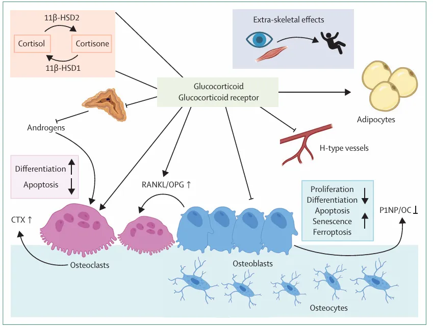
Figure 2: Pathogenesis of glucocorticoid induced osteoporosis
Pharmacokinetic and pharmacodynamic property of zoledronic acid
An antiresorptive drug and, more generally, a bisphosphonate is zoledronate (or zoledronic acid). The US Food and Drug Administration (FDA) approves zoledronate, both as prophylaxis and therapy of osteoporosis in men and postmenopausal women, and in glucocorticoid-induced osteoporosis, Paget disease of bone, hypercalcemia of malignancy, multiple myeloma, and bone metastasis. Zoledronate is an injectable drug that is usually prescribed to treat most metabolic bone diseases such as benign bone disease as well as malignant bone disease. Since zoledronate is administered intravenously, it is also able to be used in those patients who are not able to take or are contraindicated with oral bisphosphonates.
Objectives:
Pharmacokinetics
Absorption: After administration, Zoledronate mean plasma concentrations reach their peak [7] Zoledronate plasma concentration has a triphasic or three-phase drop with an immediate decrease in plasma concentration at the end of the infusion to a minimum of less than 1% of the maximum plasma concentration (Cmax) at 24 hours. The drug presents a short disposition and long terminal elimination with low levels of plasma between days 2-28 following infusion with a terminal elimination half-life of 146 hours. There is dose proportionality of pharmacokinetics between 2-16 mg dosage range, and low accumulation on repeat administration.
Distribution: The protein binding of Zoledronate in human plasma is 28 to 53 percent. The in vitro tests prove that zoledronate has the highest affinity to bind to hydroxyapatite in mineralized bone, out-of-place of alendronate, ibandronate, or risedronate. This increased binding affinity of zoledronate to mineralized bone is the reason why it is a long acting drug. Bone takes in about 55 percent of the given dose of zoledronate and releases it gradually into the bloodstream. The rest 45 percent of the dosage is excreted as a whole through urine [8]
Metabolism and excretion; Zol is not metabolized in human beings; it is excreted as it is by the kidney. On average, 39 percent of the dose of Zol administered is excreted in urine over the first 24 hours [9] Remaining portion of the dose is assumed to be attached to the bone and is released back into the circulation very slowly. Zol is cleared through the participation of creatinine clearance (CrCL). There is however no adjustment of dosage to mild (50-80 mL/min) to moderate renal impairment (35-50 mL/min) compared to normal renal functioning, and Zol is not used in patients with severe renal impairment (,35 mL/min) due to lack of data in this group [10]
Pharmacodynamic Property of Zoledronic Acid
A third-generation bisphosphonate containing nitrogen, zoledronic acid suppresses osteoclast activity and stops bone resorption [11]. Because the duration of action is long and individuals are unlikely to have serious side effects from overdosing, the therapeutic window is broad. The risks of electrolyte imbalances, renal impairment, osteonecrosis of the jaw, atypical femur fractures, bronchoconstriction, hepatic impairment, hypocalcaemia, and embryo-fatal toxicity should be discussed with patients. [12,13,14].
Mechanism of action
Zoledronic acid is a very effective nitrogen containing bisphosphonate that finds extensive use in the management of glucocorticoid-induced osteoporosis (GIOP). It binds itself to bone crystals of hydroxyapatite at a high affinity and is concentrated at locations of active bone remodeling. When bones are resorbed, osteoclasts absorb the zoledronic acid on the surface of the bones. The drug causes the inhibition of farnesyl pyrophosphate synthase (FPPS) in the mevalonate pathway that is central to the production of isoprenoid lipids within farnesyl pyrophosphate and geranylgeranyl pyrophosphate when the drug is present inside the osteoclasts. Blockage of this pathway averts the prenylation of small GTP-binding proteins (Ras, Rho, Rac, and Rab) required in the organization and regulation of the cytoskeleton and vesicular trafficking and activity of osteoclasts in resorbing bone. Consequently, the ability of the osteoclast to carry out its functions is compromised and the ruffled borders are lost and the ability to settle on the bone is lost. Zoledronic acid also inhibits the osteoclast differentiation process through RANKL by inhibiting downstream signal transduction mechanisms of NF-kB, JNK and NFATc1, which in turn inhibits the development of mature osteoclasts(Fig 3). Moreover, the drug also causes osteoclast apoptosis mediated by the activation of caspase and the distortion of intracellular signaling pathways. Because glucocorticoids stimulate osteoclasts and reduce the survival of osteoblasts, zoledronic acid opposes these effects by greatly suppressing bone resorption and stabilizing bone remodeling. It, therefore, increases bone mineral density and minimizes greatly the risk of vertebral and non-vertebral fractures in patients on long-term glucocorticoid therapy [15].
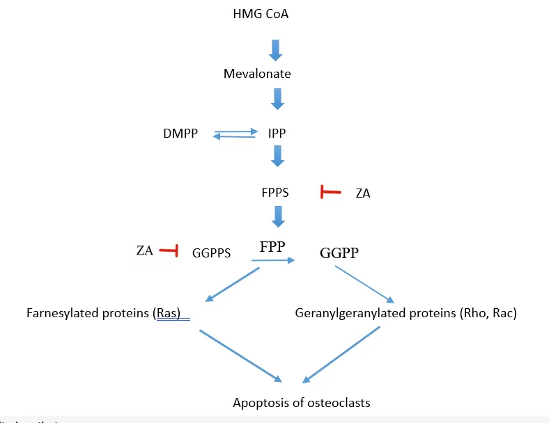
Figure 3: Mechanism of Zolendronic acid inhibit osteoclasts
Signalling pathways related to osteoporosis
A growing amount of research indicates ZA prevents osteoclast differentiation in vitro via different mechanisms, including blocking the receptor activator of nuclear factor κB ligand (RANKL)/receptor activator of nuclear factor κB (RANK) pathway, the non-canonical Wnt/Ca2+/calmodulin-dependent protein kinase II (CaMKII) pathway, and stopping macrophages from differentiating into osteoclasts [16,17]
Zoledronic Acid-Mediated Inhibition of Osteoclastogenesis
Osteoclast differentiation is significantly influenced by the RANKL/RANK signalling pathway. While researching the cDNA sequence of dendritic cells, Anderson et al. came across RANK. The sole known agonist of the RANKL receptor is RANK, a type I transmembrane homotrimer with 616 amino acids [18,19]. The combined action of RANK and RANKL may promote the differentiation and production of osteoclasts as well as the development of their precursor cells. Simultaneously, the combined action of RANK and RANKL may promote osteoclast development and maturation while inhibiting osteoclast metabolism and apoptosis. [20]. RANKL, sometimes known as osteoclast differentiation factor, is a member of the tumour necrosis factor (TNF) family. It is thought that RANKL, a type II homotrimer protein, is essential to the activation and proliferation of osteoclasts. It has a significant impact on osteoclast differentiation and development as well as bone resorption and bone formation [21]. RANKL 1, RANKL 2, and RANKL 3 were found to be the three subtypes of RANKL. Although these three subtypes exhibit slight structural variations, they can all facilitate the proliferation and differentiation of osteoclasts, subsequently impacting bone resorption and formation that were once thought to be promoted by changes in the RANKL/OPG ratio. In their study, they also considered the OPG/RANKL interaction as a way to maintain bone homeostasis [22]. The disruption of the physiological balance of the RANK/RANKL pathway also leads to the development of bone metastases and the pathological remodelling of cancer [23,24,25]. During the creation of osteoclasts, it is thought that RANKL binds to RANK in the progenitors of osteoclasts. This complex is thought to attract TNF receptor-associated factors (TRAFs), especially TRAF6, a sensitive indicator of osteoclast activity that is expressed by mature osteoclasts [26]. By increasing the phosphorylation of inhibitor of kappa B alpha (I-B alpha) and its subsequent degradation, translocation, and phosphorylation of downstream p65 [1 C-Jun N-terminal kinase (JNK), extracellular signal-regulated kinase (Erk), and p38 MAPK [27], it stimulates phosphatidylinositol 3-kinase (PI3K)/protein kinase B (Akt)/mTOR signalling. Suppression of JNK prevents RANKL-stimulated osteoclast differentiation, downregulation of Erk prevents osteoclast precursors from merging, and activation of p38MAPK significantly promotes early osteoclast maturation [28,29]. Numerous studies have shown that by blocking the RANKL/RANK pathway, ZA prevents bone loss brought on by increased osteoclast development and activity [27,30,31]. ZA inhibits osteoclast development by inhibiting RANKL and TNF-a. Additionally, by encouraging the deubiquitinating of TRAF6, the phosphorylation of tyrosine, and the nuclear translocation of p65, ZA is known to block NFATc1 and c-fos and deactivate the NF-kB pathway [32,33]. Leucine-rich repeat-containing G-protein-coupled receptor 4 (LGR4, also called GPR48) is another RANKL receptor. It competes with RANK to bind RANKL and is crucial negative feedback to the RANK-RANKL signalling pathway, which suppresses RANK-TRAF6 signalling to negatively regulate osteoclast differentiation and bone resorption (Fig 4)
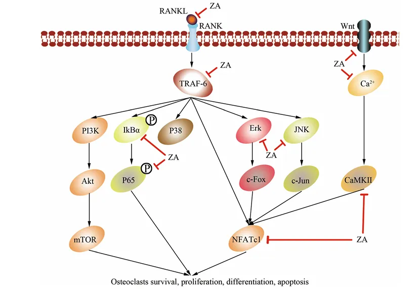
Fig 4 Signalling pathway of osteoporosis and the pathway which is inhibited by zoledronic acid (34,35)
Another pathway is the Wnt signalling pathway that plays a crucial role in osteoclast differentiation. Wnt signalling pathway can be classified into two; canonical and non-canonical. The non-canonical Wnt/Ca2+/CaMKII pathway facilitates osteoclast differentiation in association with the traditional Wnt signalling pathway, which facilitates bone development [36,37,38]. Increase of intracellular Ca2+ in the cells following osteoclast stimulation, subsequently induces the interaction of calmodulin with CaMKII, consequently leading to the expression of NFATc1 and TRAP, and ultimately the differentiation of osteoclasts. ZA reduces the amounts of non-canonical Wnt proteins, Wnt5a and CaMKII, which results in the apparent outcome of inhibiting osteoclast differentiation. Macrophage colony stimulating factor (M-CSF) is also known as colony stimulating factor-1 (CSF-1). M-CSF is a homodimeric glycoprotein, a cytokine, which can be produced and released by osteoblasts, stromal cells and T lymphocytes [39,40]. M-CSF can inhibit osteoclastic apoptosis, upregulate the RANKL-RANK interaction at the osteoclastic surfaces, increase the sensitivity of RANKL to RANK, and promote osteoclastic differentiation. Recent studies indicate that most researchers hold the view that osteoclast differentiation may only take place when both RANKL and M-CSF are co-stimulated. The combination of M-CSF and RANKL can substitute the process of transforming monocytes and macrophages into osteoclasts that are caused by the osteoclasts. Also, RANKL and M-CSF can be co-activated by the ERK, Akt, and c-fos signalling pathways to facilitate osteoclast differentiation and maturation [41,42]. ZA suppresses the differentiation of osteoclasts and the migration of macrophages by inhibiting the function, clustering, and migration of osteoclast precursors, and macrophages [43,44]. This is supported by the fact that the increase in RANK mRNA caused by RANK L is suppressed by ZA in the presence of M-CSF leading to osteoclastic differentiation [45]. However, the data of the interconnection between ZA and M-CSF is insufficient to date, which can be another possible direction of ZA against osteoclastogenesis. MAPK/JNK Signalling Pathway
MAPK/JNK Signaling Pathway
Mitogen-activated protein kinase (MAPK) signaling pathway and especially c-Jun N-terminal kinase (JNK) pathway are important in the control of bone remodeling by activating, sustaining, and differentiating the osteoclasts. The three main kinase regimes activated by extracellular stimuli, including cytokines, growth factors and mechanical stress, are MAPK signaling [extracellular signal-regulated kinase (ERK)], p38 MAPK, and JNK [46]. These pathways are closely linked with osteoclastogenesis and bone resorption in the bone tissue. In the differentiation of osteoclasts, the interaction of receptor activator of nuclear factor-kB ligand (RANKL) with the receptor RANK on osteoclast precursors causes intracellular signaling networks using adaptor proteins including tumor necrosis factor receptor-associated factor-6 (TRAF6). Activation of TRAF6 then triggers MAPK cascades, such as ERK, p38 and JNK which activate down streaming transcription factors such as activator protein-1 (AP-1) and c- Jun [47]. Activation of these transcriptional factors leads to expression of osteoclast-specific genes such as tartrate-resistant acid phosphatase (TRAP), cathepsin K, and matrix metalloproteinases that are required in osteoclast maturation and bone resorptive activity [62]. In osteoporosis caused by glucocorticoids, the balance between bone resorption and formation is disrupted following a long period of exposure to glucocorticoids. Glucocorticoids raise the amounts of RANKL and lower osteoprotegerin (OPG) levels and, as a consequence, contribute to the rise of osteoclastogenesis and the activation of downstream signaling pathways, including NF-kB and MAPK/JNK [48]. The JNK pathway is also activated, stimulating further the osteoclast differentiation by the c-Jun phosphorylation, which stimulates bone resorption. The glucocorticoids also inhibit osteoblast growth and differentiation, which ultimately causes decreased bone formation and bone frailty [49]. It has been established that zoledronic acid has the ability to inhibit phosphorylation of JNK and other MAPK proteins; this inhibits the activation of transcription factor that is involved in osteoclast differentiation as well as bone resorption [50]. In addition, MAPK/JNK inhibition by zoledronic acid induces the down-regulation of osteoclast-related regulated gene and enhances the apoptosis of osteoclasts reducing bone resorption, and the increase of bone mineral density during the glucocorticoid-induced osteoporosis [51]. These processes allow zoledronic acid to normalize the bone formation and bone resorption and is of key importance in the prevention and treatment of glucocorticoid-related bone loss.
Bone Remodelling
Cellular and molecular processes of bone remodeling are significant to the phenomenon of osteoporosis. The rate and degree of replacement control the bone gain or loss rates, the distribution of the bone, in the three-dimensional form, and the quantity of the bone in the body. Organization of the remodeling system has the following significant impact [52]. organization of the remodeling system has such significant impact. Multivariate factors such as hormones, cytokines, paracrine factors, and gene expression, control the bone remodeling system. In addition, recent studies have found out that there is an osteocyte-independent process in the bone-modelling regulation [53]. Knowledge of the bone remodelling system helps to determine possible targets of treatment. Fracture healing is one of the important aspects of normal bone remodelling and several animal and human studies have indicated that bone marrow-derived mesenchymal stem cells might be used to increase bone repair. It is not clear whether these cells stimulate fracture healing by differentiating into osteoblasts or by secreting paracrine factors that attract blood vessels and perivascular stem cells. Among these, the CD34+ cells are endothelial and hematopoietic cell-specific and have been demonstrated to contribute towards other bone repair models [54]. The results of the study conducted by David et al. were the results at 6 months and 24 months of treatment with teriparatide or zoledronic acid. Zoledronic acid is an anti-remodelling, teriparatide a pro-remodelling anabolic, agent; treatment with teriparatide has been shown to increase bone formation compared to zoledronic acid [55].
Molecular Mechanisms in Osteoclast and Osteoblast Differentiation
Osteoclast precursors cells (OCPs) are hematopoietic stem cell descendants. NFATc1 or nuclear factor of activated T-cells, cytoplasmic 1, is a master transcription factor that activates osteoclastogenic genes, which govern many osteoclast-associated genes, including TRAP, cathepsin K, osteoclast-associated receptor, and matrix metalloproteinase-9 (MMP-9). RANKL, that is, secreted by the OCPs surface binds the OCPs membrane and liberates the nuclear factor kappa-B, c-Fos or microphthalmia-associated transcription factor.MMP-9 activates the proteolysis of histone H3-N terminus as a major process of gene regulation causing osteoclastic-related genes to be expressed. Target gene localization of MMP-9 is facilitated by H3K27me1 by G9a [56]. One of the main processes in the osteoclast formation is myeloid lineage multinucleation. Eric et al. revealed an IFN γ-dependent tryptophanyl-tRNA synthetase-truncated action to support such a process in vitro, which indicated the possibility of the IFN γ /mini-TrpRS signaling axis in osteoporosis pathophysiology. The IFN γ caused aggregation of monocytes in their study resulting in the formation of multinuclear giant cells which were also parallel to the significant upregulation of mini-TrpRS. The aggregation of monocytes and the following formation of multinucleation induced by IFN γ is inhibited by blockage of mini TrpRS in their study [57]. mesenchymal stem cells (MSCs) differentiate to osteoblasts under the influence of bone morphogenic protein (BMP), Wnt, Hedgehog and Notch signaling pathways. Runt-related transcription factor 2 is a major transcription factor; it takes part in such pathways [56]. In bone resorption, MSCs are recruited by processes in which release and activation of matrix TGF-β occurs. Morphine dysregulation of TGF-b signaling distorts bone remodeling and leads to bone disorders. The parathyroid hormone (PTH) balances out the BMP, TGF-β and Wnt signaling of MSCs, thereby instructing MSC differentiation [58]. The recent research findings suggest that osteocytes regulate the bone formation and the bone resorption rates through the synthesis of RANKL and sclerostin [59]. RANKL is a TNF family member that controls the formation, activity and survival of osteoclasts. TNF is a family of receptors that block the action of RANKL on bone resorption by its mechanism of osteoprotegerin (OPG). Numerous hormonal and therapeutic agents have since been initiated to treat osteoporosis by modifying the activities of RANKL and OPG. Zoledronic acid as an anti-RANKL antibody is called Denosumab and this exhibited good control of bone resorption [60]. Normal bone metabolism is influenced by extracellular matrix (ECM) proteins. The balance between bone formation and resorption is obtained through the ability of osteoclast and osteoblast to regulate mechanical, physical and chemical properties of bone tissue and ECM through a coupling mechanism between the two cells via paracrine, autocrine and endocrine factors. IGF-1, DCN, and ON proteins control the production and formation of the type I collagen that is the primary bone-forming material. TGF-β and IGF-1 regulate the coupling, recruitment, maturation of OBs and OCs and the formation of the matrix. DCN performs the same activity on focal adhesion of osteoblast to the matrix, and OPN and BSP-2 play similar roles in the adhesion of osteoclast to ECM. OCN is an attractant to OBs and OCs which is chemo-attractant [61]. Hypomethylating the promoter of particular osteogenic genes (RUNX2 and OCN) in BMSCs will differentiate them into osteoblasts, whereas hypomethylating the promoter of the adipose tissue-related gene (PPAR-2 γ) in ASCs will change them into adipocytes. According to the recent studies, DNA methylation and post-translational modification of histones are epigenetically received by the bone remodeling processes. The DNA status of methylation regulates genes that are relevant to the differentiation of osteoblasts and osteoclasts, including RANK/RANKL/OPG, RUNX2, OSX, OCN, ALP, and Wnt signatures [62].
In-Vitro Models for Studying Zoledronic Acid in Glucocorticoid-Induced Osteoporosis
Cell culture models on in-vitro investigations of the molecular pathways and pharmacological action of zoledronic acid on osteoporosis induced by glucocorticoids have been popular. Such models enable the determination of drug effects on osteoblast differentiation, osteoclast formation, and bone remodeling signaling pathways to be controlled [63]
The molecular pathways of Zoledronic Acid in the regulation of osteoclast differentiation and bone resorption are in-vitro studies that are widely applied. A commonly utilized experimental model is the murine macrophage cell line RAW264.7 which is used as an osteoclast precursor cell model. These are cultured on a-modified Eagle medium (a-MEM) with the addition of fetal bovine serum at standard culture conditions (37 ºC and 5% CO2) (64,65). The differentiation of the cells is induced by the stimulation of the receptor activator of nuclear factor-kB ligand (RANKL), which promotes differentiation of RAW264.7 cells into multinucleated osteoclasts that can resorb bone. Two most important cytokines, namely macrophage colony-stimulating factor (M-CSF) and RANKL, are the main regulators of the differentiation process, as they enhance the survival and differentiation of osteoclast precursors via intracellular signaling pathways (NF-kB and MAPK pathways) [66]. In order to assess the inhibitory action of zoledronic acid on the osteoclastogenesis, RAW264.7 cells are incubated with various concentrations of zoledronic acid during the stimulation of the RANKL. The number of osteoclasts formed is determined by the staining of tartrate-resistant acid phosphatase (TRAP) that determines the presence of TRAP-positive multinucleated osteoclasts. Further, the bone resorptive activity is assessed with resorption pit assays that are conducted on dentine or bone pieces. It has also been demonstrated that the treatment with zoledronic acid can greatly decrease the numbers of TRAP-positive osteoclasts and prevent the formation of bone resorption pits, which can suggest the prevention of osteoclast differentiation and bone resorptive activity [67,68,69]. Additional molecular testing is done by reverse transcription-quantitative polymerase chain reaction (RT-qPCR) to measure the expression of the osteoclast-specific genes. The total RNA is isolated in RAW264.7 cells to determine the treatment of RANKL along with or without zoledronic acid, and converted to complementary DNA (cDNA) which will be analyzed with the help of quantitative PCR [70]. The levels of gene expression of osteoclast markers nuclear factor of activated T cells cytoplasmic 1 (NFATc1), c-Fos, dendritic cell-specific transmembrane protein (DC-STAMP), tartrate-resistant acid phosphatase (TRAP), receptor activator of nuclear factor-kB (RANK) as well as calcitonin receptor (CTR) are determined in relation to the housekeeping gene of GAPDH by the 2-DDCq method [71]. Moreover, the western blot technique is conducted to check the protein expression of signaling molecules of osteoclastogenesis. The cellular proteins are harvested with the help of RIPA buffer and then SDS-PAGE and consequent transfer to PVDF membranes. The antibodies against signaling proteins, including phosphorylated JNK, ERK, p38, Iκ-βα, p65, NFATc1, c-Fos and c-Jun are then used to probe the membranes. This is analyzed to define the inhibition of zoledronic acid on the NF-kB and MAPK/JNK pathway activation in osteoclast differentiation [72,73]. These in-vitro experiments have shown that zoledronic acid is a potent inhibitor of RANKL-induced osteoclastogenesis by inhibiting both canonical and non-canonical signaling pathways, such as NF-kB and JNK signaling, and suppressing downstream transcription factors such as c-Jun, c-Fos and NFATc1 needed to promote osteoclast formation and bone resorption [74,75] and caspase-3 (an early and broad promoter of osteoclastic different [76,77]
Zoledronic Acid has been extensively investigated in-vitro to determine the cellular impact of Zoledronic Acid on bone-forming cells and to comprehend its involvement in bone remodeling in the course of treating osteoporosis. In experimental model, human osteoblast-like cell line like MG- 63 and G- 292 is typically used as they have characteristics of mature osteoblasts and have often been utilized in the study of osteoblast proliferation and differentiation. Such cells are grown in special growth media such as minimal essential media or McCoy 5A media with addition of fetal bovine serum, penicillin, and streptomycin under usual conditions of incubation of 37 ºC and 5 -CO2 [80]. To assess how zoledronic acid affects the functions of osteoblasts, the drug is prepared as a stock solution and made in various concentrations of the culture medium which is of different micromolar concentrations. Multi-well plates with the cells are seeded and incubated with different concentrations of zoledronic acid over a specific duration of time, usually 48-72 h. It is then evaluated by the cell proliferation through the XTT assay method which is a technique that measures cell viability and proliferation based on the metabolic activity [78]. Chromatin condensation and fragmentation tests with the help of fluorescent dyes like Hoechst dye are used to examine apoptosis or programmed cell death. Zoledronic acid is then used and the cells are incubated with the fluorescent dye and observed under a fluorescence microscope to identify any morphological changes that accompany apoptosis. It was shown that an increase in the levels of zoledronic acid causes the osteoblast-like cells to undergo apoptosis in a dose-dependent effect which means that, when the osteoblasts are overexposed to the drug, the survival of the osteoblasts may endanger [78,79,80]. Zoledronic acid is studied on osteoblast migration by means of scratch or wound-healing test. This technique involves a scratch being made between the monolayer of the cell with a sterile pipette tip and time dependencies of cell migration into the scratched area were observed under microscopy. The fact that the number of migrating cells has declined with zoledronic acid treatment suggests that the osteoblast migration and recruitment have been identified as inhibited by the replacement [78]. Osteoblast differentiation and mineralization in the matrix can also be tested through Alizarin Red-S staining where calcium deposits formed during the formation of the bone matrix are detected. Following several days of treatment, the cells are fixed and stained and the strength of mineralized nodules are viewed under a microscope. Other biochemical determinations are carried out to determine osteogenic markers of type I collagen (COL-1), osteocalcin (OCN), and alkaline phosphatase (ALP) by ELISA based methods. These are osteoblast differentiation and osteo-bone- matrix formation indicators [78,81]. Analysis of zoledronic acid molecular effects on osteogenic signaling pathways is also carried out through gene expression. The expression level of genes related to bone formation such as RUNX2, SMAD proteins, collagen type I and bone matrix proteins are also determined by extracting the total RNA out of treated cells and analyzing it through RT-PCR arrays. Such studies can be used in deciding whether zoledronic acid affects osteoblast differentiation on a genetic basis [78,81]. Generally, the findings of these in-vitro experiments suggest that micromolar levels of zoledronic acid have the potential of mitigating osteoblast proliferation, migration and matrix mineralization mainly by triggering the process of apoptosis and reduction in cell survival. The differentiation potential of osteoblasts is however relatively unaffected and so it is presumed that the loss of bone matrix formation is chiefly because of a loss of osteoblast viability and not because of suppressed osteogenic differentiation.
In vitro cultures of osteoblast or mesenchymal stem cells are subjected to glucocorticoid like dexamethasone to simulate the condition of glucocorticoid-induced osteoporosis in vivo. Glucocorticoids in high concentrations delay the proliferation of osteoblasts, cause apoptosis and inhibit bone formation. Such effects are linked to downregulation of growth factors like vascular endothelial growth factor (VEGF) and osteoclastogenic signaling by the expression of the growth factor RANKL. The model can help determine the protective properties of zoledronic acid and other anti-resorptive medications against the bone loss induced by glucocorticoids [82,83,84]. This is the in-vitro system that is highly employed in testing the protective actions of anti-resorptive drugs like Zoledronic Acid in bone loss induced by glucocorticoids. It has been demonstrated that zoledronic acid can inhibit the osteoblast apoptosis, inhibit osteoclastogenesis, and resume bone-forming activities by regulating bone remodeling signaling pathways such as NF-kB and MAPK pathways. As such, the models of osteoblast damage induced with dexamethasone give a valid experimental model to investigate the molecular processes and therapeutic possibilities of zoledronic acid in glucocorticoid-induced osteoporosis [85,86].
INVIVO MODELS
Most of the studies examining the pathophysiology of glucocorticoid-induced osteoporosis (GIOP), and the efficacy of anti- resorptive drugs like Zoledronic Acid, have been conducted using animal models. Rats and mice are the most common models used since they have a high bone turnover in addition to the ability to respond to exposure to glucocorticoids in a similar fashion as human bone metabolism [87]. The glucocorticoids used in these models include prednisolone, methylprednisolone, or dexamethasone which should be administered via subcutaneous, oral, or intraperitoneal delivery over a few weeks to promote bone loss. Prolonged exposure to glucocorticoids causes a loss in bone mineral density (BMD), inhibition of osteoblast activity, amplified osteoblast apoptosis and amplified bone resorption by osteoclasts [88]. The rat models come in handy especially on the structural and biochemical shift in the bone during the GIOP. Prolonged glucocorticoid treatment in rats causes loss of trabecular bone, drop in bone formation rate and bone microarchitecture. In such models, zoledronic acid treatment has been found to be quite useful in enhancing bone mineral density, preventing osteoclast activity and retaining trabecular bone structure. Histomorphometry studies show a decline in the number of osteoclasts and an increase in bone volume after inserting zoledronic acid, which indicates a protective effect of zoledronic acid against the deteriorative functions of glucocorticoids on bone tissue [89,90]. The molecular mechanisms regulating the bone loss in response to glucocorticoids are also commonly studied through mouse models. These models have been especially applied in the study of gene expression and signalling pathways that are involved in bone remodelling. Glucocorticoid-administered mice exhibit reduced osteogenic gene expression: RUNX2 and osteocalcin and greater osteoclast differentiation and bone resorption gene RANKL. Also, the models can be used to assess the protective properties of zoledronic acid at the molecular level, such as inhibition of osteoclast formation, and various other pathways, like NF-KB and MAPK which mediate bone resorption [89,91].
Other preclinical models besides rodent models have also been employed to examine human bone physiology using larger animal models like rabbit and sheep. The models prove very effective in assessing the long-term outcomes of using glucocorticoids on bone strength, bone mineral density, and the risk of fractures. It has been demonstrated that treatment with Zoledronic Acid can save bone microarchitecture, bone strength, and bone resorption indicators in a significant way [92], when using these models. This data encourages translational applicability of preclinical models and gives a great case to the clinical administration of zoledronic acid in family bone loss prevention in the presence of glucocorticoids [93]. On the whole, the experimental platform of glucocorticoid-induced osteoporosis in in-vivo models is a predictable research tool that can be used to research the mechanism of bone loss as well as the effectiveness of anti-osteoporotic medications. These models assist in the explanation of the effects of glucocorticoids on bone formation, the stimulation of osteoclast activity, and the decrease in bone quality. More to the point, they also offer good preclinical data that indicate that zoledronic acid is capable of preventing the loss of bone mass and enhancing bone strength in the presence of glucocorticoids, thereby supporting the use of zoledronic acid in the clinical management of the glucocorticoid-induced osteoporosis [94].
Translational relevance
The results of the experimental work on animal and cell models strongly justify the potential in the therapeutic use of Zoledronic Acid in the prevention of the bone loss induced by glucocorticoids. In preclinical research, it has been shown that zoledronic acid is a potent inhibitor of osteoclasts, maintains bone micro-architecture and enhances bone strength in glucocorticoid-treated animals. These findings have been effectively developed into clinical practice, with zoledronic acid currently being applied extensively in the prevention and treatment of glucocorticoids induced osteoporosis in patients undergoing corticosteroid based long-term therapy [95]. Thus, preclinical models will continue to serve as helpful resources to gain insights about drug mechanisms and establish better therapeutic methods to manage osteoporosis.
CONCLUSION
Osteoporosis caused by glucocorticoids is a significant adverse event of prolonged corticosteroid treatment, which is a decrease in the activity of osteoblasts, increased bone-forming cells-apoptosis, and bone resorption mediated by osteoclasts. The therapeutic potential of the Zoledronic Acid in bone loss prevention by glucocorticoids, both in in-vitro cell culture systems and in-vivo animal models, has been demonstrated in preclinical studies. The experimental data indicates that zoledronic acid is capable of inhibiting osteoclast differentiation, preventing the various signalling pathways like NF-KB and MAPK, and maintaining bone microarchitecture, bone mineral density and bone strength in the presence of glucocorticoids. The close relationship between cellular, molecular, and animal-based results shows the translational nature of their results and the potential to utilize zoledronic acid as a potent anti-resorptive agent, which prevents and treats glucocorticoid-induced osteoporosis. Future studies on the ways of improved dosing, long term safety and combination with other therapies can enhance its clinical effects and increase its therapeutic practice.
REFERENCES


Sushmitha S., Abarnadevika A.*, G. Ariharasivakumar, Keerthana S., Praveen Kumar S., Evania Helen M. D., Preclinical Evaluation Of Zoledronic Acid In Glucocorticoid-Induced Osteoporosis: Mechanisms, Efficacy, And Translational Relevance, Int. J. Sci. R. Tech., 2026, 3 (9), 564-579. https://doi.org/10.5281/zenodo.22959196
 10.5281/zenodo.22959196