View Article

Abstract

Background: Pithecellobium dulce (Fabaceae), commonly known as Manila tamarind or jungle jalebi, is a medicinally used plant containing several reported secondary metabolites. The present study investigated the preliminary phytochemical profile of its seed extract and evaluated its in vitro cytotoxic effect against the human colorectal carcinoma cell line HCT-116. Methods: Seeds were powdered and extracted by a Soxhlet procedure as described in the project thesis. Preliminary qualitative tests were performed for glycosides, steroids, saponins, polysaccharides and lipids. Cytotoxicity was assessed using the MTT assay after 24 h exposure to concentrations ranging from 3.90 to 250 µg/mL. Results: Qualitative observations were reported as positive for anthraquinone glycosides, digitoxose-related glycosides, saponins, starch and lipids; the steroid test was reported as positive in the inference column despite the absence of the stated colour reaction. Recalculation of the supplied triplicate absorbance values after blank correction gave cell viability estimates of approximately 79.9–89.5% across the tested concentrations, relative to the blank-corrected vehicle control. Thus, the raw absorbance data indicate limited cytotoxicity within the tested range and do not establish an IC50 at or below 250 µg/mL. Conclusion: The seed extract contains qualitative phytochemical signals warranting further characterization. Its apparent effect on HCT-116 viability was modest in the supplied dataset; confirmatory experiments with validated controls, clarified extraction details and complete statistical output are required.

Keywords

Pithecellobium dulce, seed extract, phytochemical screening, HCT-116, MTT assay, cytotoxicity

Introduction

× Popup Image

Cancer is a group of diseases characterized by abnormal cell growth resulting from genetic and molecular alterations. Colorectal cancer develops in the large intestine and may progress locally or metastasize to distant organs. Conventional treatment options include surgery, chemotherapy, radiotherapy and, in selected cases, targeted therapy or immunotherapy. The search for bioactive compounds from medicinal plants remains an important area of pharmacological investigation. [13-24]

Pithecellobium dulce (Roxb.) Benth., a member of the family Fabaceae, is commonly known as Manila tamarind, Madras thorn or jungle jalebi. It is native to tropical regions of the Americas and is naturalized in India. Different plant parts have been used traditionally for gastrointestinal complaints, inflammation, fever, dental ailments and other conditions. The seeds have been reported to contain glycosides, glycolipids, polysaccharides, steroids, saponins, lipids and phospholipids. [2-3,5-12,28-32]

Previous work has investigated the phytochemical and pharmacological properties of P. dulce. Alhamed and colleagues reported that seed extract affected colorectal cancer cell responses, including apoptosis, cell-cycle progression and migration, in HCT-116 cells [1]. Other reports describe saponins and related constituents from the plant [2], while broader reviews summarize its nutritional and therapeutic uses [3].

Despite these reports, plant extracts require careful chemical characterization and reproducible biological evaluation. The present work therefore aimed to screen the phytochemical constituents of P. dulce seeds and assess the cytotoxic response of HCT-116 cells to the seed extract using an in vitro MTT assay. [29-32,53-60]

MATERIALS AND METHODS

2.1 Plant material and preparation

P. dulce seeds were selected from mature pods, cleaned to remove adhering debris, dried and ground into a fine powder. The thesis describes collection from mature, healthy pods and recommends seeds from 10–20-year-old mother trees. A voucher specimen number, collection location, collection date and botanical authentication details were not provided in the source document and should be added before submission.

2.2 Extraction procedure

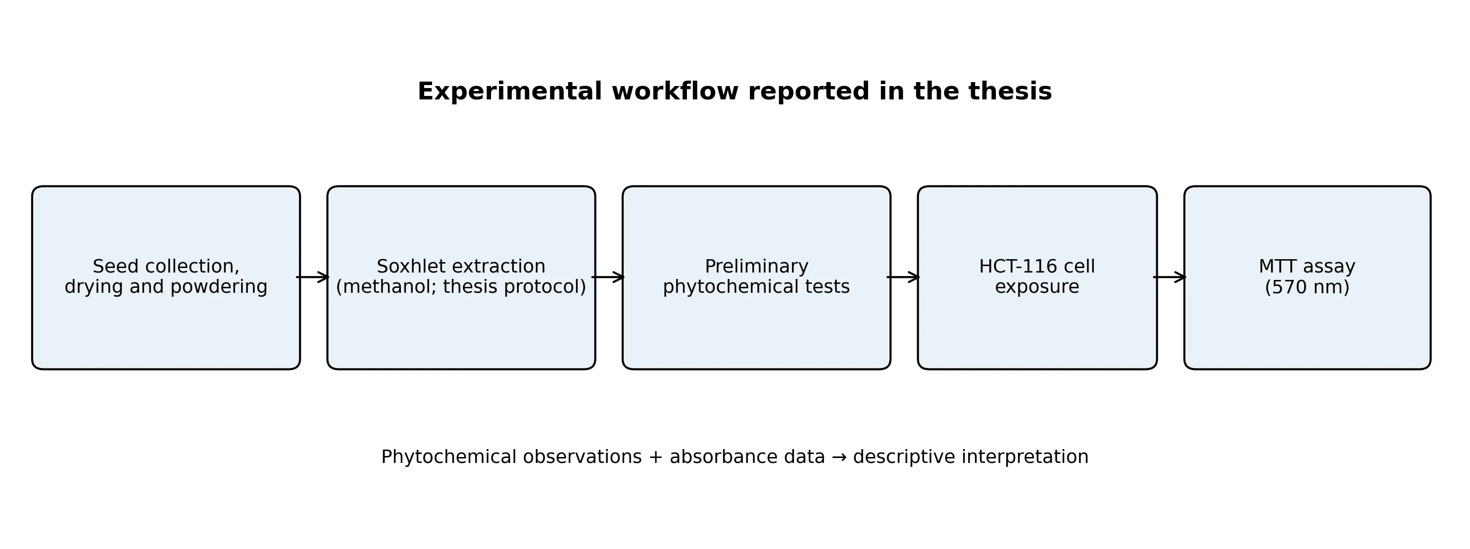
The project thesis describes Soxhlet extraction using 10 g of powdered plant material and 250 mL of methanol, with an extraction period of approximately 3–4 h. The solvent is also described in the thesis as methanol (80:20, v/v) and elsewhere as a hydroalcoholic solvent. Because these descriptions are not fully consistent, the actual solvent composition and whether water was used must be confirmed by the investigators. The extract yield, storage conditions and reconstitution solvent were not reported.

2.3 Preliminary phytochemical screening

Qualitative screening was reported for glycosides, steroids, saponins, polysaccharides and lipids. The tests listed in the thesis included Borntrager’s test and Keller–Killiani test for glycosides; Salkowski test for steroids; foam test and Liebermann–Burchard test for saponins; iodine test for starch/polysaccharides; and emulsion test for lipids. Observations and interpretations are summarized in Table 1. [54-60]

2.4 Cell line and MTT assay

The biological evaluation used the human colorectal carcinoma cell line HCT-116. The thesis lists Dulbecco’s Modified Eagle Medium (DMEM), fetal bovine serum (FBS), antibiotic solution, trypsin–EDTA, phosphate-buffered saline (PBS), MTT and dimethyl sulfoxide (DMSO) among the materials. Cells were seeded in 96-well plates and incubated at 37°C in a humidified atmosphere containing 5% CO2. The protocol section reports a cell suspension of 1 × 10^5 cells/mL and 100 µL per well, whereas the results section reports a seeding density of 1 × 10^4 cells per well. This discrepancy requires confirmation. [41-49]

After an initial incubation of 24–48 h, cells were exposed to serial two-fold dilutions of extract (250, 125, 62.5, 31.25, 15.62, 7.81 and 3.90 µg/mL) for 24 h. The thesis describes addition of 10 µL MTT solution (5 mg/mL), followed by a further incubation of approximately 2–4 h (the assay description specifies 3 h). Formazan crystals were dissolved using 100 µL DMSO, and absorbance was measured at 570 nm. Each concentration was tested in technical triplicate. Vehicle, positive, negative and blank controls were included in the recorded data. [4,34-40,50-53]

2.5 Data handling

The thesis provides triplicate absorbance readings for each concentration, vehicle control and blank. For transparent re-analysis, the mean blank absorbance (0.058) was subtracted from each individual sample and vehicle reading. Cell viability was calculated as: (mean blank-corrected sample absorbance / mean blank-corrected vehicle-control absorbance) × 100. The mean blank-corrected vehicle-control absorbance was 0.292. This calculation is a re-analysis of the raw values supplied in the thesis and is distinguished from the thesis’s separately reported viability percentages.

RESULTS

3.1 Preliminary phytochemical screening

The qualitative screening observations recorded in the thesis are presented in Table 1. Borntrager’s test produced a pink-to-red ammoniacal layer, interpreted as anthraquinone glycosides. Keller–Killiani testing produced a reddish-brown layer that reportedly turned bluish-green, interpreted as the presence of digitoxose-related glycosides. Foam formation and the reported colour change in the Liebermann–Burchard test were interpreted as evidence of saponins. The iodine test was recorded as positive for starch, and the emulsion test produced a milky-white cloudy emulsion, indicating lipids.

Table 1: Qualitative phytochemical screening observations reported in the thesis

Constituent group Test Recorded observation Interpretation in thesis
Glycosides Borntrager’s test Pink-to-red ammoniacal layer Anthraquinone glycosides present
Glycosides Keller–Killiani test Reddish-brown layer changing to bluish-green Digitoxose-related glycosides present
Steroids Salkowski test Purple/red colour reportedly not observed Inference column states steroids present; result is internally inconsistent
Saponins Foam test Foam formation Saponins present
Saponins Liebermann–Burchard test Red/pink changing to deep bluish-green Saponins reported present
Polysaccharides Iodine test Blue/black or purple colour Starch reported present
Lipids Emulsion test Milky-white cloudy emulsion Fats/oils present

3.2 MTT assay and cell viability

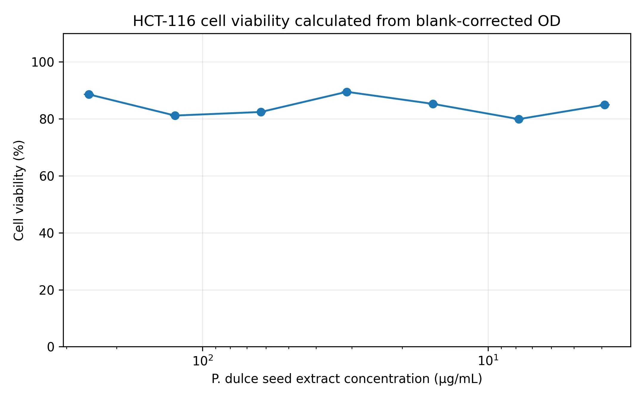
The source thesis provides triplicate absorbance readings at 570 nm for seven extract concentrations. The mean blank absorbance was 0.058, calculated from the three blank readings (0.057, 0.055 and 0.062). The mean blank-corrected vehicle-control absorbance was 0.292. Recalculated viability values are shown in Table 2.

Table 2: Re-analysis of triplicate absorbance readings and calculated HCT-116 cell viability

Extract (µg/mL) OD 1 OD 2 OD 3 Mean corrected OD SD Viability (%)
250 0.314 0.339 0.297 0.259 0.021 88.6
125 0.285 0.309 0.291 0.237 0.012 81.2
62.5 0.287 0.298 0.311 0.241 0.012 82.4
31.25 0.294 0.359 0.305 0.261 0.035 89.5
15.62 0.302 0.337 0.282 0.249 0.028 85.3
7.81 0.292 0.307 0.275 0.233 0.016 79.9
3.9 0.303 0.322 0.293 0.248 0.015 84.9

Across the tested concentrations, calculated cell viability ranged from approximately 79.9% to 89.5%. The lowest calculated viability (79.9%) occurred at 7.81 µg/mL, while the highest (89.5%) occurred at 31.25 µg/mL. The response was not consistently concentration-dependent. Since viability remained above 50% at all tested concentrations, an IC50 was not reached within the tested range; the available data are compatible with an IC50 greater than 250 µg/mL, but a precise IC50 cannot be estimated from these observations.

Table 3: Control absorbance values at 570 nm as recorded in the thesis

Control OD 1 OD 2 OD 3 Mean OD (reported)
Positive control 0.045 0.046 0.045 0.05
Negative control 0.364 0.356 0.347 0.36
Blank 0.057 0.055 0.062 0.06
Vehicle control 0.373 0.330 0.347 0.35

figure

Figure 1: HCT-116 cell viability calculated from the thesis’s raw triplicate absorbance values after blank correction. Values are means of three technical replicates; no inferential statistics are shown.

3.3 Experimental workflow
figure

Figure 2: Overview of the experimental workflow described in the thesis.

DISCUSSION

The preliminary phytochemical tests suggest the presence of several classes of constituents in the P. dulce seed extract, particularly glycosides, saponins, polysaccharides and lipids. These observations are consistent with the broad phytochemical profile attributed to P. dulce in the literature. Saponins from P. dulce have previously been isolated and characterized, supporting interest in this class of plant metabolites [2]. However, qualitative colour and foam tests are preliminary indicators only; they do not establish the identity, purity or concentration of individual compounds. [2-3,8-12,54-60]

The steroid result requires particular attention. The thesis records that the expected purple or red colour was not observed in the Salkowski test, but its inference column states that steroids were present. This contradiction should be resolved by repeating the test or correcting the record based on original laboratory notes. Similarly, the thesis conclusion lists alkaloids, flavonoids, tannins and phenolic compounds, although corresponding test procedures and observations are not presented in the results tables supplied. These constituents should not be described as experimentally confirmed by the present screening unless supporting data are available.

The MTT assay is a colorimetric method that estimates metabolic activity through the conversion of tetrazolium salt to formazan by metabolically active cells [4]. Re-analysis of the raw absorbance readings supplied in the thesis produced viability values between 79.9% and 89.5%. These values indicate a relatively modest reduction in metabolic viability compared with the vehicle control. The minimum viability was observed at 7.81 µg/mL rather than at the highest concentration, and the data therefore do not show a clear monotonic concentration-response relationship. [4,34-40,50-53]

The thesis narrative separately reports cell viability values of 66–74% and states that the highest inhibition occurred at 7.81 µg/mL. Those statements do not agree with the raw triplicate absorbance values when recalculated using the stated blank-correction approach. The thesis also contains differing descriptions of cell seeding density and solvent composition. For this manuscript, the raw absorbance values have been retained and transparently recalculated; the narrative values have not been substituted because their calculation cannot be reconstructed from the information provided.

Prior research has reported anticancer-related effects of P. dulce seed extract in colorectal cancer models, including apoptosis and changes in cell-cycle and migration outcomes [1]. The present dataset, however, contains only an MTT viability endpoint and does not include apoptosis assays, cell-cycle analysis, migration studies, molecular markers or normal-cell comparisons. Consequently, the current results should be interpreted as preliminary in vitro screening and not as evidence of a defined anticancer mechanism or therapeutic efficacy. [1,5-12,27-32]

5. LIMITATIONS

The study has several limitations. First, the plant collection details, authentication record, extract yield and exact solvent composition are not supplied. Second, the thesis contains inconsistencies in cell seeding density, assay description and reported viability values. Third, only technical triplicates are provided, with no independent biological replicate information. Fourth, although one-way ANOVA is mentioned, the F statistic, degrees of freedom, exact p values and post hoc test outputs are absent. Fifth, the phytochemical screening is qualitative and does not quantify or structurally identify active constituents. Finally, no normal colon cell line was used to assess selectivity.

CONCLUSION

Pithecellobium dulce seed extract showed qualitative evidence of glycosides, saponins, starch-related polysaccharides and lipids in the screening tests recorded in the thesis. Recalculation of the supplied MTT absorbance data indicated HCT-116 cell viability of approximately 79.9–89.5% across 3.90–250 µg/mL, suggesting modest cytotoxic activity under the reported conditions. An IC50 was not reached within the tested concentration range. Further work should standardize the extraction method, repeat the MTT assay with independent biological replicates, resolve the discrepancies in the original data and include mechanistic and selectivity studies before stronger anticancer claims are made.

REFERENCES

  1. Alhamed AS, Alqinyah M, Alghaith AF, Algahtani MM, Alqahtani F, Nasr FA, et al. Phytochemical analysis and anticancer activity of the Pithecellobium dulce seed extract in colorectal cancer cells. Open Chem. 2023;21:20230362. doi:10.1515/chem-2023-0362.
  2. Yoshikawa K, Suzaki Y, Tanaka M, Arihara S, Nigam SK. Three acylated saponins and a related compound from Pithecellobium dulce. J Nat Prod. 1997;60 (12):1269-1274. doi:10.1021/np9703555.
  3. Murugesan S, Kumar Lakshmanan D, Arumugam V, Alexander RA. Nutritional and therapeutic benefits of medicinal plant Pithecellobium dulce (Fabaceae): a review. J Appl Pharm Sci. 2019;9:130-139. doi:10.7324/JAPS.2019.90718.
  4. Mosmann T. Rapid colorimetric assay for cellular growth and survival: application to proliferation and cytotoxicity assays. J Immunol Methods. 1983;65 (1-2):55-63. doi:10.1016/0022-1759 (83)90303-4.
  5. Megala J, Geetha A. Acute and sub-acute toxicity study of hydroalcoholic fruit extract of Pithecellobium dulce. Nat Prod Res. 2012;26 (12):1167-1171. doi:10.1080/14786419.2011.562206.
  6. Dhanisha SS, Drishya S, Guruvayoorappan C. Traditional knowledge to clinical trials: a review on nutritional and therapeutic potential of Pithecellobium dulce. J Basic Clin Physiol Pharmacol. 2021;33 (2):133-142. doi:10.1515/jbcpp-2020-0166.
  7. Kulkarni KV, Jamakhandi VR. Medicinal uses of Pithecellobium dulce and its health benefits. J Pharmacogn Phytochem. 2018;7 (2):700-704.
  8. Sahu NP, Mahato SB. Anti-inflammatory triterpene saponins of Pithecellobium dulce: characterization of an echinocystic acid bisdesmoside. Phytochemistry. 1994;37 (5):1425-1427. doi:10.1016/S0031-9422 (00)90425-4.
  9. Nigam SK, Gopal M, Uddin R, Yoshikawa K, Kawamoto M, Arihara S. Pithedulosides A-G, oleanane glycosides from Pithecellobium dulce. Phytochemistry. 1997;44 (7):1329-1334. doi:10.1016/S0031-9422 (96)00725-X.
  10. Kumar M, Govindrajan J, Nyola NK. Antihyperglycemic potential of saponin-enriched fraction from Pithecellobium dulce Benth. seed extract. Pharmacogn Res. 2017;9 (Suppl 1):S23-S26. doi:10.4103/pr.pr_18_17.
  11. Bagchi S, Jayaram KK. Studies on water soluble polysaccharides from Pithecellobium dulce (Roxb.) Benth. seeds. Carbohydr Polym. 2016;138:215-221. doi:10.1016/j.carbpol.2015.11.018.
  12. Hameed A, Srivastav Y, Afaq H, Hashmi A, Ahmad MI. Outline of the pharmacological and phytochemical profile of the Jungle Jalebi (Pithecellobium dulce). Asian Plant Res J. 2024;12 (2):22-27. doi:10.9734/aprj/2024/v12i2245.
  13. Sung H, Ferlay J, Siegel RL, Laversanne M, Soerjomataram I, Jemal A, et al. Global cancer statistics 2020: GLOBOCAN estimates of incidence and mortality worldwide for 36 cancers in 185 countries. CA Cancer J Clin. 2021;71 (3):209-249. doi:10.3322/caac.21660.
  14. Bray F, Laversanne M, Sung H, Ferlay J, Siegel RL, Soerjomataram I, et al. Global cancer statistics 2022: GLOBOCAN estimates of incidence and mortality worldwide for 36 cancers in 185 countries. CA Cancer J Clin. 2024;74 (3):229-263. doi:10.3322/caac.21834.
  15. Siegel RL, Miller KD, Wagle NS, Jemal A. Cancer statistics, 2023. CA Cancer J Clin. 2023;73 (1):17-48. doi:10.3322/caac.21763.
  16. Dekker E, Tanis PJ, Vleugels JLA, Kasi PM, Wallace MB. Colorectal cancer. Lancet. 2019;394 (10207):1467-1480. doi:10.1016/S0140-6736 (19)32319-0.
  17. Arnold M, Sierra MS, Laversanne M, Soerjomataram I, Jemal A, Bray F. Global patterns and trends in colorectal cancer incidence and mortality. Gut. 2017;66 (4):683-691. doi:10.1136/gutjnl-2015-310912.
  18. Brenner H, Kloor M, Pox CP. Colorectal cancer. Lancet. 2014;383 (9927):1490-1502. doi:10.1016/S0140-6736 (13)61649-9.
  19. Keum N, Giovannucci E. Global burden of colorectal cancer: emerging trends, risk factors and prevention strategies. Nat Rev Gastroenterol Hepatol. 2019;16 (12):713-732. doi:10.1038/s41575-019-0189-8.
  20. Hanahan D. Hallmarks of cancer: new dimensions. Cancer Discov. 2022;12 (1):31-46. doi:10.1158/2159-8290.CD-21-1059.
  21. Hanahan D, Weinberg RA. Hallmarks of cancer: the next generation. Cell. 2011;144 (5):646-674. doi:10.1016/j.cell.2011.02.013.
  22. Fearon ER, Vogelstein B. A genetic model for colorectal tumorigenesis. Cell. 1990;61 (5):759-767. doi:10.1016/0092-8674 (90)90186-I.
  23. Kinzler KW, Vogelstein B. Lessons from hereditary colorectal cancer. Cell. 1996;87 (2):159-170. doi:10.1016/S0092-8674 (00)81333-1.
  24. Dekker E, Rex DK. Advances in CRC prevention: screening and surveillance. Gastroenterology. 2018;154 (7):1970-1984. doi:10.1053/j.gastro.2018.01.069.
  25. Fujita K, Kubota Y, Ishida H, Sasaki Y. Irinotecan, a key chemotherapeutic drug for metastatic colorectal cancer. World J Gastroenterol. 2015;21 (43):12234-12248. doi:10.3748/wjg.v21.i43.12234.
  26. Bailly C. Irinotecan: 25 years of cancer treatment. Pharmacol Res. 2019;148:104398. doi:10.1016/j.phrs.2019.104398.
  27. Green DR, Reed JC. Mitochondria and apoptosis. Science. 1998;281 (5381):1309-1312. doi:10.1126/science.281.5381.1309.
  28. Safarzadeh E, Shotorbani SS, Baradaran B. Herbal medicine as inducers of apoptosis in cancer treatment. Adv Pharm Bull. 2014;4 (Suppl 1):421-427. doi:10.5681/apb.2014.062.
  29. Newman DJ, Cragg GM. Natural products as sources of new drugs over the nearly four decades from 01/1981 to 09/2019. J Nat Prod. 2020;83 (3):770-803. doi:10.1021/acs.jnatprod.9b01285.
  30. Atanasov AG, Zotchev SB, Dirsch VM, Supuran CT. Natural products in drug discovery: advances and opportunities. Nat Rev Drug Discov. 2021;20 (3):200-216. doi:10.1038/s41573-020-00114-z.
  31. Cragg GM, Newman DJ. Natural products: a continuing source of novel drug leads. Biochim Biophys Acta. 2013;1830 (6):3670-3695. doi:10.1016/j.bbagen.2012.08.008.
  32. Buyel JF. Plants as sources of natural and recombinant anti-cancer agents. Biotechnol Adv. 2018;36 (2):506-520. doi:10.1016/j.biotechadv.2018.02.002.
  33. Kinghorn AD, Pan L, Fletcher JN, Chai H. The relevance of higher plants in lead compound discovery programs. J Nat Prod. 2011;74 (6):1539-1555.
  34. Tada H, Shiho O, Kuroshima K, Koyama M, Tsukamoto K. An improved colorimetric assay for interleukin 2. J Immunol Methods. 1986;93 (2):157-165. doi:10.1016/0022-1759 (86)90183-3.
  35. Denizot F, Lang R. Rapid colorimetric assay for cell growth and survival: modifications to the tetrazolium dye procedure giving improved sensitivity and reliability. J Immunol Methods. 1986;89 (2):271-277. doi:10.1016/0022-1759 (86)90368-6.
  36. Gerlier D, Thomasset N. Use of MTT colorimetric assay to measure cell activation. J Immunol Methods. 1986;94 (1-2):57-63. doi:10.1016/0022-1759 (86)90215-2.
  37. Hansen MB, Nielsen SE, Berg K. Re-examination and further development of a precise and rapid dye method for measuring cell growth/cell kill. J Immunol Methods. 1989;119 (2):203-210. doi:10.1016/0022-1759 (89)90396-3.
  38. Berridge MV, Herst PM, Tan AS. Tetrazolium dyes as tools in cell biology: new insights into their cellular reduction. Biotechnol Annu Rev. 2005;11:127-152. doi:10.1016/S1387-2656 (05)11004-7.
  39. Riss TL, Moraves RA, Smith SW, et al. Cell viability assays. In: Markossian S, Grossman A, Brimacombe K, et al., editors. Assay Guidance Manual. Bethesda (MD): Eli Lilly & Company and the National Center for Advancing Translational Sciences; 2016.
  40. van Meerloo J, Kaspers GJL, Cloos J. Cell sensitivity assays: the MTT assay. Methods Mol Biol. 2011;731:237-245. doi:10.1007/978-1-61779-080-5_20.
  41. Riss TL, Moravec RA. Use of multiple assay endpoints to investigate the effects of compounds on cell viability and proliferation. Assay Drug Dev Technol. 2004;2 (1):51-62. doi:10.1089/154065804322966315.
  42. Vega-Avila E, Pugsley MK. An overview of colorimetric assay methods used to assess survival or proliferation of mammalian cells. Proc West Pharmacol Soc. 2011;54:10-14.
  43. Freshney RI. Culture of Animal Cells: A Manual of Basic Technique and Specialized Applications. 7th ed. Hoboken: Wiley-Blackwell; 2016.
  44. Doyle A, Griffiths JB, editors. Cell and Tissue Culture: Laboratory Procedures. Chichester: John Wiley & Sons; 1998.
  45. Masters JR. Human cancer cell lines: fact and fantasy. Nat Rev Mol Cell Biol. 2000;1 (3):233-236. doi:10.1038/35043103.
  46. Klebe RJ, Chen TR, Ruddle FH. Mapping of a human genetic regulatory mechanism: the gene for galactokinase is located on chromosome 17. Proc Natl Acad Sci U S A. 1970;66 (4):1220-1227.
  47. Brattain MG, Fine WD, Khaled FM, Thompson J, Brattain DE. Heterogeneity of malignant cells from a human colonic carcinoma. Cancer Res. 1981;41 (5):1751-1756.
  48. Ahmed SA, Gogal RM Jr, Walsh JE. A new rapid and simple non-radioactive assay to monitor and determine the proliferation of lymphocytes: an alternative to [3H]thymidine incorporation assay. J Immunol Methods. 1994;170 (2):211-224. doi:10.1016/0022-1759 (94)90396-4.
  49. Borenfreund E, Puerner JA. Toxicity determined in vitro by morphological alterations and neutral red absorption. Toxicol Lett. 1985;24 (2-3):119-124. doi:10.1016/0378-4274 (85)90046-3.
  50. Skehan P, Storeng R, Scudiero D, Monks A, McMahon J, Vistica D, et al. New colorimetric cytotoxicity assay for anticancer-drug screening. J Natl Cancer Inst. 1990;82 (13):1107-1112. doi:10.1093/jnci/82.13.1107.
  51. Carmichael J, DeGraff WG, Gazdar AF, Minna JD, Mitchell JB. Evaluation of a tetrazolium-based semiautomated colorimetric assay: assessment of chemosensitivity testing. Cancer Res. 1987;47 (4):936-942.
  52. Stockert JC, Horobin RW, Colombo LL, Blázquez-Castro A. Tetrazolium salts and formazan products in Cell Biology: viability assessment, fluorescence imaging, and labeling perspectives. Acta Histochem. 2018;120 (3):159-167. doi:10.1016/j.acthis.2018.02.005.
  53. Singleton VL, Orthofer R, Lamuela-Raventós RM. Analysis of total phenols and other oxidation substrates and antioxidants by means of Folin-Ciocalteu reagent. Methods Enzymol. 1999;299:152-178. doi:10.1016/S0076-6879 (99)99017-1.
  54. Harborne JB. Phytochemical Methods: A Guide to Modern Techniques of Plant Analysis. 3rd ed. London: Chapman & Hall; 1998.
  55. Trease GE, Evans WC. Pharmacognosy. 16th ed. London: Saunders Elsevier; 2009.
  56. Sofowora A. Medicinal Plants and Traditional Medicine in Africa. 3rd ed. Ibadan: Spectrum Books; 2008.
  57. Kokate CK, Purohit AP, Gokhale SB. Pharmacognosy. 50th ed. Pune: Nirali Prakashan; 2014.
  58. Edeoga HO, Okwu DE, Mbaebie BO. Phytochemical constituents of some Nigerian medicinal plants. Afr J Biotechnol. 2005;4 (7):685-688.
  59. Evans WC. Trease and Evans Pharmacognosy. 16th ed. Edinburgh: Saunders Elsevier; 2009.
  60. World Health Organization. Quality Control Methods for Herbal Materials. Geneva: WHO; 2011.

Reference

  1. Alhamed AS, Alqinyah M, Alghaith AF, Algahtani MM, Alqahtani F, Nasr FA, et al. Phytochemical analysis and anticancer activity of the Pithecellobium dulce seed extract in colorectal cancer cells. Open Chem. 2023;21:20230362. doi:10.1515/chem-2023-0362.
  2. Yoshikawa K, Suzaki Y, Tanaka M, Arihara S, Nigam SK. Three acylated saponins and a related compound from Pithecellobium dulce. J Nat Prod. 1997;60 (12):1269-1274. doi:10.1021/np9703555.
  3. Murugesan S, Kumar Lakshmanan D, Arumugam V, Alexander RA. Nutritional and therapeutic benefits of medicinal plant Pithecellobium dulce (Fabaceae): a review. J Appl Pharm Sci. 2019;9:130-139. doi:10.7324/JAPS.2019.90718.
  4. Mosmann T. Rapid colorimetric assay for cellular growth and survival: application to proliferation and cytotoxicity assays. J Immunol Methods. 1983;65 (1-2):55-63. doi:10.1016/0022-1759 (83)90303-4.
  5. Megala J, Geetha A. Acute and sub-acute toxicity study of hydroalcoholic fruit extract of Pithecellobium dulce. Nat Prod Res. 2012;26 (12):1167-1171. doi:10.1080/14786419.2011.562206.
  6. Dhanisha SS, Drishya S, Guruvayoorappan C. Traditional knowledge to clinical trials: a review on nutritional and therapeutic potential of Pithecellobium dulce. J Basic Clin Physiol Pharmacol. 2021;33 (2):133-142. doi:10.1515/jbcpp-2020-0166.
  7. Kulkarni KV, Jamakhandi VR. Medicinal uses of Pithecellobium dulce and its health benefits. J Pharmacogn Phytochem. 2018;7 (2):700-704.
  8. Sahu NP, Mahato SB. Anti-inflammatory triterpene saponins of Pithecellobium dulce: characterization of an echinocystic acid bisdesmoside. Phytochemistry. 1994;37 (5):1425-1427. doi:10.1016/S0031-9422 (00)90425-4.
  9. Nigam SK, Gopal M, Uddin R, Yoshikawa K, Kawamoto M, Arihara S. Pithedulosides A-G, oleanane glycosides from Pithecellobium dulce. Phytochemistry. 1997;44 (7):1329-1334. doi:10.1016/S0031-9422 (96)00725-X.
  10. Kumar M, Govindrajan J, Nyola NK. Antihyperglycemic potential of saponin-enriched fraction from Pithecellobium dulce Benth. seed extract. Pharmacogn Res. 2017;9 (Suppl 1):S23-S26. doi:10.4103/pr.pr_18_17.
  11. Bagchi S, Jayaram KK. Studies on water soluble polysaccharides from Pithecellobium dulce (Roxb.) Benth. seeds. Carbohydr Polym. 2016;138:215-221. doi:10.1016/j.carbpol.2015.11.018.
  12. Hameed A, Srivastav Y, Afaq H, Hashmi A, Ahmad MI. Outline of the pharmacological and phytochemical profile of the Jungle Jalebi (Pithecellobium dulce). Asian Plant Res J. 2024;12 (2):22-27. doi:10.9734/aprj/2024/v12i2245.
  13. Sung H, Ferlay J, Siegel RL, Laversanne M, Soerjomataram I, Jemal A, et al. Global cancer statistics 2020: GLOBOCAN estimates of incidence and mortality worldwide for 36 cancers in 185 countries. CA Cancer J Clin. 2021;71 (3):209-249. doi:10.3322/caac.21660.
  14. Bray F, Laversanne M, Sung H, Ferlay J, Siegel RL, Soerjomataram I, et al. Global cancer statistics 2022: GLOBOCAN estimates of incidence and mortality worldwide for 36 cancers in 185 countries. CA Cancer J Clin. 2024;74 (3):229-263. doi:10.3322/caac.21834.
  15. Siegel RL, Miller KD, Wagle NS, Jemal A. Cancer statistics, 2023. CA Cancer J Clin. 2023;73 (1):17-48. doi:10.3322/caac.21763.
  16. Dekker E, Tanis PJ, Vleugels JLA, Kasi PM, Wallace MB. Colorectal cancer. Lancet. 2019;394 (10207):1467-1480. doi:10.1016/S0140-6736 (19)32319-0.
  17. Arnold M, Sierra MS, Laversanne M, Soerjomataram I, Jemal A, Bray F. Global patterns and trends in colorectal cancer incidence and mortality. Gut. 2017;66 (4):683-691. doi:10.1136/gutjnl-2015-310912.
  18. Brenner H, Kloor M, Pox CP. Colorectal cancer. Lancet. 2014;383 (9927):1490-1502. doi:10.1016/S0140-6736 (13)61649-9.
  19. Keum N, Giovannucci E. Global burden of colorectal cancer: emerging trends, risk factors and prevention strategies. Nat Rev Gastroenterol Hepatol. 2019;16 (12):713-732. doi:10.1038/s41575-019-0189-8.
  20. Hanahan D. Hallmarks of cancer: new dimensions. Cancer Discov. 2022;12 (1):31-46. doi:10.1158/2159-8290.CD-21-1059.
  21. Hanahan D, Weinberg RA. Hallmarks of cancer: the next generation. Cell. 2011;144 (5):646-674. doi:10.1016/j.cell.2011.02.013.
  22. Fearon ER, Vogelstein B. A genetic model for colorectal tumorigenesis. Cell. 1990;61 (5):759-767. doi:10.1016/0092-8674 (90)90186-I.
  23. Kinzler KW, Vogelstein B. Lessons from hereditary colorectal cancer. Cell. 1996;87 (2):159-170. doi:10.1016/S0092-8674 (00)81333-1.
  24. Dekker E, Rex DK. Advances in CRC prevention: screening and surveillance. Gastroenterology. 2018;154 (7):1970-1984. doi:10.1053/j.gastro.2018.01.069.
  25. Fujita K, Kubota Y, Ishida H, Sasaki Y. Irinotecan, a key chemotherapeutic drug for metastatic colorectal cancer. World J Gastroenterol. 2015;21 (43):12234-12248. doi:10.3748/wjg.v21.i43.12234.
  26. Bailly C. Irinotecan: 25 years of cancer treatment. Pharmacol Res. 2019;148:104398. doi:10.1016/j.phrs.2019.104398.
  27. Green DR, Reed JC. Mitochondria and apoptosis. Science. 1998;281 (5381):1309-1312. doi:10.1126/science.281.5381.1309.
  28. Safarzadeh E, Shotorbani SS, Baradaran B. Herbal medicine as inducers of apoptosis in cancer treatment. Adv Pharm Bull. 2014;4 (Suppl 1):421-427. doi:10.5681/apb.2014.062.
  29. Newman DJ, Cragg GM. Natural products as sources of new drugs over the nearly four decades from 01/1981 to 09/2019. J Nat Prod. 2020;83 (3):770-803. doi:10.1021/acs.jnatprod.9b01285.
  30. Atanasov AG, Zotchev SB, Dirsch VM, Supuran CT. Natural products in drug discovery: advances and opportunities. Nat Rev Drug Discov. 2021;20 (3):200-216. doi:10.1038/s41573-020-00114-z.
  31. Cragg GM, Newman DJ. Natural products: a continuing source of novel drug leads. Biochim Biophys Acta. 2013;1830 (6):3670-3695. doi:10.1016/j.bbagen.2012.08.008.
  32. Buyel JF. Plants as sources of natural and recombinant anti-cancer agents. Biotechnol Adv. 2018;36 (2):506-520. doi:10.1016/j.biotechadv.2018.02.002.
  33. Kinghorn AD, Pan L, Fletcher JN, Chai H. The relevance of higher plants in lead compound discovery programs. J Nat Prod. 2011;74 (6):1539-1555.
  34. Tada H, Shiho O, Kuroshima K, Koyama M, Tsukamoto K. An improved colorimetric assay for interleukin 2. J Immunol Methods. 1986;93 (2):157-165. doi:10.1016/0022-1759 (86)90183-3.
  35. Denizot F, Lang R. Rapid colorimetric assay for cell growth and survival: modifications to the tetrazolium dye procedure giving improved sensitivity and reliability. J Immunol Methods. 1986;89 (2):271-277. doi:10.1016/0022-1759 (86)90368-6.
  36. Gerlier D, Thomasset N. Use of MTT colorimetric assay to measure cell activation. J Immunol Methods. 1986;94 (1-2):57-63. doi:10.1016/0022-1759 (86)90215-2.
  37. Hansen MB, Nielsen SE, Berg K. Re-examination and further development of a precise and rapid dye method for measuring cell growth/cell kill. J Immunol Methods. 1989;119 (2):203-210. doi:10.1016/0022-1759 (89)90396-3.
  38. Berridge MV, Herst PM, Tan AS. Tetrazolium dyes as tools in cell biology: new insights into their cellular reduction. Biotechnol Annu Rev. 2005;11:127-152. doi:10.1016/S1387-2656 (05)11004-7.
  39. Riss TL, Moraves RA, Smith SW, et al. Cell viability assays. In: Markossian S, Grossman A, Brimacombe K, et al., editors. Assay Guidance Manual. Bethesda (MD): Eli Lilly & Company and the National Center for Advancing Translational Sciences; 2016.
  40. van Meerloo J, Kaspers GJL, Cloos J. Cell sensitivity assays: the MTT assay. Methods Mol Biol. 2011;731:237-245. doi:10.1007/978-1-61779-080-5_20.
  41. Riss TL, Moravec RA. Use of multiple assay endpoints to investigate the effects of compounds on cell viability and proliferation. Assay Drug Dev Technol. 2004;2 (1):51-62. doi:10.1089/154065804322966315.
  42. Vega-Avila E, Pugsley MK. An overview of colorimetric assay methods used to assess survival or proliferation of mammalian cells. Proc West Pharmacol Soc. 2011;54:10-14.
  43. Freshney RI. Culture of Animal Cells: A Manual of Basic Technique and Specialized Applications. 7th ed. Hoboken: Wiley-Blackwell; 2016.
  44. Doyle A, Griffiths JB, editors. Cell and Tissue Culture: Laboratory Procedures. Chichester: John Wiley & Sons; 1998.
  45. Masters JR. Human cancer cell lines: fact and fantasy. Nat Rev Mol Cell Biol. 2000;1 (3):233-236. doi:10.1038/35043103.
  46. Klebe RJ, Chen TR, Ruddle FH. Mapping of a human genetic regulatory mechanism: the gene for galactokinase is located on chromosome 17. Proc Natl Acad Sci U S A. 1970;66 (4):1220-1227.
  47. Brattain MG, Fine WD, Khaled FM, Thompson J, Brattain DE. Heterogeneity of malignant cells from a human colonic carcinoma. Cancer Res. 1981;41 (5):1751-1756.
  48. Ahmed SA, Gogal RM Jr, Walsh JE. A new rapid and simple non-radioactive assay to monitor and determine the proliferation of lymphocytes: an alternative to [3H]thymidine incorporation assay. J Immunol Methods. 1994;170 (2):211-224. doi:10.1016/0022-1759 (94)90396-4.
  49. Borenfreund E, Puerner JA. Toxicity determined in vitro by morphological alterations and neutral red absorption. Toxicol Lett. 1985;24 (2-3):119-124. doi:10.1016/0378-4274 (85)90046-3.
  50. Skehan P, Storeng R, Scudiero D, Monks A, McMahon J, Vistica D, et al. New colorimetric cytotoxicity assay for anticancer-drug screening. J Natl Cancer Inst. 1990;82 (13):1107-1112. doi:10.1093/jnci/82.13.1107.
  51. Carmichael J, DeGraff WG, Gazdar AF, Minna JD, Mitchell JB. Evaluation of a tetrazolium-based semiautomated colorimetric assay: assessment of chemosensitivity testing. Cancer Res. 1987;47 (4):936-942.
  52. Stockert JC, Horobin RW, Colombo LL, Blázquez-Castro A. Tetrazolium salts and formazan products in Cell Biology: viability assessment, fluorescence imaging, and labeling perspectives. Acta Histochem. 2018;120 (3):159-167. doi:10.1016/j.acthis.2018.02.005.
  53. Singleton VL, Orthofer R, Lamuela-Raventós RM. Analysis of total phenols and other oxidation substrates and antioxidants by means of Folin-Ciocalteu reagent. Methods Enzymol. 1999;299:152-178. doi:10.1016/S0076-6879 (99)99017-1.
  54. Harborne JB. Phytochemical Methods: A Guide to Modern Techniques of Plant Analysis. 3rd ed. London: Chapman & Hall; 1998.
  55. Trease GE, Evans WC. Pharmacognosy. 16th ed. London: Saunders Elsevier; 2009.
  56. Sofowora A. Medicinal Plants and Traditional Medicine in Africa. 3rd ed. Ibadan: Spectrum Books; 2008.
  57. Kokate CK, Purohit AP, Gokhale SB. Pharmacognosy. 50th ed. Pune: Nirali Prakashan; 2014.
  58. Edeoga HO, Okwu DE, Mbaebie BO. Phytochemical constituents of some Nigerian medicinal plants. Afr J Biotechnol. 2005;4 (7):685-688.
  59. Evans WC. Trease and Evans Pharmacognosy. 16th ed. Edinburgh: Saunders Elsevier; 2009.
  60. World Health Organization. Quality Control Methods for Herbal Materials. Geneva: WHO; 2011.

Photo
Anantha Kumar S.
Corresponding author

Department of Pharmaceutics, St. Mariam College of Pharmacy, Pudur, Tirunelveli – 627851, Tamil Nadu, India

Photo
A. Ashiya Banashir
Co-author

Department of Pharmaceutics, St. Mariam College of Pharmacy, Pudur, Tirunelveli – 627851, Tamil Nadu, India

Photo
Nancy Prabha P.
Co-author

Department of Pharmaceutics, St. Mariam College of Pharmacy, Pudur, Tirunelveli – 627851, Tamil Nadu, India

Photo
Praveen Kumar P.
Co-author

Department of Pharmaceutics, St. Mariam College of Pharmacy, Pudur, Tirunelveli – 627851, Tamil Nadu, India

Photo
Rexton Leon L.
Co-author

Department of Pharmaceutics, St. Mariam College of Pharmacy, Pudur, Tirunelveli – 627851, Tamil Nadu, India

Photo
Soundarya M.
Co-author

Department of Pharmaceutics, St. Mariam College of Pharmacy, Pudur, Tirunelveli – 627851, Tamil Nadu, India

Anantha Kumar S., A. Ashiya Banashir, Nancy Prabha P., Praveen Kumar P., Rexton Leon L., Soundarya M., Research Paper “ Phytochemical Screening and in Vitro Cytotoxicity Evaluation of Pithecellobium Dulce Seed Extract Against HCT-116 Cells ”, Int. J. Sci. R. Tech., 2026, 3 (11), 877-884. https://doi.org/10.5281/zenodo.24300412

More related articles
Development and Evaluation of Sprayable Nanoemulsi...
Anjali Sahu, Sneha Singh, Rajesh Kumar Nema, Gyanesh Kumar Sahu, ...
Screening and Early Diagnosis of Ovarian Cancer: A...
Ashlesha Chavhan, Vishal Bhoye, Sani Gaikwad, Prachi Gaikwad, Poo...
A Systematic Study On Phytochemical Screening And Antioxidant Potential Of Sanse...
Anusha Maitreya, Solanki Hitesh, Pandya Himanshu, Jaradi Parthkumar...
Bioactive Compounds and Biological Activities of Jacquemontia Pentanthos (Jacq.)...
Nainesh Modi , Riya Kadia , Milan Dabhi , Meet Dhokiya, Dweipayan Goswami ...
Phytochemical Screening, Anticoagulation And In Vitro Anti-Inflammatory Activity...
Sushmita S. Pawar, Vaishnavi N. Kathar, Ankita M. Sahani...
Related Articles
Systematic Biological Evaluation of the Starbeam™ IVUS Imaging Catheter: Evide...
Shinde Divya, Sharma Rahul, Sharma Mehul, Pandya Kamna, Minocha Dr. Pramodkumar, Ladumor Rahul, Koth...
Design, Synthesis, Characterization And In Vitro Antileishmanial Evaluation Of 2...
Dipraj Kochak, Shikha Nagle, Dishant Gupta, Archana Tiwari...
Novel Pyridopyrimidine Derivatives As Anticancer Leads: Rational Design, Synthes...
Baswaraju Macha, Vijay Raparthi, Swetha Malyala, Chandraprakash Bayya...
Molecular Docking And In Vitro Anticonvulsant Evaluation Of Newly Synthesized Su...
Anil Manohar Kambale, Prathamesh Sachin Ranjane, Pankaj Bhoja Kalel, Jisan Dastagir Tamboli, Bhavana...
Development and Evaluation of Sprayable Nanoemulsion For Skin Cancer Using 5- Fl...
Anjali Sahu, Sneha Singh, Rajesh Kumar Nema, Gyanesh Kumar Sahu, Ayushi Khadatkar, Aparna Tiwari...
More related articles
Development and Evaluation of Sprayable Nanoemulsion For Skin Cancer Using 5- Fl...
Anjali Sahu, Sneha Singh, Rajesh Kumar Nema, Gyanesh Kumar Sahu, Ayushi Khadatkar, Aparna Tiwari...
Screening and Early Diagnosis of Ovarian Cancer: An Updated Review...
Ashlesha Chavhan, Vishal Bhoye, Sani Gaikwad, Prachi Gaikwad, Pooja Rasal, Kiran Kambale...
Development and Evaluation of Sprayable Nanoemulsion For Skin Cancer Using 5- Fl...
Anjali Sahu, Sneha Singh, Rajesh Kumar Nema, Gyanesh Kumar Sahu, Ayushi Khadatkar, Aparna Tiwari...
Screening and Early Diagnosis of Ovarian Cancer: An Updated Review...
Ashlesha Chavhan, Vishal Bhoye, Sani Gaikwad, Prachi Gaikwad, Pooja Rasal, Kiran Kambale...