

We use cookies to ensure our website works properly and to personalise your experience. Cookies policy
Meril Medical Innovations Private Limited, Bilakhia House, Survey no.879, Muktanand Marg, Chala, Vapi, Dist-Valsad, Gujarat, 396191, India.
Background: The STARBEAM™ Intravascular Ultrasound (IVUS) Imaging Catheter is a sterile, single-use, blood-contacting intravascular device designed for use with the STARBEAM™ OCT+IVUS dual imaging system to generate high-resolution ultrasound images of blood vessels. As a limited-duration, blood-contacting device, biological safety evaluation is required in accordance with ISO 10993 and U.S. FDA guidelines. Objective: To evaluate and demonstrate the biocompatibility and biological safety of the STARBEAM™ IVUS Imaging Catheter through a risk-based preclinical testing program. Methods: A comprehensive assessment was conducted based on the device’s nature and duration of contact. Testing included in-vitro cytotoxicity (L929 cells), sensitization, intracutaneous irritation, acute systemic toxicity, and pyrogenicity. Hemocompatibility (hemolysis, platelet activation, coagulation, leukocyte activation, complement activation) and genotoxicity (Ames test and in-vitro chromosomal aberration assay) were also evaluated. Results: The device showed no cytotoxicity (cell viability >70%), no sensitization, irritation, systemic toxicity, or pyrogenic effects. It was non-hemolytic and demonstrated no adverse effects on coagulation, platelets, leukocytes or complement activation. No mutagenic or clastogenic potential was observed. Conclusion: The STARBEAM™ IVUS Imaging Catheter met all biological safety endpoints and is considered biocompatible for its intended intravascular use, supporting regulatory submission in accordance with ISO 10993 and U.S. FDA requirements.
Intravascular imaging devices are increasingly utilized in interventional cardiology to provide real-time visualization of vascular architecture and guide therapeutic decision making. Among these technologies, intravascular ultrasound (IVUS) catheters enable high-resolution cross-sectional imaging of coronary arteries, supporting accurate lesion assessment, stent sizing, and procedural optimization. Given their direct and transient contact with circulating blood and vascular endothelium, such devices must demonstrate a well-characterized biological safety profile prior to clinical application.
Although biocompatibility testing frameworks are well established under the ISO 10993 series, biological responses to medical devices remain material and design-specific. Variations in polymer composition, manufacturing processes, sterilization methods, surface characteristics, and device configuration may influence cytotoxic, immunological, hematological, or genotoxic outcomes. Therefore, biological safety data generated for one intravascular device cannot be assumed to apply to another, even when devices share similar intended uses. For newly developed imaging catheters, uncertainties may exist regarding potential local tissue reactions, systemic toxicity, thrombogenicity, or complement activation arising from blood contact and device manipulation within the vasculature.
The STARBEAM™ IVUS Imaging Catheter is a sterile, single-use, rapid-exchange intravascular ultrasound device designed for coronary imaging. The device incorporates a rotating drive cable and a 50 MHz ultrasonic transducer housed within a catheter shaft that includes a distal imaging window, telescoping section and a radiopaque markers for the precise positioning. Its intravascular and blood-contacting nature, combined with controlled rotational and pullback mechanisms during imaging, necessitates comprehensive evaluation of potential interactions with vascular tissue and circulating blood components. To date, no published biological safety data are available for this specific device configuration and material composition.
Accordingly, this study was undertaken to address the need for a systematic, risk-based biological evaluation of the STARBEAM™ IVUS Imaging Catheter. The objective was to characterize potential cytotoxic, sensitization, irritation, systemic, pyrogenic, hemocompatibility, and genotoxic risks associated with the device in accordance with internationally accepted regulatory standards, including ISO 10993 and guidance from the U.S. Food and Drug Administration. By generating device-specific preclinical evidence, this investigation aims to resolve existing safety uncertainties, support risk assessment, and establish the biological safety profile required for regulatory submission and clinical use [1].
DEVICE DESCRIPTION
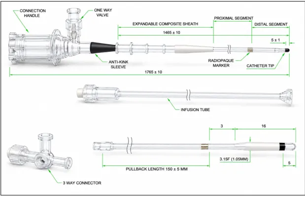
The IVUS Imaging Catheter is a sterile, single-use, rapid-exchange intravascular catheter intended for use with the STARBEAM™ OCT + IVUS dual imaging system to provide intravascular ultrasound imaging of blood vessels. The device consists of an imaging core incorporating a flexible, rotating drive cable with a 50 MHz ultrasonic transducer positioned at the distal tip to generate high-resolution cross-sectional images, and a catheter body comprising a distal imaging window section, a proximal shaft section, and a telescoping section. The effective working length of the catheter is defined by the distal and proximal sections, while the telescoping section allows controlled linear movement of the imaging core by up to 15 cm during imaging. The catheter is equipped with radiopaque markers to support fluoroscopic visualization and accurate positioning, as well as insertion depth markers to assist in estimating advancement and is compatible with a 0.014-inch guidewire for delivery to the target vessel’s flush port with an integrated one-way valve is provided to allow flushing with heparinized saline to maintain appropriate acoustic coupling and support image. Rotation and retraction of the catheter are controlled by the Patient Interface Unit (PIU) to acquire cross-sectional images during vessel scanning.
MARKED IMAGES
Figure 1: Component Identification Diagram for IVUS Imaging Catheter
Before the widespread clinical adoption of Optical Coherence Tomography (OCT), Intravascular Ultrasound (IVUS) was the primary intravascular imaging modality used to complement conventional angiography. While angiography provides a two-dimensional silhouette of the vessel lumen, IVUS enables real-time cross-sectional visualization of the vessel wall, lumen dimensions, plaque burden and lesion architecture. Today, IVUS is the preferred imaging modality for initial assessment of many complex lesions due to its greater tissue penetration and ability to image without the need for blood clearance.
Representative IVUS images demonstrating pre and post procedure/treatment is incorporated below for comparative evaluation and better visualization of the observed changes. IVUS image looks like a circular black lumen surrounded by gray vessel tissue, with bright areas indicating dense structures such as fibrous tissue, calcium, or stent struts. Therefore, IVUS provides better penetration into deeper vessel structures.
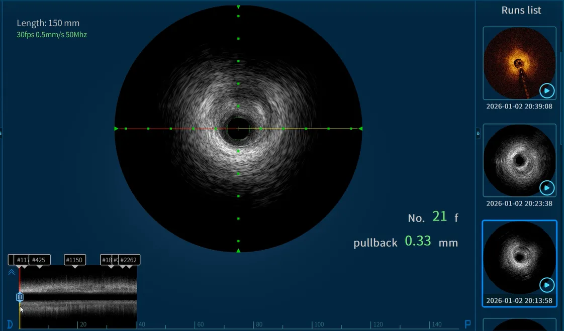
Figure 2: IVUS Image Pre-procedure/Treatment
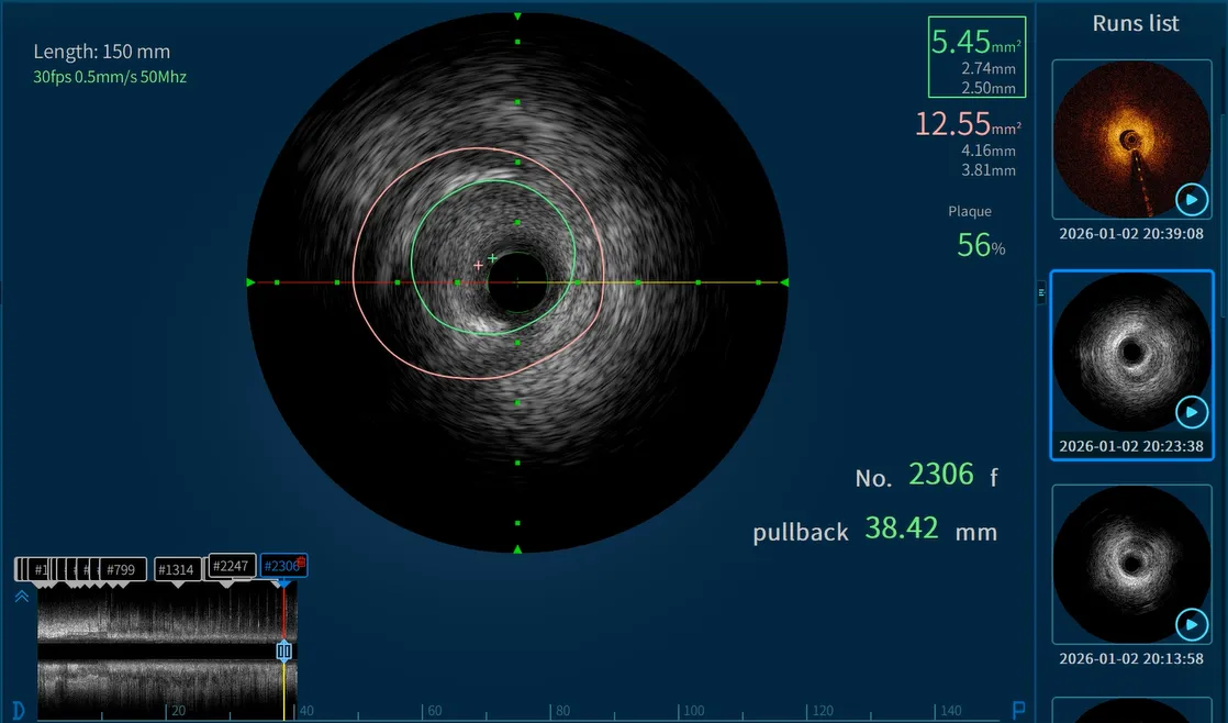
Figure 3: IVUS Image Post Procedure/Treatment
INTENDED USE
The STARBEAM™ IVUS Imaging Catheter with the STARBEAM™ OCT+IVUS Dual Imaging System, is intended for ultrasound examination of coronary intravascular pathology only. Intravascular ultrasound imaging is indicated in patients who are candidates for transluminal coronary interventional procedures.
Table 1: Size Matrix of IVUS Imaging Catheter
|
Sr. No. |
Parameters |
Dimensional Requirements (mm) |
|
1. |
The length of the imaging catheter |
1765±10 |
|
2. |
Active Length |
1465±10 |
|
3. |
The distal end of the catheter is separated from the outer edge of the radiopaque marker |
5±1 |
|
4. |
Expansion part length |
150±5 |
MATERIALS AND METHODS
Table 2: Component Description of IVUS Imaging Catheter
|
Sr. No. |
Name of Components |
Function |
Materials |
|
1. |
Proximal Segment |
The section closer to the connection handle, providing structural support and housing essential transmission components. It offers stiffness for improved handling |
Polyether Ether Ketone (PEEK) |
|
2. |
Distal Segment |
Section near the catheter tip, designed to be flexible for maneuvering through arteries. It contains imaging components responsible for capturing high resolution ultrasound images. |
Polyethylene (PE) |
|
3. |
Radiopaque Marker |
Visible under fluoroscopy, it helps position the catheter accurately within the artery for precise imaging. |
Platinum-iridium |
|
4. |
Connection Handle |
Provides the interface for connecting the catheter to the imaging system and infusion system. It allows the operator to control the catheter during the procedure, manage imaging settings, and handle movement within the vessel. |
Polycarbonate (PC) |
|
5. |
One-way Valve |
Ensures unidirectional fluid flow (contrast agent or saline) while preventing backflow, maintaining system integrity, and ensuring proper infusion during imaging |
Polycarbonate (PC) |
|
6. |
Anti-kink Sleeve |
Prevents kinking or bending of the catheter, ensuring smooth navigation through arteries and preventing obstruction of fluid flow or damage to internal components. |
Medical silicone |
|
7. |
Expandable Composite Sheath |
Protects the catheter's internal components and expands to accommodate catheter movement. It facilitates smooth insertion and reduces trauma to the vessel walls |
Polyamide (PA), Polycarbonate (PC), and silicone |
|
8. |
Catheter Tip |
Contains the IVUS ultrasound transducer, which emits and receives sound waves to generate real-time cross-sectional images of the artery. The tip is designed to be atraumatic to minimize vessel trauma |
Polyvinylpyrrolidone (PVP) |
|
9. |
Three-way Valve (Connector) |
Allows simultaneous management of multiple inputs/outputs, including fluid infusion (saline/contrast), pressure monitoring, and connection to the imaging system |
Polycarbonate (PC) |
|
10. |
Infusion Tube |
Delivers contrast agents, saline, or other necessary fluids for image enhancement and clearing the catheter tip for uninterrupted operation. |
Thermoplastic Polyurethane (TPU) |
|
11. |
Syringe 3 ml |
Used for controlled, precise injection of saline or contrast during catheter. |
Polycarbonate (PC) |
|
12. |
Syringe 10 ml |
Used for controlled, precise injection of saline or contrast during catheter. |
Polypropylene (PP) |
|
13. |
Sterile Bag for PIU |
Used to cover the Patient Interface Unit and maintaining a sterile field during the procedure. |
Polyethylene (PE) |
Applications of IVUS Imaging Catheter
Intravascular Ultrasound (IVUS) imaging catheters are used to visualize the inside of blood vessels and assess vessel architecture in real time. During coronary stent implantation, IVUS helps determine the correct stent size and verifies complete stent expansion, reducing the risk of complications.
1. Coronary Artery Disease Assessment
IVUS is widely used to evaluate coronary artery lesions by:
Example: A patient with intermediate coronary stenosis (40–70% narrowing) on angiography undergoes IVUS to determine the true severity of the lesion and guide treatment decisions.
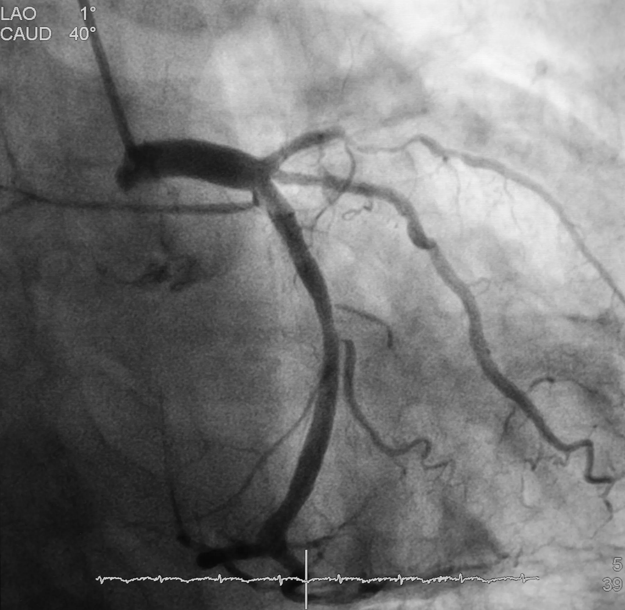
Figure 4: Angiographic Image Demonstrating Coronary Artery Lesions
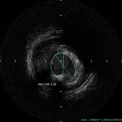
Figure 5: IVUS Image Illustrating Coronary Artery Lesions
2. Percutaneous Coronary Intervention (PCI) Guidance
IVUS helps optimize stent implantation by:
Example: During implantation of a drug-eluting stent in the left anterior descending artery, IVUS confirms adequate stent expansion and apposition to the vessel wall.
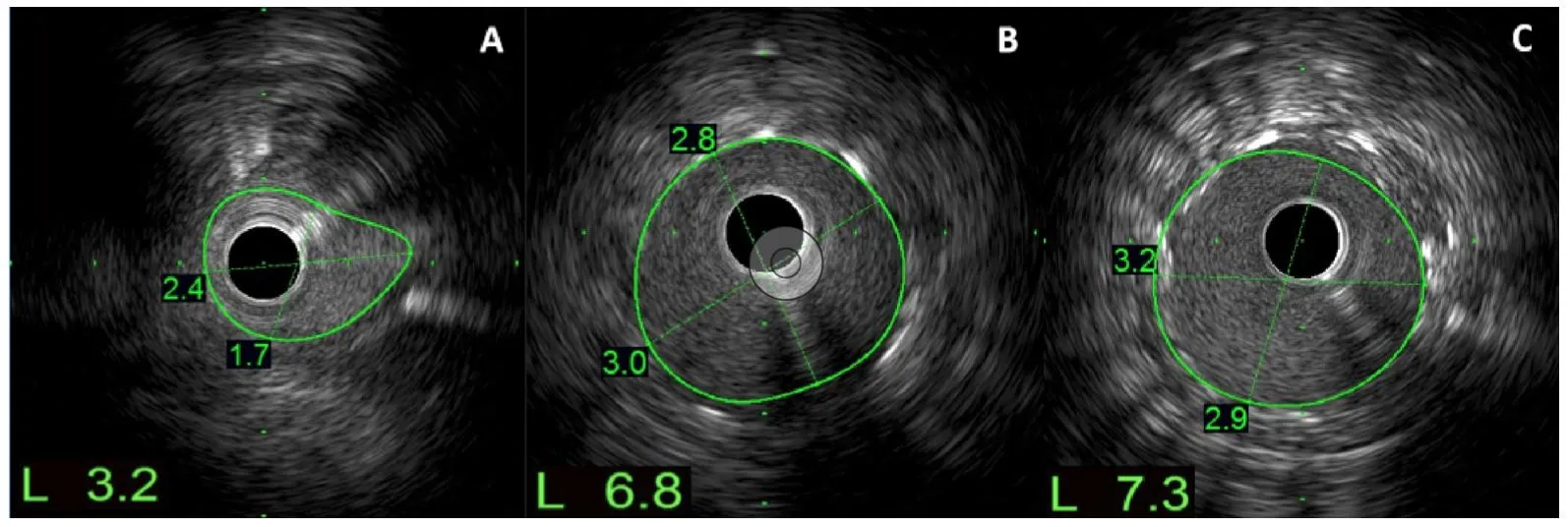
Figure 6: IVUS Image During PCI.
3. Assessment of In-Stent Restenosis (ISR)
IVUS can determine the cause of restenosis, such as:
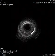
Figure 7: Pre-Procedural IVUS Image
Example: A patient develops recurrent angina one year after stent implantation. IVUS identifies under-expanded stent segments causing restenosis.
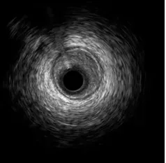
Figure 8: Post-Procedural IVUS Image
4. Evaluation of Stent Thrombosis
IVUS helps identify mechanisms responsible for thrombosis, including:
Example: Acute stent thrombosis occurring shortly after PCI is investigated using IVUS to identify procedural factors contributing to thrombus formation.
5. Characterization of Atherosclerotic Plaque
IVUS can differentiate various plaque components:
Example: Pre-interventional IVUS imaging demonstrates extensive circumferential calcification, prompting the use of atherectomy before stent placement.
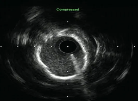
Figure 9: IVUS Image of Atherosclerotic Plaque
6. Left Main Coronary Artery Evaluation
Angiography may underestimate disease severity in the left main coronary artery, but IVUS provides:
Example: An ambiguous left main lesion on angiography is evaluated using IVUS to determine whether revascularization is required.
7. Research and Preclinical Studies
IVUS is extensively used in animal studies (especially porcine models) for evaluating:
Example: In a porcine coronary model, IVUS is performed at baseline and follow-up to quantify lumen area, vessel area, plaque burden, and neointimal growth after implantation of a bioresorbable scaffold.
EXPERIMENTAL DESIGN
Cytotoxicity Test:
In-vitro cytotoxicity test was performed using the L929 mouse fibroblast cell line in accordance with ISO 10993-5. Cells were cultured under standard conditions, harvested using trypsin-EDTA and seeded into 96-well
plates at a density of 1 × 10â´ cells/well. Plates were incubated at 37°C with 5% COâ‚‚ for ~24 h to form a semi-confluent monolayer. Test item extracts were prepared in MEM (Minimum Essential Medium) with serum using an extraction ratio 6 cm²/ml at 37±1°C for ~24 h as per ISO 10993-12. Cells were exposed to the neat extract (100%) and serial dilutions (50%, 25%, 12.5%, and 6.25%), along with controls for ~24 h. Following exposure, cells were examined microscopically and cytotoxicity was quantified using the MTT {3-(4, 5-dimethylthiazol-2-yl)-2, 5-diphenyl tetrazolium bromide} assay. Formazan crystals were solubilized with isopropanol and absorbance was measured at 570 nm to determine cell viability [4, 6].
Skin Sensitization Test:
A guinea pig maximization test was conducted using male Dunkin–Hartley guinea pigs in accordance with ISO 10993-10. Animals were acclimatized, randomized into four groups (polar and non-polar vehicle controls and corresponding test extract groups) and maintained under controlled environmental conditions with standard feed and water. Polar (normal saline) and non-polar (sesame oil) extracts of the test item were prepared at an extraction ratio of 6 cm²/ml at 50 ± 2°C for ~72 h as per ISO 10993-12. Intradermal induction was performed on day 0 using FCA with vehicle or test extracts, followed by topical induction on day 7 after SDS pretreatment. A challenge application was carried out on day 21 using undiluted extracts applied topically to untreated flank sites. Skin reactions were observed to evaluate the sensitization potential of the test item [13, 14].
Skin Irritation Test:
An intracutaneous reactivity study was conducted in female New Zealand White rabbits in accordance with ISO 10993-23. Animals were acclimatized, housed individually under controlled environmental conditions and identified as per SOPs. The dorsal skin was clipped prior to dosing. Polar (normal saline) and non-polar (sesame oil) extracts of the test item were prepared using an extraction ratio of 6 cm²/ml at 50 ± 2°C for ~72 h as per ISO 10993-12. Each rabbit received intra-cutaneous injections of the 200 µl/site of polar and non-polar test extracts and corresponding vehicle controls at five sites each. Animals were observed for local skin reactions to assess the intracutaneous irritation potential of the test item [16, 17, 18].
Acute Systemic Toxicity Test:
An acute systemic toxicity study was conducted in male Swiss Albino mice in accordance with ISO 10993-11. Animals were acclimatized, randomized into four groups (polar and non-polar vehicle controls and corresponding test extract groups), and maintained under controlled environmental conditions with standard feed and water. Polar (normal saline) and non-polar (sesame oil) extracts of the test item were prepared using an extraction ratio of 6 cm²/mL at 50 ± 2°C for approximately 72 h as per ISO 10993-12. Extracts were used without modification within 24 h of preparation. Each group of mice (n=5) received a single dose of the respective extract or vehicle control on day 1. Polar extracts were administered intravenously and non-polar extracts were administered intra-peritoneal at a dose volume of 50 ml/kg body weight. Animals were observed for systemic toxic effects to evaluate the acute systemic toxicity potential of the test item [19,20].
Pyrogenicity Test:
A rabbit pyrogen test was conducted using three healthy, nulliparous, non-pregnant female New Zealand White rabbits (Oryctolagus cuniculus), weighing 1.90–2.05 kg, sourced from a CCSEA-registered facility. Rabbits were selected as they are the recommended species for pyrogen testing as per ISO, USP, EP, BP, IP and US FDA guidelines. Animals were acclimatized for 6 days and housed individually under controlled environmental conditions (temperature: 19.0–22.8 °C; relative humidity: 42–66%; 12 h light/dark cycle), with ad libitum access to certified feed and RO-processed drinking water. A sham test was performed during acclimatization to ensure suitability of animals. Polar extracts of the test item were prepared using normal saline at an extraction ratio 6cm²/ml and incubated at 50 ± 2°C for approximately 72 h with continuous agitation, in accordance with ISO 10993-12. The extract was used without modification within 24 h of preparation. Prior to dosing, animals were fasted overnight and baseline rectal temperatures were recorded. The test extract, pre-warmed to 37°C, was administered intravenously via the marginal ear vein at a dose volume of 10 ml/kg body weight using pyrogen-free syringes and needles. The intravenous route was selected in accordance with United States Pharmacopeia requirements for pyrogen testing [20, 21].
Hemocompatibility Test
In-Vitro Hemolysis test (Direct Contact):
Rabbit (New Zealand White, Male) blood anticoagulated with sodium citrate was collected from three animals, pooled and used for the study. Plasma free hemoglobin was determined using Drabkin’s reagent, and only blood with plasma hemoglobin < 2 mg/ml was used. Total blood hemoglobin was measured and the blood was diluted with CMF-PBS to achieve a final hemoglobin concentration of approximately 10 mg/ml. A hemoglobin standard curve was prepared using Drabkin’s reagent and absorbance was measured at 540 nm. The hemolysis test was performed using the direct contact method as per ISO 10993-4 and ASTM F756. The test item was cut into small pieces and exposed to diluted rabbit blood at a surface area to volume ratio of 6 cm²/ml. Blank, negative control (HDPE) and positive control (SWI) were included in triplicate. Samples were incubated at 37°C for 3 hours with intermittent mixing. After incubation, samples were centrifuged and the supernatant was reacted with Drabkin’s reagent. Absorbance was measured at 540 nm, hemoglobin concentration was calculated from the standard curve, and percent hemolysis was determined after blank correction [27, 28].
In-Vitro Hemolysis test (Indirect Contact):
Citrated rabbit (New Zealand White, Male) blood collected from three animals was pooled and used for the study. Plasma free hemoglobin was determined using Drabkin’s reagent and only blood with plasma hemoglobin < 2 mg/mL was used. Total blood hemoglobin was measured and the blood was diluted with CMF-PBS to achieve a final hemoglobin concentration of approximately 10 mg/ml. A hemoglobin standard curve was prepared using Drabkin’s reagent and absorbance was measured at 540 nm. Test item extracts were prepared as per ISO 10993-12 using CMF-PBS at an extraction ratio of 6cm²/ml and incubated at 50°C for approximately 72 hours with continuous agitation. HDPE extract served as the negative control, SWI as the positive control and CMF-PBS as the blank. For the indirect contact hemolysis test, diluted blood was mixed with test item extract, negative control extract, positive control and blank at a ratio of 1:7 (blood: extract) in triplicate and incubated at 37°C for 3 hours with intermittent mixing. After incubation, samples were centrifuged and the supernatant was reacted with Drabkin’s reagent. Absorbance was measured at 540 nm, hemoglobin concentration was calculated from the standard curve, and blank-corrected percent hemolysis and hemolytic index were determined [27, 28].
Platelet Count and PF4 Estimation:
Fresh citrated human whole blood from three healthy male donors was pre-screened, pooled and used within 8 hours of collection. Test items, negative reference control (HDPE), positive control (glass beads) and negative control were exposed to human blood at a ratio of 12 cm²/ml. Prior to exposure, CaClâ‚‚ (10 mm) and heparin (2 U/ml) were added to the blood. Samples were incubated for 1 hour at 37°C with shaking, followed by addition of EDTA (5 mm) to terminate reactions. Hematology parameters were analyzed using an automated hematology analyzer. For platelet activation assessment, plasma was separated by centrifugation and platelet factor-4 (PF4) levels were quantified using a commercial ELISA kit. Plasma samples were diluted 1:200 and PF4 concentration was determined from a standard curve prepared according to the manufacturer’s instructions [28, 29].
In-vitro Assessment of Partial Thromboplastin Time, Thrombin and Fibrin Formation:
Sodium-citrated human plasma from healthy male donors (HBV and HIV negative) was used as the test system in accordance with ISO 10993-4. Test items were aseptically cut and exposed to plasma at a surface area to volume ratio of 6 cm²/ml. Blank, negative reference control (HDPE) and positive control (glass beads) were included in triplicate. All samples were incubated at 37°C for 15 minutes with agitation. After exposure, plasma was analyzed for coagulation parameters. Partial thromboplastin time (PTT) was measured using a coagulation analyzer following activation with RBC reagent and calcium chloride. The remaining plasma was used for fibrin and thrombin–antithrombin (TAT) complex estimation using commercially available ELISA kits. Absorbance was measured at 450 nm and concentrations were calculated from standard curves prepared according to the manufacturer’s instructions [30, 31].
Complete Blood Count by Leukocyte Activation Test of Human Blood:
Fresh citrated human blood from three healthy male donors (23–28 years) was pooled and used within 8 hours of collection. Finished medical device samples, along with negative and positive controls, were prepared under aseptic conditions as per ISO 10993-4 and exposed to human blood at a surface area–to–blood ratio of 12 cm²/ml. Blood was anticoagulated with CaClâ‚‚ and heparin, incubated with the test and control materials for 1 hour at 37°C under shaking conditions and reactions were stopped using EDTA. Complete blood count was analyzed using a hematology analyzer. Plasma obtained after centrifugation was used to assess leukocyte activation by measuring neutrophil elastase levels using a validated ELISA method, with standards run in duplicate and samples in triplicate [34, 35].
Complement Activation Test by using ELISA method:
Freshly fractionated human serum was used as the test system for complement activation assessment in accordance with ISO 10993-4. Finished device samples were aseptically prepared, cut into contact-relevant portions and extracted in human serum at a surface area to volume ratio of 6cm²/ml at 37°C for 60 minutes. The serum extracts, along with appropriate vehicle, positive and negative controls, were diluted (1:40 and 1:200) as required. Complement activation (SC5b-9) was quantified using the MicroVue™ Complement SC5b-9 plus ELISA kit following the manufacturer’s instructions. Absorbance was measured at 450 nm using a microplate reader [34, 35].
Genotoxicity
Bacterial Reverse Mutation Test:
The test item (<0.5 mm thickness) was extracted in water at 50°C for 72 h with agitation (6 cm²/ml, 80 RPM) to prepare polar extracts. S. typhimurium strains (TA98, TA100, TA102, TA1535 and TA1537) were cultured in nutrient broth and confirmed viable. Test item extracts and controls were evaluated using a dose range-finding (DRF) study and a limit test via the plate incorporation method, both with and without metabolic activation (5% S9 mix). Plates were incubated at 37°C for 48 h, and revertant colonies were counted to assess cytotoxicity and mutagenicity [22, 23, 24, 25].
In-Vitro Mammalian Chromosomal Aberration Test using Human Peripheral Blood Lymphocytes:
Human peripheral blood lymphocytes from healthy donors were used as the test system. Test item extracts were prepared in RPMI 1640 with 10% FBS and 1% antibiotics at a ratio of 6 cm²/ml and incubated at 37°C.
Test item extracts were prepared in RPMI 1640 with 10% FBS and 1% antibiotics at a ratio of 6 cm²/ml and incubated at 37°C for ~72 h with continuous agitation. Vehicle control (media alone) and positive controls (Mitomycin-C without S9, Cyclophosphamide with S9) were included. Blood cultures were set up in growth medium (RPMI 1640 + 20% FBS + 2% PHA-M + 1% antibiotics) and incubated at 37°C, 5% COâ‚‚. Cultures were treated with test item extract, vehicle, or positive control for short-term (3–6 h) and long-term (~24 h) exposures, with or without metabolic activation (S9 mix). Colchicine was added before harvesting. Cells were harvested, treated with hypotonic KCl, and fixed with Carnoy’s fixative. Slides were prepared, air-dried, stained with 5% Giemsa, and cover slipped. Mitotic index (MI) was determined by scoring 1000 cells per culture at 40X magnification to assess cytotoxicity. Only cells with normal chromosome number (44–48) and good morphology were evaluated [26].
OBSERVATIONS
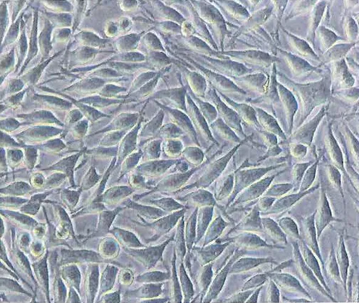
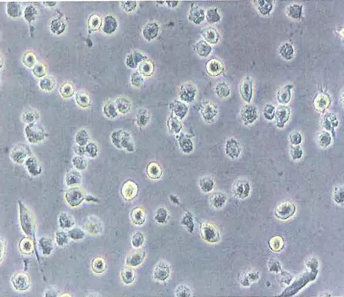
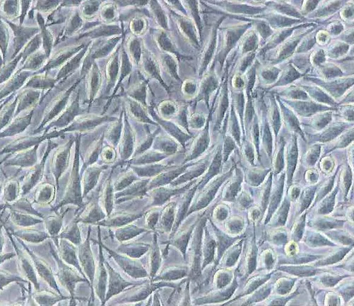
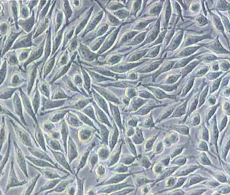
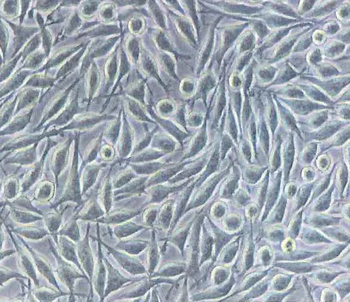
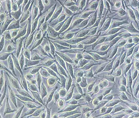
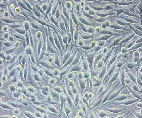
Cytotoxicity Test: Microscopic examination revealed cellular responses including changes in morphology, vacuolization, detachment, cell lysis, and membrane integrity. Cellular reactivity was graded from 0 to 4 based on qualitative morphological criteria. A numerical grade greater than 2 was considered indicative of cytotoxicity. During the MTT assay, viable cells reduced MTT to purple formazan crystals, which were solubilized and visually assessed. Reduced color intensity corresponded to decreased cell viability. Quantitative analysis was performed by measuring optical density at 570 nm. Percent cell viability was calculated relative to vehicle
and blank controls. A test sample was considered cytotoxic, if cell viability was less than 70% of the vehicle control. Acceptance criteria were met as follows: untreated control optical density was ≥ 0.2 with ≤ 15% variation between replicates; the positive control showed ≥ Grade 3 cytotoxic response; and the vehicle control showed Grade 0 response. Based on qualitative grading, visual assessment and quantitative cell viability results, the data were found to be valid and acceptable for evaluation. The spectrophotometric analysis of the culture well plate was resulted in numerical optical density at 570 nm. The average of the replicate value was calculated.
% cell viability was calculated using following equation:
% cell viability =OD570e – OD570b/ OD570c – OD570b × 100
Where,
OD570e: The mean value of the measured optical density of the extracts of the test sample, negative control or positive control.
OD570c: The mean value of the measured optical density of the vehicle control.
OD570b: The mean value of the measured optical density of the blank control.
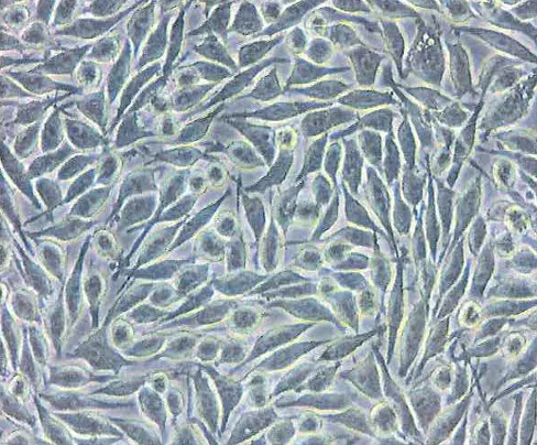
Figure 2: Representative Experimental Photographs of L929 Cell Line
|
|
|
|
Vehicle Control : Reactivity (None) Grade: 0 |
Positive Control (ZDEC) : Reactivity (Severe) Grade: 4 |
|
|
|
|
Negative Control (HDPE), Reactivity (None) Grade: 0 |
Test Item D0: 100%, Reactivity (None) Grade: 0 |
|
|
|
|
Test Item D1: 50%, Reactivity (None) Grade: 0 |
Test Item D2: 25%, Reactivity (None)Grade: 0 |
|
|
|
|
Test Item D3: 12.5%, Reactivity (None) Grade: 0 |
Test Item D4: 6.25%, Reactivity (None) Grade: 0 |
Skin Sensitization Test:
All animals were observed twice daily for mortality and morbidity and once daily for clinical signs throughout the study period. No treatment related mortality or morbidity was observed during the course of the study. Individual body weights were recorded prior to treatment (day 0) and at the end of the observation period (day 24). Body weight data
were expressed as Mean ± SD and showed no treatment-related adverse effects. Dermal reactions were evaluated at intra-dermal induction sites (sites A and C) on days 1 and 2 (approximately 25 h and 49 h post-induction) to assess the local effects of Freund’s Complete Adjuvant (FCA). Site B was observed at the same time points and additionally prior to topical induction on day 6 to assess the irritant potential of the test item. Challenge sites of both test and control animals were examined at approximately 25 h and 49 h following patch removal. Dermal responses were assessed and scored according to the Magnusson and Kligman grading scale (Grades 0–3). The results obtained during the challenge phase were used for the final interpretation of the sensitization potential. The study met the acceptance criteria, as the positive control produced a clear sensitization response within the test facility, and no sensitization reactions were observed in the vehicle control–treated animals. Accordingly, the study was considered valid. Based on the observations, the sensitization rate (%) was calculated with the formula as mentioned below:
Sensitization Rate (%) = No. of animals with positive reaction /No. of tested animal×100
Skin Irritation Test:
All animals were monitored twice daily for mortality and morbidity and once daily for clinical signs throughout the study period. Individual body weights were recorded prior to treatment (day 1) and at the end of the final observation period, with no treatment-related adverse effects observed. Injection sites were examined immediately after dosing and at approximately 23 h, 47 h and 71 h post-injection for evidence of local tissue reactions such as erythema and oedema. Dermal reactions were scored according to the standardized intracutaneous reaction grading system. Any additional adverse changes at the injection sites were recorded and reported. Acceptance criteria were met, as all injection sites treated with polar and non-polar vehicle controls showed Grade 0 and ≤Grade 1 dermal reactions, respectively. For evaluation, erythema and oedema scores at all observation time points were totaled and averaged per animal and per group. The final test extract score was calculated by subtracting the corresponding vehicle control score. The study requirements were satisfied as the mean score difference between the test item and vehicle control was zero. At study termination, all animals were humanely euthanized using an intravenous overdose of thiopentone sodium.
Acute Systemic Toxicity Test:
All animals were monitored twice daily for mortality and morbidity and once daily for clinical signs throughout the study period, and no treatment-related mortality, morbidity, or clinical abnormalities were observed. Individual body weights were recorded on day 1 (prior to treatment) and on days 2, 3, 4 and 7. Body weight data, expressed as Mean ± SD, showed no treatment-related effects. Clinical pathology evaluations (hematology and clinical chemistry) were not performed, as neither the test item–treated groups nor the vehicle control groups exhibited any signs of adverse effects. At the end of the observation period, all animals were humanely euthanized by COâ‚‚ asphyxiation followed by exsanguination and subjected to a complete gross necropsy, including examination of external surfaces, natural orifices and thoracic and abdominal cavities. No gross pathological lesions were observed, and therefore histopathological examination was not performed.
Pyrogenicity Test:
All animals were monitored for mortality and morbidity twice on day 1 of treatment and observed once daily for clinical signs on the same day. No treatment-related mortality, morbidity or clinical abnormalities were observed. Individual body weights were recorded prior to treatment on day 1 and showed no abnormalities. Baseline body temperature for each rabbit was recorded 30 minutes prior to treatment and used as the control value. Post-treatment body temperature was measured at 30-minute intervals up to 3 hours following injection of the polar test item extract. No abnormal temperature variations attributable to the test item were observed. Upon completion of the test, all animals were returned to the animal facility without further investigation.
Hemocompatibility Test
In-Vitro Hemolysis test (Direct & Indirect Contact):
Percent hemolysis for the test item, positive control and negative control was calculated based on the ratio of supernatant hemoglobin concentration to total hemoglobin concentration. Supernatant hemoglobin concentration was derived from the sample absorbance multiplied by the slope and a factor of two. Total hemoglobin concentration was based on the total diluted blood hemoglobin. In cases where absorbance at 540 nm exceeded 2, it was considered a procedural or background issue, and the test was repeated if necessary. Blank-corrected percent hemolysis was calculated by adjusting sample absorbance with blank and diluted blood absorbance values. The hemolytic index was determined by subtracting the mean blank corrected hemolysis of the negative control from that of the test item or positive control. A hemolytic index of less than 2% was considered non-hemolytic, 2–5% slightly hemolytic and greater than 5% hemolytic. If the mean hemolytic index was below 5% but any replicate exceeded 5%, the test was repeated with double the number of test items. All acceptance criteria were met.
% Hemolysis = Supernatant hemoglobin concentration /Total hemoglobin concentration in tube ×100
Hemolytic Index = % Mean Blank Corrected Hemolysis of test item or positive control – % Mean Blank Corrected Hemolysis Negative control.
Platelet Count and PF4 Estimation:
Platelet count analysis was performed using duplicate readings per sample and mean values were reported. Platelet counts of the test item, negative reference control and positive control were normalized to the negative control. The test item showed a platelet count of 107.53% relative to the negative control, the negative reference control showed 114.12%, and the positive control showed a reduced value of 52.36%. PF4 activity was visually confirmed
by color change from blue to yellow after substrate and stop solution addition. Biochemical analysis indicated PF4 activation in the test item group was comparable to the negative control and lower than the positive control. The assay was considered valid, as replicate values for the test item and negative reference control were within ±20% of the mean and the controls showed expected responses.
A/B × 100 = C
Where,
A = Average count (platelet) of the test item replicate
B = Average count (platelet) of negative control (untreated blood)
C = Percentage (%) of negative control
In-vitro Assessment of Partial Thromboplastin Time, Thrombin and Fibrin Formation:
The partial thromboplastin time for the test item, negative control, negative reference control and positive control was evaluated by calculating the mean clotting time for each group and expressing the results as a percentage relative to the negative control.
The percentage of the test item compared to the negative control was 100.71% while the negative reference control showed a value of 97.44% and the positive control showed a reduced clotting time relative to the negative control. Mean and standard deviation values were calculated for all groups. Statistical analysis was performed using one-way ANOVA followed by Tukey’s post hoc test. No statistically significant difference was observed between the test item and the negative control indicating no effect of the test item on partial thromboplastin time.
Complete Blood Count by Leukocyte Activation Test of Human Blood:
The complete blood count (CBC) parameters of the test item were within the range of the negative control and negative reference control, with no test item–related changes observed in hematological parameters. Neutrophil elastase estimation, based on absorbance measurements at 450 nm and standard curve analysis, showed low levels of neutrophil elastase formation in the test item group compared to the positive control.
Complement Activation Test by using ELISA method:
After addition of the substrate, a blue color developed, which changed to yellow upon addition of the stop solution, indicating completion of the assay reaction. Duplicate readings for standards, controls and test samples were averaged after subtraction of blank values. A standard curve was prepared by plotting absorbance against corresponding standard concentrations and the concentrations of the vehicle control, negative control, positive control and test item were determined from the best-fit line, taking dilution factors into account. The highest level of complement activation was observed in the positive control while the lowest was observed in the negative control. Percent complement activation of the test item was calculated relative to the positive control. Mean and standard deviation values were calculated for all groups. Statistical analysis using ANOVA followed by Tukey’s post-hoc test showed no significant difference between the test item and the negative control at both dilutions (1:40 and 1:200). Therefore, the test item was considered non-activating.
Genotoxicity
Bacterial Reverse Mutation Test:
During the dose range finding (DRF) and limit studies, revertant colonies on each plate were manually counted and the background bacterial lawn was examined microscopically. Revertant colony counts for the neat extract (100%) along with the concurrent vehicle and positive controls were recorded and are presented in tabular form, including individual plate counts, mean values, standard deviations, and observations on background lawn growth. The assay was considered acceptable, as normal background bacterial growth was observed in the vehicle control and a significant increase in revertant colony frequency was produced by the positive control.
In-Vitro Mammalian Chromosomal Aberration Test using Human Peripheral Blood Lymphocytes:
All slides from test item treated cultures and concurrent controls were independently coded prior to microscopic evaluation to ensure blinded analysis. Microscopic examination was performed using 40X and 100X objectives to assess the number of metaphases and chromosomal aberrations respectively. Following completion of observations, the slides were decoded for result evaluation. Cytotoxicity was assessed by counting a minimum of 1000 cells per culture across different microscopic fields, and the number of metaphases was recorded for calculation of the mitotic index (MI). In the main experiment, a total of 300 well-spread metaphases per concentration were analyzed, with approximately equal distribution between duplicate cultures. Structural chromosomal aberrations including chromatid and chromosome-type aberrations and their subtypes were recorded along with their frequencies. Slides were also examined for numerical aberrations such as polyploidy and endo-reduplication. Individual culture data were summarized in tabular form. Mitotic index and relative mitotic index (RMI) were calculated using standard formulas, and results were expressed as Mean ± SD. Statistical analysis of structural chromosomal aberrations was performed using ANOVA with comparison to concurrent vehicle and positive controls. A statistically significant increase in chromosomal aberrations was observed in the positive control groups whereas no statistically significant increase was observed in the test item treated groups compared to the vehicle control across all experimental conditions. The assay met all acceptability criteria, as vehicle controls were within historical ranges, positive controls produced expected statistically significant responses, cell proliferation in solvent controls was appropriate and sufficient numbers of cells and concentrations were analyzed. Based on fulfillment of acceptability criteria and absence of statistically significant or dose-related increases in chromosomal aberrations, the test item was considered negative for induction of chromosomal aberrations under the conditions of this study.
(MI %) = Number of mitotic cells/ Total number of cells scored × 100
RMI (%) = (Test Concentration MI) / (Solvent Control MI) × 100
RESULT AND DISCUSSION
Cytotoxicity Test:
Microscopic examination showed no morphological changes in cells treated with the undiluted (100%) and all diluted extracts (50%, 25%, 12.5%, and 6.25%) corresponding to a reactivity grade of 0. Vehicle and negative controls showed no reactivity while the positive control exhibited severe cytotoxicity. Systemic cell seeding error was within acceptable limits (4.81%). In the MTT assay, purple formazan formation was observed in all test item extracts and controls except the positive control. Cell viability for the undiluted extract was 93.52% and all diluted concentrations showed viability above 70%.
Skin Sensitization Test:
No mortality, morbidity or abnormal clinical signs were observed in any animals during the study. All animals showed normal body weight gain by day 24 compared to day 0. No treatment-related dermal reactions were observed at injection site B during induction or after challenge. Dermal reactions such as erythema observed at injection sites A and C were attributed to the local effect of Freund’s Complete Adjuvant and not to the test item. No dermal reactions were observed following challenge application. The average dermal reaction grade for test group animals was 0, with a sensitization rate of 0%, indicating no skin sensitization. Re-challenge was not required as no equivocal responses were noted. All animals were humanely euthanized at study termination.
Skin Irritation Test:
No mortality, morbidity, or abnormal clinical signs were observed in any animals during the study. Body weights remained within the normal range from day 1 to the end of the observation period. No dermal reactions such as erythema or oedema were observed at the injection sites immediately or at approximately 23 h, 47 h, and 71 h following administration of polar and non-polar extracts or vehicle control.
Acute Systemic Toxicity Test:
No mortality, morbidity or abnormal clinical signs were observed in any animals treated with polar or non-polar extracts of the test item or their respective vehicle controls throughout the study period. All animals showed a gradual and normal increase in body weight up to day 7, with no statistically significant differences between groups as determined by one way ANOVA at a 95% confidence interval. Gross pathological examination revealed no abnormalities in any treated animals.
Pyrogenicity Test:
No mortality, morbidity, or abnormal clinical signs were observed in animals treated with the test item extract throughout the study period. All animals remained normal. Body weights were within the required range (>1.5 kg; 1.89754–2.05117 kg) as per United States Pharmacopeia requirements. Individual rabbit body temperatures measured at different intervals showed a difference of less than 0.5°C compared to respective control animals, meeting USP acceptance criteria.
Hemocompatibility Test
In-Vitro Hemolysis test (Direct Contact):
The hemolytic index of the test item IVUS Imaging Catheter, determined by the direct contact method, was −0.010% (< 2%) and was therefore graded as non-hemolytic whereas the positive control showed a hemolytic index of 101.540% and was graded as hemolytic.
In-Vitro Hemolysis test (Indirect Contact):
The hemolytic index of the test item IVUS Imaging Catheter, determined by the indirect contact method, was −0.660% (< 2%) and was therefore graded as non-hemolytic while the positive control showed a hemolytic index of 98.460% and was graded as hemolytic.
Platelet Count and PF4 Estimation:
Based on the results, it was observed that the platelet count of test item treated with blood was within the range of the negative reference control material (that is 80–120% of the negative control blood) and the test item was 55.17% above that of the positive control material. The Platelet Factor 4 ELISA showed almost similar result in test group as compared to the negative reference control and negative control in human plasma.
In-vitro Assessment of Partial Thromboplastin Time, Thrombin and Fibrin Formation:
The partial thromboplastin time of the test item ‘IVUS Imaging Catheter’ was found to be within the acceptable range of the negative reference control. The test item group exhibited Fibrin and TAT formation similar to that of the negative control and negative reference control respectively while a lower level was observed compared to the positive control. No statistically significant difference in partial thromboplastin time was observed between the test item and either the negative control and negative reference control.
Complete Blood Count by Leukocyte Activation Test of Human Blood:
The CBC of the test item was found to be within the range of negative control and negative reference control. Neutrophil Elastase estimation showed low levels of Neutrophil Elastase formation in test item group as compared to positive control. Based on the results, it was observed that the CBC and Neutrophil Elastase formation of the test item treated blood was within the acceptable range of the negative control and negative reference control material.
Complement Activation Test by using ELISA method:
The test item was incubated with serum to evaluate the potential of complement activation components. The analysis was performed on the basis of the ELISA assay as the quantitative evaluation. In negative control, it was observed that the complement activation was non-reactive during exposure whereas the positive control had produced a significant effect on the complement system activation in the assay. The complement system activation was not observed for the test item.
Genotoxicity
Bacterial Reverse Mutation Test:
In the dose range finding study, neat extracts (100%) of the test item showed normal background bacterial lawn and revertant colony counts comparable to the vehicle control and lower than the positive control, in all five S. typhimurium strains (TA98, TA100, TA102, TA1535 and TA1537), both in the presence (5% v/v S9 mix) and absence of metabolic activation, indicating no cytotoxicity. As no cytotoxicity was observed, the same plate count data were considered for the limit study to assess mutagenicity. No increase in revertant colonies was observed under any test condition and therefore a confirmatory experiment was not required.
In-Vitro Mammalian Chromosomal Aberration Test using Human Peripheral Blood Lymphocytes:
The vehicle control was considered acceptable as found within the range of historical control database available for the same assay whereas concurrent positive controls were considered acceptable as induced responses that are compatible with the assay requirements. The results obtained with the test item did not show any increase in the incidence of cells with aberrant chromosomes in all the tested conditions. There was no increase in numerical aberrations in the conditions tested.
CONCLUSION
A comprehensive biological evaluation of the STARBEAM™ IVUS Imaging Catheter was conducted in accordance with the applicable ISO 10993 series standards, OECD guidelines, ASTM standards, and relevant US FDA guidance documents to determine its safety for intended clinical use. The in-vitro cytotoxicity assessment using L929 mouse fibroblast cells demonstrated that the device extracts were non-cytotoxic, with greater than 70% cell viability across all tested concentrations and 93.52% viability at 100% extract, corresponding to grade zero (0) reactivity. Sensitization studies in Dunkin Hartley guinea pigs confirmed that the device was non-sensitizing when evaluated with both polar and non-polar extracts. Intracutaneous reactivity testing in New Zealand White rabbits revealed no evidence of erythema or edema, indicating that the device was non-irritant. Acute systemic toxicity studies in Swiss albino mice showed no mortality, clinical signs of toxicity, or gross pathological findings following administration of the extracts via intravenous and intraperitoneal routes. Pyrogenicity testing demonstrated temperature variations of less than 0.5°C compared to controls, confirming the device to be non-pyrogenic. Hemocompatibility assessment by both direct and indirect hemolysis methods established that the device was non-hemolytic. Genotoxicity evaluation, including the bacterial reverse mutation (Ames) assay using five Salmonella typhimurium strains with and without metabolic activation and the in-vitro chromosomal aberration assay in human lymphocytes revealed no evidence of mutagenic or clastogenic effects. Additionally, complement activation testing using human serum showed no activation of the complement system.
Collectively, these findings demonstrated that the STARBEAM™ IVUS Imaging Catheter exhibits a favorable biocompatibility profile and meets the biological safety requirements outlined in the ISO 10993 framework for its intended medical application.
COI (Conflict of Interest):
All the authors are an employee of Meril Medical Innovations Private Limited, Vapi, Gujarat – India and have no conflict of interest related to this study.
REFERENCES

Minocha Dr. Pramodkumar, Kothwala Dr. Deveshkumar, Pandya Kamna, Shinde Divya, Kadam Aniket, Ladumor Rahul, Sharma Rahul, Sharma Mehul, Systematic Biological Evaluation of the Starbeam™ IVUS Imaging Catheter: Evidence from In-Vitro and In-Vivo Studies, Int. J. Sci. R. Tech., 2026, 3 (10), 597-613. https://doi.org/10.5281/zenodo.23260539
 10.5281/zenodo.23260539