

We use cookies to ensure our website works properly and to personalise your experience. Cookies policy
Computer Science Engineering, VIt Vellore, Tamil Nadu, India
Theranostics links disease characterization with treatment selection and response assessment, creating a framework in which diagnostic information directly informs individualized therapy. Recent advances in artificial intelligence (AI), machine learning (ML), deep learning, radiomics, radiogenomics, and multimodal data integration are expanding this concept from target verification toward quantitative prediction of diagnosis, prognosis, therapeutic response, dose, and toxicity. This review examines how AI can connect imaging, electronic health records, genomics, transcriptomics, proteomics, pharmacogenomics, and treatment response data across a unified theranostic workflow. Evidence from ophthalmology, dermatology, pathology, cardiology, oncology, drug response prediction, radiopharmaceutical therapy, and personalized dosing demonstrates that ML models can extract clinically useful patterns from complex datasets and support patient level stratification. In nuclear medicine, AI assisted segmentation, quantitative imaging, and dosimetry may shorten analysis time and facilitate patient-specific radiopharmaceutical therapy. In precision oncology, radiomics and radiogenomics can associate imaging phenotypes with molecular characteristics and treatment outcomes, while computational drug response models can prioritize therapies for molecularly defined disease. However, clinical translation remains constrained by dataset shift, limited external validation, data leakage, class imbalance, algorithmic bias, interpretability, privacy, interoperability, regulatory uncertainty, and insufficient prospective evidence. Future progress will require multicenter datasets, standardized data pipelines, transparent reporting, calibrated uncertainty, explainable models, federated or privacy preserving learning, prospective clinical trials, and continuous post deployment monitoring. AI driven theranostics is therefore best viewed as a decision support ecosystem in which computational prediction complements, rather than replaces, clinical expertise and experimentally validated treatment pathways.
Theranostics is a patient centered concept in which diagnostic characterization is directly linked to therapeutic selection and subsequent assessment of treatment response. The approach is particularly established in molecular imaging and nuclear medicine, where a diagnostic ligand can identify target expression and a related therapeutic ligand can deliver treatment to the same biological target. The central principle is not simply that diagnosis and therapy occur in the same pathway, but that information obtained at the diagnostic stage changes the therapeutic decision for an individual patient. This creates a natural setting for artificial intelligence because modern theranostic workflows generate high dimensional imaging, molecular, clinical, and longitudinal outcome data that are difficult to integrate using conventional rule based approaches [1]. AI encompasses computational methods capable of learning patterns from data, while ML refers to algorithms that learn relationships between input variables and outcomes. Deep learning extends this approach through multilayer neural networks that can learn hierarchical representations directly from complex data such as images, waveforms and text. In medicine, the value of these methods is increasingly demonstrated in image classification, segmentation, prognosis, clinical prediction, and treatment response modeling. Large scale studies have shown clinically relevant performance in retinal disease, skin cancer, breast cancer screening, pathology and electrocardiography [2]. These developments are important for theranostics because accurate target identification and quantitative phenotyping are prerequisites for individualized treatment. The growing availability of multimodal datasets creates an additional opportunity. A patient may simultaneously have CT, MRI, PET or SPECT images; pathology; laboratory measurements; genomic and transcriptomic profiles; medication history; treatment exposure; and longitudinal outcomes. AI can potentially combine these heterogeneous sources to estimate disease probability, identify biological subgroups, predict response, estimate toxicity and support treatment selection. Deep learning applied to electronic health records has demonstrated that large longitudinal records can be represented directly for prediction of multiple clinical events Similarly, radiomics has shown that quantitative imaging features can capture tumor characteristics beyond routine visual assessment [3]. The most useful way to conceptualize AI driven theranostics is as an integrated workflow rather than a single algorithm. The workflow begins with patient level data acquisition and quality control. Imaging data are reconstructed, normalized and segmented; molecular and clinical data are curated; and missingness, batch effects and site specific differences are addressed. AI models then generate quantitative features or predictions. These outputs can support diagnostic classification, target confirmation, risk stratification, treatment selection, dose estimation and response monitoring. New outcome data can subsequently be used to recalibrate or retrain models under controlled governance. This creates a feedback structure in which diagnostic information influences treatment and treatment outcomes improve future prediction [4].
The distinction between prediction and decision making is essential. An ML model may estimate the probability of treatment response, but a clinical decision also depends on contraindications, patient preference, toxicity risk, organ function, prior therapy, treatment availability and guideline recommendations. Therefore, an AI system should provide interpretable evidence, uncertainty estimates and relevant supporting variables rather than an unexplained command. In high consequence settings such as radiopharmaceutical therapy, individualized dosimetry and treatment planning must remain linked to physical and biological constraints [5]. The integration of AI into theranostics is also broader than oncology. The same principles can be applied to infectious diseases, cardiometabolic disorders, neurological disease and inflammatory disorders whenever diagnostic biomarkers can be connected to targeted intervention. However, oncology and nuclear medicine provide particularly clear examples because disease heterogeneity, molecular targets, imaging biomarkers and dose response relationships are central to treatment selection[6].
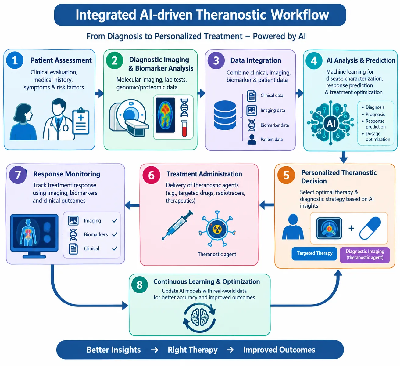
Figure 1. Integrated AI driven theranostic workflow
2. SCIENTIFIC BASIS AND CLINICAL RATIONALE
Theranostics is particularly suitable for computational medicine because the diagnostic and therapeutic stages generate linked patient specific information. Molecular imaging can demonstrate whether a biological target is present, estimate its spatial distribution, and provide a baseline for treatment planning. When the same or a closely related molecular target is used for therapy, the imaging phenotype can become a quantitative treatment selection variable rather than a purely descriptive finding. AI can combine target expression, lesion burden, organ function, prior treatment history, laboratory values, and imaging derived measurements to characterize the therapeutic window for an individual patient. A clinically useful AI enabled theranostic system should therefore be viewed as a decision support pipeline rather than a single prediction algorithm. The pipeline may include image acquisition and reconstruction, artifact correction, registration of serial examinations, organ and lesion segmentation, quantitative uptake measurement, radiomic feature extraction, dosimetric estimation, response prediction, toxicity prediction, and longitudinal outcome analysis. Each component has different error sources, and errors can propagate from one stage to the next. For example, an inaccurate lesion boundary can alter measured activity concentration, which can subsequently alter estimated absorbed dose and the model's treatment recommendation [7]. Another important principle is multimodality integration. PET, SPECT, CT, MRI, pathology, laboratory data, genomics, transcriptomics, and electronic health records contain complementary information. Multimodal machine learning can theoretically learn relationships between molecular phenotype, anatomical structure, tumor heterogeneity, and clinical outcome. However, missing data are common in real clinical datasets. Robust systems should explicitly model missingness and uncertainty rather than treating absent measurements as normal values. The scientific foundation of theranostics is target verification. A diagnostic biomarker is clinically useful when it identifies a biological property that is relevant to treatment. Molecular imaging provides an advantage over a single tissue sample because it can evaluate target distribution across the body and reveal spatial heterogeneity. This is important in cancer, where different lesions within the same patient can show different molecular characteristics [8]. Radiopharmaceutical therapy demonstrates the principle clearly. In somatostatin receptor positive neuroendocrine tumors, lutetium 177 DOTATATE has demonstrated clinical benefit, while PSMA directed radioligand therapy has produced important outcomes in metastatic prostate cancer . These therapies depend on appropriate target expression and patient selection. AI can assist by quantifying target uptake, tumor burden and heterogeneity across many lesions. Patient specific dosimetry adds a second biological and physical layer. Dosimetry estimates the absorbed radiation dose delivered to tumors and organs at risk. The same activity administered to two patients does not necessarily produce the same absorbed dose because anatomy, biodistribution and clearance differ. Their integration can reveal associations between imaging patterns and molecular characteristics that may be difficult to infer visually. This can support noninvasive phenotyping and patient stratification, but it requires careful control of image acquisition, preprocessing and external validation [9].
3. AI AND MACHINE LEARNING METHODS IN THERANOSTICS
|
Sr. No. |
AI Method |
Principle |
Application in Theranostics |
Major Advantages |
|
1 |
Machine Learning (ML) |
Data driven algorithms learn patterns from clinical, molecular and imaging datasets |
Disease diagnosis, patient stratification, treatment response prediction and biomarker identification |
Handles complex datasets and supports personalized treatment [10] |
|
2 |
Deep Learning (DL) |
Uses multilayer neural networks to automatically extract complex features from data |
Medical image analysis, tumor detection, drug response prediction and disease classification |
High performance in image and high dimensional data analysis [11] |
|
3 |
Artificial Neural Networks (ANNs) |
Mimics interconnected biological neurons to identify nonlinear relationships |
Prediction of therapeutic response, toxicity and disease progression |
Effective for nonlinear and complex relationships [12] |
|
4 |
Convolutional Neural Networks (CNNs) |
Extracts spatial and hierarchical features from images |
Tumor segmentation, cancer detection, histopathological image analysis and molecular imaging |
Excellent for image recognition and classification [13] |
|
5 |
Recurrent Neural Networks (RNNs) |
Processes sequential and time dependent information |
Analysis of longitudinal patient data, treatment response and disease progression |
Suitable for temporal and sequential datasets [14] |
|
6 |
Long Short Term Memory (LSTM) |
Advanced RNN architecture designed to retain long term information |
Prediction of treatment response, patient outcomes and longitudinal clinical events |
Handles long term dependencies effectively [15] |
|
7 |
Support Vector Machine (SVM) |
Identifies an optimal decision boundary between data classes |
Cancer classification, biomarker discovery and diagnostic prediction |
Effective with high dimensional datasets and smaller sample sizes [16] |
|
8 |
Random Forest (RF) |
Combines multiple decision trees to improve classification or prediction |
Biomarker identification, disease classification and therapeutic response prediction |
Robust, relatively interpretable and resistant to overfitting [17] |
|
9 |
Clustering Algorithms |
Groups similar observations without predefined labels |
Patient stratification, tumor subtype identification |
Useful for discovering hidden patterns [18] |
Table 1: AI and Machine Learning Methods in Theranostics
Summarizes major approaches and their theranostic applications. The important principle is that algorithm selection should follow the clinical question, data structure, sample size, interpretability requirement and validation strategy rather than being driven only by algorithmic novelty. Model development also requires careful separation of training, validation and testing data. Patient level splitting is essential because multiple images or visits from the same patient can otherwise leak information across datasets. External validation is more informative than an internal random split when the objective is clinical translation. Calibration should be assessed alongside discrimination because a model that ranks patients correctly can still produce misleading probabilities [18].
4. AI FOR PRECISION DIAGNOSIS AND QUANTITATIVE IMAGING
AI can support precision diagnosis at several levels: image reconstruction, lesion detection, lesion classification, quantitative measurement, differential diagnosis, and integrated reporting. In PET and SPECT, machine learning can be used to improve image quality from low count acquisitions, reduce noise, identify artifacts, and accelerate reconstruction. These applications may be particularly valuable when acquisition time, administered activity, or patient motion limits image quality. However, image enhancement algorithms should be validated against quantitative standards because visually attractive images can potentially alter measured uptake or obscure small lesions. Automated segmentation is one of the most important enabling technologies. Accurate delineation of tumors and organs allows calculation of metabolic tumor volume, total lesion activity, standardized uptake measures, organ activity, and spatially resolved dose. Segmentation algorithms can also reduce repetitive manual work and improve consistency between observers. Nevertheless, small lesions, low contrast lesions, heterogeneous uptake, postoperative anatomy, and lesions close to physiologically active organs remain challenging. Human review and correction remain important when segmentation affects treatment decisions [19]. AI supported quantitative imaging can also transform longitudinal assessment. Instead of comparing two scans only by visual impression, software can register serial images, match corresponding lesions, calculate changes in uptake and volume, and track lesion level trajectories. This enables a more granular view of treatment response, including mixed response in which some lesions regress while others remain stable or progress. Such information may be useful for adaptive therapy and for identifying patients who require a change in management. Medical imaging is one of the most established applications of AI. Deep learning has been used for classification, detection, segmentation, image reconstruction, denoising, registration and quantitative measurement. In diabetic retinopathy, deep learning achieved high diagnostic performance from retinal photographs . In retinal OCT, deep learning has been evaluated for diagnosis and referral . In breast imaging evaluation has demonstrated the potential of AI assisted screening Pathology provides another example. Whole slide images contain extremely large amounts of visual information, and weakly supervised learning can reduce the need for exhaustive pixel level annotation [20].
5. RADIOMICS AND RADIOGENOMICS
Radiomics converts medical images into large numbers of quantitative descriptors reflecting intensity, shape, texture, spatial relationships, and higher order patterns. Feature families can describe first order intensity distributions, shape characteristics, gray level cooccurrence relationships, runlength patterns, neighborhood dependence, and filtered or transformed image information. Machine learning can then identify combinations of features associated with diagnosis, molecular phenotype, treatment response, progression, or survival. Radiomics is highly sensitive to technical factors. Scanner manufacturer, acquisition protocol, reconstruction method, voxel dimensions, segmentation variability, preprocessing, and intensity discretization can change extracted features. Therefore, reproducibility requires standardized imaging protocols, feature quality assessment, test retest studies, harmonization, and independent validation. A feature that is statistically significant in one dataset should not automatically be interpreted as a stable biological biomarker [21]. Radiogenomics extends this concept by linking imaging phenotypes with genomic, transcriptomic, epigenomic, or other molecular information. The attraction is that imaging can provide a noninvasive and spatially distributed phenotype, whereas tissue sequencing provides molecular information that may be limited by sampling. Integrated models may help characterize tumor heterogeneity and infer biological processes that are difficult to observe from either imaging or genomics alone. Radiomics converts medical images into quantitative descriptors of intensity, shape, texture and spatial relationships. Aerts et al. demonstrated that radiomic features could characterize tumor phenotype and provide prognostic information. This approach is attractive for theranostics because it can provide information about tumor heterogeneity and disease phenotype without requiring additional invasive sampling. Radiogenomics links imaging phenotypes with genomic or molecular characteristics. It is especially relevant when a molecular biomarker is for treatment selection but tissue sampling is limited or incomplete [22].
6. AI FOR PERSONALIZED THERAPY AND DRUG RESPONSE PREDICTION
Personalized therapy requires estimation of both expected benefit and expected harm. AI models can combine demographic characteristics, disease stage, molecular markers, imaging phenotype, previous treatment exposure, laboratory variables, and patient specific factors to estimate response or toxicity. In drug development, machine learning can assist target identification, molecular property prediction, virtual screening, drug target interaction prediction, and optimization of candidate compounds. These approaches can shorten exploratory cycles, but computational predictions still require experimental and clinical confirmation. In theranostics, response prediction can be linked directly to the molecular target used for treatment. A model may estimate whether target positive disease is likely to respond, whether heterogeneous target expression may lead to incomplete treatment, or whether baseline tumor burden and organ reserve indicate a narrow therapeutic window. For radiopharmaceutical therapy, response prediction can potentially incorporate uptake intensity, lesion volume, spatial heterogeneity, absorbed dose, and prior cycle response rather than relying on a single imaging variable [23]. Adaptive treatment is a further extension of personalized therapy. After each treatment cycle, updated imaging and laboratory information can be used to revise the estimated response probability and toxicity risk. In principle, treatment intensity, cycle timing, or continuation can then be reconsidered using the patient's evolving data. Such adaptive approaches require prospective validation because retrospective associations do not demonstrate that changing therapy according to an algorithm improves outcomes. Personalized therapy aims to match an intervention with the biological and clinical characteristics of an individual patient. ML can integrate molecular alterations, imaging phenotype, previous treatment, laboratory values, pharmacogenomic variables and outcomes to estimate treatment response. Large pharmacogenomic studies have identified relationships between genomic features and drug sensitivity. Drug response prediction can be performed using patient derived molecular features or experimental cancer cell line datasets. Models may estimate sensitivity to individual agents, identify drug combinations or prioritize candidates for laboratory testing. Graph based approaches can represent molecular structures and drug target relationships . Protein structure prediction can also improve the computational representation of potential targets [24,25].
7. AI IN RADIOPHARMACEUTICAL THERAPY AND PERSONALIZED DOSIMETRY
Personalized dosimetry is one of the most clinically important areas for AI in radiotheranostics. The absorbed dose to a tumor or normal organ depends on activity distribution, biological clearance, physical decay, tissue mass, and time integrated activity. Traditional workflows may require several imaging time points and substantial manual processing. AI can assist registration, segmentation, activity quantification, time activity curve estimation, and dose map prediction, potentially reducing the time required for patient specific dosimetry. AI based dosimetry can operate at several spatial scales. Organ level approaches estimate average absorbed dose to organs, whereas lesion level and voxelbased methods preserve spatial heterogeneity. Voxel based approaches are particularly relevant when tumor dose is highly nonuniform because two lesions with the same mean dose may have different internal dose distributions. Deep learning models may estimate dose distributions from limited imaging time points, but their predictions should be compared with reference dosimetry and accompanied by uncertainty estimates [26]. Radiopharmaceutical therapy is a high value application of AI because the diagnostic and therapeutic stages can produce quantitative information about the same biological target. Personalized dosimetry estimates radiation absorbed by tumors and normal organs and may help optimize treatment intensity. Traditional dosimetry can require serial SPECT/CT imaging, image registration, segmentation, activity quantification, timeactivity curve fitting and computational dose calculation. These steps can be labor intensive. AI may reduce the burden through automated segmentation, image reconstruction, activity estimation and prediction of dose distributions [27,28].
8. AI FOR NANOTHERANOSTICS AND DRUG DISCOVERY
Nanotheranostics combines diagnostic and therapeutic functions within engineered nanoscale systems. Nanocarriers may be designed to transport drugs, imaging agents, radionuclides, nucleic acids, or combinations of these payloads. AI can support nanocarrier design by learning relationships between formulation variables and properties such as particle size, surface charge, encapsulation efficiency, stability, release kinetics, biodistribution, and cellular uptake. Machine learning can also help optimize formulation variables through design of experiments strategies and Bayesian optimization. Instead of evaluating every possible combination of materials and process parameters experimentally, models can prioritize formulations that are predicted to provide desirable properties. This can reduce experimental burden and identify nonlinear relationships that are difficult to detect using one factor at a time experimentation. Nevertheless, model predictions must remain linked to physicochemical characterization and biological testing [29,30]. For theranostic nanoparticles, AI may eventually integrate imaging, pharmacokinetics, molecular targeting, and treatment response to identify formulations suited to specific biological environments. Important translational barriers include manufacturing reproducibility, scale up, batch to batch consistency, long-term stability, toxicity assessment, regulatory characterization, and reproducible biodistribution. Computational optimization cannot substitute for these experimental and regulatory requirements [31,32]. Nanotheranostics combines diagnostic imaging and therapy within nanoscale systems. A nanoparticle may carry a therapeutic payload together with an imaging probe, enabling delivery and monitoring within a single platform. AI can assist formulation development by learning relationships among particle size, surface charge, drug loading, encapsulation efficiency, release kinetics and biological behavior. Drug discovery is another important area. Machine learning can predict drug target interactions, physicochemical properties and toxicity. Graph neural networks are suitable for molecular structures, while protein structure prediction can improve the representation of therapeutic targets. These approaches can prioritize compounds and reduce experimental search space [35].
9. AI FOR TREATMENT RESPONSE, TOXICITY AND LONGITUDINAL MONITORING
Treatment response assessment can be improved by combining quantitative imaging with temporal data. Instead of relying only on categorical response criteria, AI can learn continuous patterns involving lesion size, uptake, metabolic activity, heterogeneity, and dose response relationships. Such models may detect subtle changes before they become obvious on conventional visual assessment. However, the model endpoint must be clearly defined because early imaging changes can reflect inflammation, altered perfusion, or transient biological effects rather than durable tumor control [36]. Toxicity prediction is equally important because precision therapy must optimize the balance between tumor control and normal tissue safety. In radiopharmaceutical therapy, relevant variables may include kidney function, marrow reserve, administered activity, cumulative absorbed dose, prior therapies, and organ specific uptake. Machine learning can combine these variables to identify patients at increased risk of adverse effects. Prospective calibration and clinically meaningful thresholds are necessary before such models can guide treatment modification [37]. Longitudinal AI systems can maintain a patient specific trajectory rather than producing a single prediction. Each new imaging examination, laboratory result, treatment cycle, and adverse event can update the computational representation of the patient. This concept supports continuous monitoring and may eventually enable digital twin like simulations. Theranostics is inherently longitudinal because the treatment pathway generates new diagnostic information. AI can analyze serial imaging and laboratory measurements to detect changes in tumor volume, target uptake, heterogeneity and disease distribution [40].
10. CLINICAL TRANSLATION, ETHICS, SAFETY AND REGULATION
Clinical translation requires more than high accuracy on a retrospective test set. Dataset shift occurs when patient populations, scanners, acquisition protocols, disease prevalence or treatment practices change. External validation should therefore test meaningful variation in setting and population. Interpretability is important in high consequence decisions. Saliency maps, feature attribution and attention visualizations can provide clues about model behavior, but they do not automatically demonstrate biological validity. Explanations should be stable, clinically meaningful and capable of helping users recognize model errors [42,43]. Fairness must also be evaluated. An algorithm trained on unrepresentative data can produce unequal performance across patient groups. The well known analysis of a healthcare risk algorithm demonstrated that a seemingly useful prediction system can reproduce systematic disparities when the target outcome is an imperfect proxy for clinical need [45]. Theranostic AI should therefore evaluate performance across relevant populations and avoid inappropriate proxies. Privacy is especially important because theranostic datasets may combine imaging, genomic and longitudinal clinical information. Federated learning and privacy preserving computation may enable collaboration without centralizing raw data, although they introduce additional technical requirements. Governance should define intended use, contraindications, human oversight, audit trails, update procedures and post deployment monitoring. TRIPOD+AI provides updated reporting recommendations for clinical prediction models, while DECIDE AI, SPIRIT AI and CONSORT AI address early clinical evaluation and clinical trial reporting WHO guidance also emphasizes ethics, governance and regulatory considerations for AI in health [49,50].
11. RESEARCH GAPS AND FUTURE PERSPECTIVES
A major research gap is the limited availability of large, high quality, longitudinal theranostic datasets that link imaging, dosimetry, treatment exposure, toxicity, molecular data, and outcomes. Many current studies are retrospective and single center. Such designs are useful for hypothesis generation but can overestimate performance because of selection bias, spectrum bias, data leakage, and site specific technical characteristics. Multicenter datasets with standardized definitions and independent test cohorts are needed [55]. Another priority is uncertainty quantification. A clinically deployed system should ideally communicate when its prediction is outside the range represented in training data. Out of distribution detection, confidence estimation, conformal prediction, and task specific uncertainty modeling are promising approaches. In dosimetry, uncertainty may arise from segmentation, activity quantification, registration, calibration, pharmacokinetic assumptions, and physical dose calculation. Reporting only a single predicted dose can therefore provide an incomplete picture. A major gap is the limited number of prospective multicenter evaluations. Many studies remain retrospective and use technical accuracy as the primary endpoint. Future studies should determine whether AI changes clinically meaningful outcomes such as diagnostic accuracy, treatment selection, dosimetry quality, workflow time, toxicity, response or survival [58,60]. External and temporal validation should become routine. A model that performs well on an internal random split may fail when applied to another institution or later patient population. Calibration and uncertainty should be reported in addition to discrimination. Models should also detect out of distribution cases and defer uncertain cases to human review. Future systems should handle missing modalities without producing unjustified confidence. Standardized data models and interoperable pipelines will be necessary. Physics informed and biology informed AI may improve robustness [63,64]. In radiopharmaceutical therapy, models can incorporate radionuclide decay, activity distribution, anatomy and dose response relationships. In pharmacology, PK PD models can constrain adaptive dosing. Such hybrid systems may be safer and more interpretable than unconstrained black box models. These systems remain experimental and require prospective validation. Generative AI may also support radioligand design, image analysis, synthetic data generation and scientific information management, but hallucination and validation remain important concerns. Future clinical platforms should include automated data quality checks, uncertainty estimation, drift detection, version control, audit logs and predefined criteria for recalibration. The objective should be a human supervised closed loop in which AI supplies quantitative evidence while clinicians retain contextual responsibility for treatment decisions [66,67].
CONCLUSION
Artificial intelligence driven theranostics represents an emerging approach that combines advanced computational methods with precision diagnosis, targeted therapy, and individualized treatment monitoring. The integration of artificial intelligence and machine learning can transform complex clinical and biomedical data into clinically useful information for patient stratification, disease characterization, treatment selection, response prediction, and toxicity assessment. In medical imaging, deep learning can support lesion detection, segmentation, quantitative image analysis, radiomics, and longitudinal assessment, while radiogenomics can connect imaging phenotypes with molecular characteristics. These capabilities may improve understanding of tumor heterogeneity and support more individualized therapeutic strategies. Radiopharmaceutical theranostics provides an important application in which artificial intelligence can assist target identification, quantitative imaging, personalized dosimetry, treatment planning, response assessment, and adaptive therapy. AI assisted dosimetry may reduce computational workload and potentially facilitate patient specific treatment optimization. Similarly, machine learning can contribute to nanotheranostic formulation, drug discovery, drug response prediction, and identification of clinically relevant biomarkers. Despite these opportunities, important challenges remain, including limited multicenter datasets, data heterogeneity, algorithmic bias, lack of external validation, interpretability, uncertainty estimation, regulatory requirements, data privacy, and integration into clinical workflows. High retrospective accuracy alone cannot establish clinical effectiveness. Prospective studies, standardized reporting, independent validation, multidisciplinary collaboration, and continuous monitoring are required for responsible implementation. Overall, AI driven theranostics has the potential to establish a more quantitative, adaptive, and patient centered model of precision medicine. Future research should focus on robust multimodal models, personalized dosimetry, explainable and trustworthy AI, privacy preserving collaboration, and prospective clinical evaluation. Successful translation will depend on demonstrating that AI supported decisions provide measurable improvements in treatment personalization, safety, efficiency, and patient outcomes.
REFERENCES

Shivika Singh*, Artificial Intelligence-Driven Theranostics: Integrating Machine Learning For Precision Diagnosis And Personalized Therapy, Int. J. Sci. R. Tech., 2026, 3 (9), 836-848. https://doi.org/10.5281/zenodo.23124389
 10.5281/zenodo.23124389