

We use cookies to ensure our website works properly and to personalise your experience. Cookies policy
Department Of Conservative Dentistry And Endodontics, Mr Ambedkar Dental College And Hospital Bangalore, India.
Root canal treatment restores the biological function of infected or necrotic teeth, but long term success also depends on appropriate post endodontic restoration. Fiber reinforced posts are frequently selected when substantial coronal tooth structure has been lost because their elastic behavior, esthetic properties, and adhesive cementation characteristics allow conservative restoration of endodontically treated teeth. However, preparation of the post space can leave residual gutta percha and root canal sealer on the dentinal walls, potentially modifying the surface available for resin cement adhesion. The composition and physicochemical behavior of the sealer may influence its penetration into dentinal irregularities, removability, interaction with dentin, and subsequent bonding of fiber posts. This review evaluates the influence of resin based, calcium hydroxide based, calcium silicate based, and mineral trioxide aggregate based sealers, with particular emphasis on AH Plus, Sealapex, BioRoot RCS, and MTA Fillapex. Evidence concerning post space preparation, residual sealer distribution, microscopic assessment, resin cement adhesion, push out bond strength, regional differences, and failure modes is critically discussed. Existing studies demonstrate considerable methodological variation in tooth selection, canal preparation, obturation techniques, post space preparation, cleaning protocols, cementation systems, storage periods, and mechanical testing. AH Plus has frequently demonstrated favorable adhesion and relatively predictable physicochemical properties, whereas some calcium silicate and MTA based sealers demonstrate different adhesion and removal characteristics. Nevertheless, the presence of residual sealer does not invariably result in reduced fiber post bonding. Recent evidence indicates that residual calcium silicate sealer may remain without producing a measurable reduction in post bond strength under particular experimental conditions. Therefore, sealer selection should be considered together with post space preparation, dentin cleaning, adhesive strategy, resin cement, and preservation of the apical seal. Further standardized clinical and laboratory investigations are required to establish clinically relevant relationships between residual sealer and long term fiber post retention.
Root canal treatment is an established method for preserving teeth affected by pulpal inflammation, necrosis, and apical disease. Long-term success depends not only on adequate chemomechanical preparation and obturation but also on an appropriate definitive coronal restoration that prevents reinfection and restores functional integrity. Endodontically treated teeth frequently exhibit substantial loss of coronal structure because of caries, trauma, previous restorations, and access preparation; consequently, remaining tooth structure, ferrule configuration, restoration design, and functional loading are important determinants of restoration survival [1,2].When insufficient coronal tooth structure remains, intracanal posts may be required primarily to retain the core restoration. Fiber-reinforced composite posts are widely used because their mechanical behavior is relatively compatible with dentin and their adhesive cementation and esthetic properties are advantageous [3,4]. Nevertheless, clinical outcomes are influenced by the quantity of remaining tooth structure and ferrule rather than by post selection alone [5,6]. Adhesion within the root canal is technically challenging because of restricted access, dentinal sclerosis, anatomical irregularities, moisture-control limitations, reduced light transmission, and regional differences in dentinal tubule characteristics [7,8]. Root canal sealers fill microscopic spaces between gutta-percha and canal walls and are available in several chemical classes, including epoxy resin-, calcium hydroxide-, zinc oxide-eugenol-, and calcium silicate-based formulations [9,10]. Their composition affects physicochemical properties and interactions with dentin. During post-space preparation, gutta-percha is removed while an adequate apical seal is maintained; however, sealer remnants may remain attached to canal walls despite mechanical or chemical removal procedures [11,12]. Such remnants may alter resin-cement penetration and the quality of the adhesive interface [13].Evidence regarding sealer-dependent effects on fiber-post bonding remains heterogeneous. AH Plus has frequently demonstrated favorable bonding, whereas lower bond-strength values have been reported with some calcium silicate-based formulations, including MTA Fillapex [14,15]. Conversely, recent studies indicate that residual calcium silicate sealer may not necessarily compromise fiber-post adhesion under appropriate cementation protocols [16]. Therefore, sealer composition, residual material, post-space cleanliness, adhesive strategy, resin cement, and root-canal region should be considered collectively when evaluating fiber-post bond strength. This review focuses particularly on AH Plus, MTA Fillapex, BioRoot RCS, and Sealapex.
2. ROOT CANAL SEALERS: CLASSIFICATION AND PROPERTIES
Root canal sealers are used together with a solid core material, most commonly gutta percha, to obtain a three dimensional obturation of the prepared canal system. An ideal sealer should demonstrate adequate flow, working time, setting behavior, dimensional stability, radiopacity, low solubility, biocompatibility, and appropriate adhesion to dentin and the core material [9,17]. No currently available sealer possesses every ideal characteristic, and the clinical selection of a sealer involves balancing physicochemical, biological, and handling properties. Resin based sealers, particularly epoxy resin based materials such as AH Plus, have been widely investigated. AH Plus contains epoxy resin components and is recognized for favorable flow, dimensional stability, radiopacity, relatively low solubility, and interaction with dentinal components [18,19]. Its adhesion to dentin has been demonstrated using push out and microscopic techniques. AH Plus generally produces favorable interfacial adaptation compared with several alternative sealers [20]. Its relatively strong adhesion can be beneficial for obturation, but the same characteristic may make residual material more difficult to remove from the post space. Calcium hydroxide based sealers were developed to provide an alkaline environment and biological effects associated with calcium hydroxide. Sealapex is a representative material in this category. Its calcium hydroxide content contributes to alkaline behavior and calcium ion release. However, its physicochemical characteristics and adhesion to dentin differ from those of epoxy resin sealers [21,22]. Studies comparing Sealapex with AH Plus have frequently reported lower adhesion values for Sealapex, although differences depend on the experimental methodology [23].Calcium silicate based sealers have become increasingly important because of their bioactive behavior, calcium ion release, alkaline pH, and potential for apatite formation. BioRoot RCS is a tricalcium silicate based sealer containing components intended to promote interaction with dentin and biological mineralization [24].
|
Characteristic |
AH Plus |
Sealapex |
MTA Fillapex |
BioRoot RCS |
|
Major category |
Epoxy resin based |
Calcium hydroxide based |
MTA based |
Calcium silicate based |
|
Main clinical characteristic |
Adhesion and dimensional stability |
Alkalinity and calcium hydroxide activity |
MTA related biological properties |
Bioactivity and calcium ion release |
|
Dentin interaction |
Strong mechanical and chemical interaction |
Generally lower than AH Plus |
Variable |
Bioactive interaction |
|
Solubility behavior |
Relatively low |
Higher than many epoxy resin sealers |
Variable |
Greater water interaction |
|
Alkaline behavior |
Moderate |
High |
High |
High |
|
Potential post space concern |
Strongly adherent remnants may remain |
Residual material may alter bonding surface |
Residual material may interfere depending on cementation |
Remnants may persist despite removal |
Table 1. Comparative characteristics of major root canal sealers relevant to fiber post restoration [25,27]
Physicochemical investigations have demonstrated that AH Plus generally exhibits favorable radiopacity, dimensional stability, and low solubility compared with several other sealers. BioRoot RCS has demonstrated calcium release, alkaline behavior, and physicochemical characteristics associated with calcium silicate based materials. MTA Fillapex and Sealapex have shown different physicochemical and adhesion profiles from AH Plus.
3. POST ENDODONTIC RESTORATION AND FIBER POSTS
Endodontically treated teeth may demonstrate considerable structural loss, but the reduction in mechanical performance cannot be attributed simply to dehydration. Loss of tooth structure from caries, trauma, cavity preparation, access preparation, and previous restorations plays an important role in determining fracture susceptibility. Consequently, restorative treatment should prioritize preservation of remaining dentin.Fiber posts are generally indicated when the remaining coronal structure cannot adequately retain the definitive restoration or core. Their modulus of elasticity is relatively close to that of dentin, which may facilitate stress distribution within the restored root [28]. Unlike rigid metallic posts, fiber posts are usually adhesively luted and may demonstrate favorable failure patterns when debonding occurs. However, fiber posts do not inherently strengthen every endodontically treated tooth, and unnecessary removal of root dentin to accommodate a post can be detrimental [29].The ferrule effect is another important factor. A circumferential band of sound tooth structure can improve resistance to functional forces and reduce the dependence of restoration stability on the post itself. Clinical and systematic review evidence indicates that remaining coronal tooth structure and ferrule configuration are major determinants of restorative performance [30].
4. POST SPACE PREPARATION AND SEALER REMNANTS
The principal objective of post space preparation is to create sufficient space for the post while preserving an adequate apical seal and maintaining sufficient radicular dentin. Excessive enlargement can weaken the root, whereas insufficient preparation may result in inadequate post adaptation and excessive cement thickness [31]. Consequently, post space preparation should be conservative and should preserve a continuous apical segment of obturation material.Gutta percha can be removed using heated instruments, Gates Glidden drills, Peeso reamers, Largo drills, rotary instruments, or specialized post preparation systems. Mechanical removal is generally efficient, but instrumentation can leave thin layers of sealer on the canal walls. Thermal techniques may soften gutta percha and facilitate removal, while mechanical techniques can produce a standardized post space. Studies have demonstrated that both the technique and amount of remaining apical gutta percha influence apical sealing [32].
5. INFLUENCE OF ROOT CANAL SEALERS ON FIBER POST ADHESION
Residual sealer can affect fiber post adhesion through several mechanisms. It may physically cover dentinal surfaces, modify surface energy, alter wettability, block dentinal tubules, interfere with adhesive infiltration, or influence polymerization of resin cement. These effects vary according to the chemical nature of the sealer and the resin cement used [33].
AH Plus has received considerable attention because its strong dentin interaction may result in persistent remnants after post space preparation. Nevertheless, several investigations have demonstrated favorable fiber post bond strength after canals have been obturated with AH Plus [34]. In one study comparing Endofill, Endosequence BC Sealer, and AH Plus, AH Plus produced higher bond strength values than the other tested sealers when fiberglass posts were cemented using resin cement. Another investigation found that AH Plus and Acroseal produced similar bond strength, whereas Endofill produced lower values [35].
MTA Fillapex has produced variable results. Comparative studies have reported lower dentin adhesion for MTA Fillapex than AH Plus. A study evaluating AH Plus, MTA Fillapex, and Epiphany SE reported higher push out values for AH Plus, followed by MTA Fillapex and Epiphany SE [36]. However, these findings should not be interpreted as proof that MTA Fillapex invariably prevents adequate post retention because cementation system, residual thickness, dentin treatment, and experimental design can substantially influence the result.
Calcium silicate based sealers have generated particular interest because of their increasing use in single cone obturation. Some early investigations reported lower push out bond strength for calcium silicate based sealers compared with AH Plus and MTA Fillapex [15]. However, later studies have produced more nuanced findings. An investigation of iRoot SP demonstrated residual material in approximately 38.9% of samples after post space preparation, while no statistically significant difference in fiber post bond strength was detected compared with AH Plus. Importantly, the study did not demonstrate adhesive failure between resin cement and dentin specifically at the sealer remnant sites.
BioRoot RCS is a calcium silicate based sealer with bioactive characteristics. Its calcium ion release and mineral interaction may influence the dentin interface differently from epoxy resin sealers [37]. Direct evidence specifically examining BioRoot RCS remnants and fiber post adhesion remains less extensive than the evidence available for AH Plus and some other bioceramic sealers. This represents an important research gap.
6. EVALUATION OF FIBER POST BOND STRENGTH
The push out test is one of the most commonly used laboratory methods for evaluating the bond strength of fiber posts. Root specimens are sectioned perpendicular to the long axis of the post, and a plunger applies a load until the post is displaced from the surrounding cement and dentin. The maximum force is divided by the bonded interfacial area to obtain a value generally expressed in MPa [38].
One important advantage of the push out method is that it allows evaluation of different root regions from the same specimen. This makes it possible to compare coronal, middle, and apical portions of the post space. Push out testing also generates relatively small specimens and can be combined with microscopic examination of failure surfaces.
However, push out methodology is sensitive to experimental variables. Slice thickness, post diameter, root canal geometry, plunger size, crosshead speed, storage conditions, sectioning technique, and calculation method can influence results. A systematic review of push out test methodology emphasized substantial variation among experimental protocols and highlighted the need for standardized testing.Regional differences are frequently observed. Coronal and middle sections may demonstrate greater bond strength than apical sections because of improved access for adhesive application, greater light transmission in some cement systems, larger dentinal tubule density, and more effective removal of smear layer and residual materials [39]. Nevertheless, individual studies may report different patterns because of differences in canal preparation, post geometry, cement, and testing methods.
Failure mode analysis provides additional information. Adhesive failure may occur between cement and dentin or between cement and post. Cohesive failure occurs within the cement, post, or dentin, whereas mixed failure contains more than one failure mechanism. Studies frequently report adhesive failure as a major failure mode, indicating that the bonded interface remains a critical weak region [40].
7. METHODS FOR IMPROVING POST SPACE CLEANLINESS AND FIBER POST ADHESION
Mechanical removal remains the primary approach for creating post space. Peeso or Largo instruments can efficiently remove gutta percha and sealer from the central canal region, but they may not contact every irregularity. Conservative preparation is therefore required to avoid unnecessary dentin removal. Studies comparing mechanical and thermal techniques have demonstrated differences in apical sealing, emphasizing that post space preparation should preserve sufficient apical filling material.
Chemical and irrigation based approaches have been investigated to remove smear layer and residual sealer. Sodium hypochlorite is primarily used for organic tissue dissolution and antimicrobial activity, while EDTA is commonly used for removal of inorganic smear layer. Ethanol based cleaning protocols have also been investigated to improve surface conditions before fiber post cementation. Ultrasonic agitation can enhance irrigation and may improve cleaning of the post space [41].
A laboratory investigation comparing different post space treatments reported that ultrasonic cleaning significantly increased fiber post push out bond strength compared with an untreated control under the conditions of that study. More recent studies have investigated ultrasonic agitation and XP Endo Finisher instruments for improving removal of sealer remnants from canals obturated with calcium silicate or epoxy resin sealers [42]. These methods may have potential, but standardized clinical protocols have not yet been established.
Adhesive and resin cement selection is equally important. Conventional resin cement systems require controlled application of an adhesive, whereas self adhesive resin cements simplify the clinical procedure but do not necessarily produce identical bonding behavior. The interaction between the cement, dentin pretreatment, post material, and residual endodontic material must be considered [43].
Post surface treatment may also improve adhesion. Silane application can promote interaction between glass fibers and resin matrix components, while mechanical or chemical modifications can increase surface area and improve wetting . However, aggressive treatment can damage the fiber post structure and should therefore be carefully controlled.
The overall strategy should combine conservative post space preparation, effective cleaning, appropriate dentin pretreatment, suitable adhesive selection, controlled cementation, and adequate post adaptation. Removing every microscopic remnant of sealer may not always be clinically achievable or necessary, but substantial continuous sealer layers should be minimized whenever possible [44].
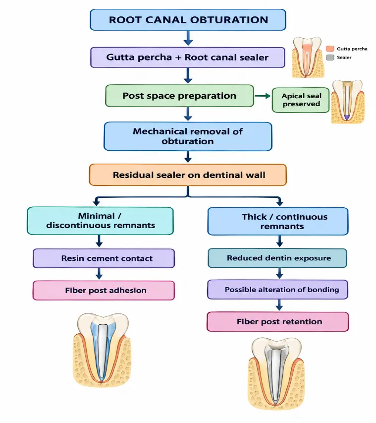
Figure 1. Proposed relationship between root canal sealer, post space preparation, residual sealer, and fiber post adhesion
The diagram illustrates that the influence of a root canal sealer on fiber post adhesion is mediated primarily through the amount, distribution, and characteristics of residual material after post space preparation rather than through sealer composition alone.
8. CRITICAL ANALYSIS OF EXISTING LITERATURE
The literature demonstrates considerable variability in conclusions regarding the relationship between root canal sealers and fiber post adhesion. Some investigations demonstrate higher bond strength following use of AH Plus, whereas other studies report no clinically meaningful difference among sealers after appropriate post space cleaning [45]. These differences are not necessarily contradictory because the experimental systems are frequently different.
Obturation technique represents another variable. Cold lateral compaction may distribute sealer differently from single cone obturation. Calcium silicate based sealers are frequently used with single cone techniques, and the quantity of sealer present in the canal may therefore be higher than with techniques that incorporate multiple gutta percha cones.
The timing of post preparation and cementation is another important factor. Immediate preparation may expose unset or incompletely matured sealer, whereas delayed preparation occurs after further setting and maturation. Studies have reported that cementation time can influence fiber post bond strength, although results differ according to the sealer and cement system [46].The type of resin cement also has a substantial effect. Conventional resin cements, self adhesive resin cements, and dual cure adhesive systems interact differently with root dentin. Consequently, an endodontic sealer that produces acceptable bonding with one resin cement may demonstrate different behavior with another. Storage conditions and aging are additional methodological variables. Immediate push out testing measures initial interfacial performance but does not necessarily represent long term behavior. Thermocycling, water storage, mechanical fatigue, and aging can modify the cement dentin interface [47].
9. RESEARCH GAPS AND FUTURE PERSPECTIVES
Several important research gaps remain. First, relatively few studies directly compare AH Plus, MTA Fillapex, BioRoot RCS, and Sealapex under the same experimental conditions. Direct comparison using identical tooth types, canal preparation, obturation techniques, post space dimensions, cleaning procedures, resin cement, storage conditions, and mechanical testing would provide more reliable evidence. Second, most available evidence concerning sealer remnants and fiber post adhesion remains laboratory based[48]. Extracted teeth cannot reproduce all clinical variables, including saliva contamination, occlusal loading, periodontal support, thermal changes, fatigue, and patient related factors. Long term clinical studies are therefore required. Third, newer calcium silicate based sealers require further investigation. Their increasing use has created a need to understand not only their biological and sealing properties but also their compatibility with post endodontic adhesive procedures. BioRoot RCS deserves particular attention because its physicochemical and bioactive behavior differs from conventional epoxy resin materials [49].Fourth, future studies should quantify residual sealer rather than simply reporting its presence or absence. Confocal microscopy, microcomputed tomography, fluorescence labeling, SEM, and advanced surface analytical techniques may provide quantitative information about remnant volume and distribution.Fifth, standardized push out protocols are required. Differences in slice thickness, root region, plunger diameter, crosshead speed, post geometry, and calculation methods can produce considerable variability [50]. A standardized methodology would improve comparison among studies.
Sixth, future research should evaluate the relationship between residual sealer and fatigue behavior rather than relying exclusively on immediate push out testing. A post interface may demonstrate acceptable initial bond strength but deteriorate after water storage, thermal cycling, and mechanical loading [51].
Finally, research should evaluate whether complete removal of sealer is actually necessary for clinical success. If a thin, discontinuous remnant does not significantly affect bonding, aggressive removal could cause unnecessary dentin loss. The clinically relevant target may therefore be controlled reduction of sealer contamination rather than absolute elimination [52].
10. CLINICAL IMPLICATIONS
The selection of a root canal sealer should take into consideration the possibility of subsequent post space preparation. AH Plus has extensive evidence supporting its physicochemical performance and dentin adhesion, and several fiber post studies have demonstrated favorable bonding after its use [53]. However, strong adhesion may also contribute to persistent remnants after post space preparation.
MTA Fillapex and Sealapex demonstrate different physicochemical and adhesive characteristics from AH Plus. Laboratory evidence has frequently shown lower dentin adhesion for these materials than for AH Plus, but this should not be interpreted as evidence that they are clinically unsuitable when a fiber post is planned [54].
BioRoot RCS and other calcium silicate based sealers have potential advantages related to bioactivity and calcium ion release. Nevertheless, clinicians should recognize that remnants may persist following post space preparation. Current evidence indicates that such remnants do not necessarily produce a measurable reduction in fiber post adhesion under every cementation protocol [55].
The clinician should prioritize preservation of root dentin, maintenance of an adequate apical seal, appropriate post length and diameter, effective post space cleaning, and proper adhesive cementation. Excessive enlargement of the canal to eliminate every visible remnant may be more damaging than leaving a clinically insignificant thin remnant [56].
Fiber post cementation should be performed using a protocol compatible with the selected adhesive and resin cement. Moisture control is important because excessive moisture can interfere with some adhesive systems, whereas excessive drying can collapse collagen structures in systems that depend on a moist dentin substrate [57].
CONCLUSION
Root canal sealers can influence post-space cleanliness and fiber-post adhesion through interactions involving sealer composition, dentinal penetration, post-space preparation, residual material, cleaning procedures, adhesive systems, resin cement, root anatomy, and testing conditions. AH Plus exhibits favorable physicochemical properties and dentin interaction, with several studies reporting satisfactory fiber-post bond strength. MTA Fillapex and Sealapex possess distinct physicochemical and adhesive characteristics, whereas BioRoot RCS is a calcium silicate-based sealer with bioactive properties. Sealer remnants frequently remain after post-space preparation, particularly in areas inaccessible to mechanical instruments. However, their presence does not necessarily compromise fiber-post adhesion. Studies involving calcium silicate-based sealers have demonstrated that residual material may remain without significantly reducing push-out bond strength under specific experimental conditions [16]. Therefore, the influence of residual sealer depends on its thickness, continuity, chemical composition, location, and interaction with the adhesive cement.
Current evidence shows considerable methodological variability related to tooth selection, canal preparation, obturation, post-space preparation, cleaning protocols, adhesive systems, resin cements, post design, aging procedures, and bond-strength testing. Future studies should incorporate standardized protocols, quantitative evaluation of sealer remnants, long-term aging, fatigue testing, and clinical outcomes. Clinically, treatment should prioritize preservation of radicular dentin, maintenance of the apical seal, reduction of substantial sealer contamination, and formation of a stable dentin–cement–fiber post interface for predictable restoration.
REFERENCES

Purvi Mehta*, Kiran J. Kasti, Ananthakrishna S., Pradeep PR., Aishwarya Bhattad, A Comprehensive Review On Influence Of Root Canal Sealers On Sealer Remnants After Post Space Preparation And Fiber Post Adhesion, Int. J. Sci. R. Tech., 2026, 3 (10), 232-242. https://doi.org/10.5281/zenodo.23126225
 10.5281/zenodo.23126225