

We use cookies to ensure our website works properly and to personalise your experience. Cookies policy
Department of Orthodontics and Dentofacial Orthopedics, Hitech Dental College and Hospital, Bhubaneswar, Odisha, India.
Artificial intelligence (AI) is reshaping endodontic practice through advances in diagnostic imaging, treatment planning, prognostic assessment, and clinical decision support. Machine learning and deep learning algorithms, particularly convolutional neural networks, have demonstrated promising capabilities in analyzing periapical radiographs and cone-beam computed tomography (CBCT) datasets for detecting periapical lesions, identifying vertical root fractures, and characterizing root canal morphology. Emerging applications include AI-assisted guided endodontics, dynamic navigation, outcome prediction, and digital education. This state-of-the-art review synthesizes evidence from PubMed/MEDLINE, Scopus, Web of Science, and Google Scholar to critically examine the diagnostic, therapeutic, prognostic, educational, and ethical dimensions of AI in endodontics. Emerging technologies, including explainable AI, radiomics, and integrated digital workflows, are discussed alongside challenges related to dataset heterogeneity, algorithmic transparency, external validation, data privacy, and medico-legal accountability. Current evidence indicates that AI may enhance diagnostic consistency, procedural precision, and individualized treatment planning. However, methodological heterogeneity, limited external validation, and a predominance of retrospective and laboratory-based studies constrain its clinical generalizability. Future translation into routine practice will require robust multicenter validation, standardized reporting, interpretable algorithms, and responsible clinician-supervised implementation. AI should therefore be regarded as an adjunct to, rather than a replacement for, clinical expertise in evidence-based, patient-centered endodontic care.
Artificial intelligence (AI) has emerged as one of the most transformative technologies in contemporary healthcare, significantly influencing diagnostic interpretation, clinical decision-making, and precision-based treatment approaches. Advances in machine learning, deep learning, and neural network architectures have enabled automated analysis of complex medical datasets with diagnostic performances approaching human expertise.1–3 Consequently, AI-assisted systems are increasingly being integrated into healthcare to improve diagnostic accuracy, workflow efficiency, predictive analytics, and evidence-based patient care.4
Dentistry has experienced substantial digital transformation with the introduction of digital radiography, cone-beam computed tomography (CBCT), intraoral scanning, and computer-assisted treatment planning technologies.5 Among dental specialties, endodontics is particularly suitable for AI integration because diagnosis and treatment planning rely heavily on radiographic interpretation and precise anatomical assessment.6 Conventional interpretation of two-dimensional and three-dimensional imaging findings may be influenced by observer variability, clinician experience, and image quality limitations.7
Accurate identification of periapical lesions, vertical root fractures, accessory canals, and complex root canal morphologies remains a major challenge in endodontic practice.8 Recent advances in AI-driven image analysis and convolutional neural networks (CNNs) have demonstrated promising accuracy in detecting pathological changes and anatomical variations from radiographic and CBCT datasets.9,10 Current applications of AI in endodontics include periapical lesion detection, root fracture identification, working length determination, canal segmentation, root canal morphology assessment, and prognostic prediction.11–13
The emergence of guided endodontics and dynamic navigation systems has further expanded the role of AI-assisted technologies in minimally invasive and precision-based endodontic therapy.14,15 In addition, AI-assisted educational platforms, simulation technologies, and conversational AI systems have shown potential in improving clinical training, treatment planning abilities, and rapid access to scientific information in endodontics.16,17
Despite these advancements, several limitations continue to restrict the routine clinical implementation of AI in endodontics. Many currently available studies are based on relatively small datasets with limited multicenter validation, while variations in imaging protocols and algorithm training methods may influence reproducibility and reliability.18 Ethical concerns related to explainability, data privacy, algorithm bias, and medico-legal accountability remain additional barriers to widespread adoption.19 Although AI systems have demonstrated considerable diagnostic and predictive capabilities, they should currently be regarded as supportive adjunctive technologies rather than replacements for clinical expertise. The expanding applications of artificial intelligence in endodontics are summarized in Figure 1.
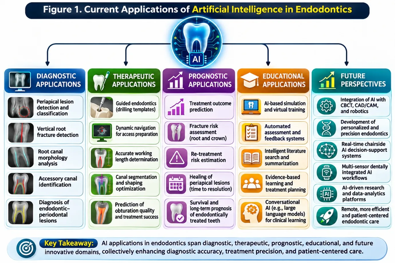
Figure 1: Current Applications of Artificial Intelligence in Endodontics.
The purpose of this state-of-the-art comprehensive review is to critically evaluate the current evidence regarding the diagnostic, therapeutic, prognostic, educational, and ethical applications of artificial intelligence in endodontics. In addition, emerging technologies including explainable AI, radiomics, AI-assisted navigation systems, and digitally integrated workflows are discussed to provide insight into future directions of precision-based endodontic care.
2. Literature Search Strategy
A comprehensive literature search was conducted to identify studies evaluating the applications of artificial intelligence in endodontics. Electronic databases including PubMed/MEDLINE, Scopus, Web of Science, and Google Scholar were searched for articles published up to March 2026. The search strategy used combinations of
keywords and Medical Subject Headings (MeSH) terms such as “artificial intelligence,” “machine learning,” “deep learning,” “convolutional neural network,” “endodontics,” “CBCT,” “periapical lesions,” “guided endodontics,” “root canal morphology,” and “prognostic prediction.”
Original research articles, systematic reviews, narrative reviews, and clinically relevant studies related to AI-assisted endodontic diagnosis, treatment planning, prognostic assessment, guided procedures, and education were included. Non-English articles, studies unrelated to endodontics, and reports lacking adequate methodological information were excluded.
Priority was given to recent high-quality studies, landmark publications, and clinically relevant investigations contributing to the current understanding of AI-assisted endodontic practice. Manual screening of reference lists was also performed to identify additional relevant studies not retrieved through electronic database searching.
3. Fundamentals of Artificial Intelligence in Endodontics
3.1 Machine Learning and Deep Learning
Artificial intelligence in endodontics is primarily based on machine learning (ML) and deep learning (DL) technologies. Machine learning refers to computational systems that learn from existing datasets and improve their performance without explicit programming.1 These systems identify patterns within clinical and radiographic data and apply them to diagnostic or predictive tasks.3
Deep learning is an advanced subset of machine learning that utilizes multilayered artificial neural networks capable of analyzing highly complex imaging datasets.1 In healthcare, deep learning models have shown remarkable accuracy in image recognition and diagnostic interpretation.2 In endodontics, these systems are increasingly used for detecting periapical lesions, identifying root canal morphology, and assisting in treatment planning.6
The growing availability of digital radiographs and CBCT imaging has accelerated the development of AI-based diagnostic systems in dentistry.5 Deep learning algorithms can analyze radiographic images rapidly and consistently, thereby reducing observer variability and improving diagnostic precision.7 Unlike conventional software systems, deep learning models continuously improve as additional datasets become available.8
The performance of AI models largely depends on the quality of training datasets and validation protocols. During algorithm development, annotated radiographic images are used to train neural networks to recognize specific anatomical or pathological features.10 Following training, these systems are validated using independent datasets to evaluate their reliability and reproducibility.11 Although current evidence demonstrates encouraging results, further multicenter validation studies remain necessary before routine clinical implementation can be widely adopted.6
3.2 Convolutional Neural Networks in Dental Imaging
Convolutional neural networks (CNNs) are among the most widely used deep learning architectures in medical and dental imaging. These models are specifically designed to process image-based datasets by automatically identifying important visual patterns and anatomical structures.1 Owing to their ability to analyze pixel-level image details with minimal human intervention, CNNs have demonstrated high diagnostic accuracy in radiology.2
In endodontics, CNN-based systems are increasingly used for interpretation of periapical radiographs and cone-beam computed tomography (CBCT) scans.6 These models can assist in detecting periapical lesions, vertical root fractures, root resorption, and complex canal anatomy with improved consistency.12 AI-assisted radiographic analysis may reduce observer variability and improve early detection of pathological conditions that are difficult to identify during routine clinical examination.13
Several studies have reported promising outcomes using CNNs for automated dental image analysis. Deep learning algorithms have shown effectiveness in detecting apical lesions and differentiating pathological from normal anatomical structures on radiographs.14 CNN-based models have also demonstrated encouraging diagnostic performance in identifying vertical root fractures and dental caries.15,16
The increasing application of CNNs reflects the growing shift toward AI-assisted diagnosis in endodontics. Although these systems are not intended to replace clinical expertise, they may serve as supportive tools that enhance diagnostic precision and clinical efficiency.6,8
3.3 CBCT and AI Integration
Cone-beam computed tomography (CBCT) has significantly improved endodontic diagnosis by providing three-dimensional visualization of dental and maxillofacial structures. Compared with conventional two-dimensional radiography, CBCT offers superior assessment of root canal morphology, periapical lesions, root fractures, and anatomical relationships.20,21 Integration of artificial intelligence with CBCT imaging has further enhanced the accuracy and efficiency of radiographic interpretation.
AI-assisted CBCT analysis mainly relies on deep learning algorithms capable of processing large volumetric imaging datasets. These systems can automatically detect pathological changes, segment root canal systems, and identify complex anatomical variations with improved consistency.12 Recent studies have demonstrated promising results in automated detection of apical periodontitis and assessment of root canal morphology using AI-based CBCT analysis.13
Segmentation represents one of the most important applications of AI in CBCT imaging. Deep learning models can separate teeth and root canal systems from surrounding anatomical structures, thereby improving diagnostic accuracy and treatment planning.18 Such technologies may also support guided endodontic procedures through enhanced virtual planning and anatomical mapping.19
Despite these advancements, important challenges remain regarding image standardization, dataset quality, and external validation of AI systems.6 Variations in CBCT resolution, imaging protocols, and annotation methods may influence algorithm reproducibility and performance. Nevertheless, AI-assisted CBCT analysis continues to represent a major advancement in digitally driven endodontic diagnosis and treatment planning.8 The major clinical applications of artificial intelligence in endodontics are summarized in Table 1.
Table 1: Applications of Artificial Intelligence in Endodontics
| Domain | AI Application | Clinical Purpose | Potential Benefits |
|---|---|---|---|
| Diagnostic Imaging | Periapical lesion detection | Identification of apical pathology | Improved diagnostic accuracy |
| Diagnostic Imaging | Vertical root fracture detection | Detection of root fractures | Reduced missed diagnosis |
| Anatomical Assessment | Root canal morphology analysis | Identification of canal variations | Better treatment planning |
| Working Length Determination | Automated apex identification | Determination of canal length | Reduced operator variability |
| Therapeutic Applications | Guided endodontics | Accurate access cavity preparation | Minimally invasive treatment |
| Therapeutic Applications | Dynamic navigation | Real-time procedural guidance | Improved procedural precision |
| Prognostic Assessment | Outcome prediction models | Prediction of healing and success | Personalized treatment planning |
| Education | AI-assisted simulation and assessment | Student training and evaluation | Enhanced learning efficiency |
4. Diagnostic Applications
4.1 Periapical Lesion Detection
Accurate detection of periapical lesions is a fundamental objective in endodontic diagnosis. Conventional radiographic interpretation may fail to identify early or small lesions because of anatomical superimposition, image quality limitations, and observer variability.7 Artificial intelligence has demonstrated considerable potential in improving detection and interpretation of periapical pathologies through automated image analysis systems.6
Deep learning algorithms, particularly convolutional neural networks (CNNs), have shown promising accuracy in detecting apical lesions on both periapical radiographs and cone-beam computed tomography (CBCT) images.12 AI-assisted systems can analyze radiographic datasets at the pixel level and identify subtle pathological changes that may not be easily visible during manual interpretation.13 This capability may improve early diagnosis and facilitate more accurate treatment planning.
Several studies have evaluated the effectiveness of AI-based lesion detection systems in endodontics. Endres et al reported successful detection of periapical disease on dental radiographs using deep learning algorithms, while Ekert et al demonstrated high diagnostic accuracy of CNN-based systems for detecting apical lesions.13,14 These findings suggest that AI-assisted diagnostic systems may serve as valuable adjuncts to conventional radiographic interpretation.
Integration of AI with CBCT imaging has further improved assessment of periapical pathology by enabling three-dimensional evaluation of lesions.12 AI-assisted CBCT analysis may reduce observer variability and improve diagnostic consistency, particularly in anatomically complex cases.6 However, most currently available AI models still require larger multicenter validation studies before routine clinical implementation can be widely recommended.11
Despite encouraging diagnostic performance, several limitations continue to affect the clinical applicability of AI-assisted lesion detection systems. Most available studies are retrospective and based on relatively small single-center datasets, which may increase the risk of overfitting and reduce external validity. Considerable heterogeneity also exists regarding imaging modalities, annotation protocols, CNN architectures, and validation methods, making direct comparison between studies difficult. Variations in radiographic quality, lesion size, and anatomical overlap may further influence algorithm performance under clinical conditions. Although CBCT-based AI systems generally demonstrate greater diagnostic sensitivity than two-dimensional radiographic models, higher radiation exposure and increased financial cost remain important limitations. Consequently, larger prospective multicenter studies and standardized validation frameworks are necessary before AI-assisted lesion detection systems can be reliably integrated into routine endodontic diagnosis.
4.2 Vertical Root Fracture Detection
Vertical root fractures (VRFs) are among the most challenging conditions to diagnose in endodontics because clinical signs and radiographic findings are often nonspecific. Conventional periapical radiographs may fail to identify fractures, especially when fracture lines are narrow or obscured by surrounding anatomical structures.7 Delayed diagnosis can lead to persistent symptoms, periodontal destruction, and eventual tooth loss.
Artificial intelligence has recently emerged as a promising adjunctive tool for VRF detection. Deep learning systems can analyze radiographic datasets and identify fracture-related patterns with greater consistency than subjective manual interpretation.6 Convolutional neural networks (CNNs) have demonstrated encouraging performance in differentiating fractured from non-fractured teeth through automated image analysis.15 Fukuda et al reported favorable diagnostic accuracy using an AI-based system for detecting vertical root fractures on panoramic radiographs.15 These findings suggest that AI-assisted imaging analysis may improve early diagnosis and reduce missed fractures during routine clinical examination, particularly in cases with subtle or inconclusive radiographic findings.
Cone-beam computed tomography (CBCT) has further enhanced the diagnostic potential of AI for VRF detection because of its ability to provide three-dimensional visualization of root structures.12 AI-assisted CBCT analysis may improve fracture identification by reducing observer variability and enhancing interpretation of anatomically complex regions.8 However, artifacts produced by metallic restorations and endodontic filling materials remain significant challenges during automated fracture detection.
Although current evidence demonstrates promising diagnostic capability, several limitations continue to restrict clinical applicability of AI-assisted VRF detection systems. Many studies rely on experimentally created fractures or idealized imaging datasets that may not accurately represent routine clinical conditions. In addition, differences in fracture orientation, tooth anatomy, imaging resolution, and validation methods may contribute to inconsistent results among studies. The lack of standardized datasets and external multicenter validation protocols further limits reproducibility and generalizability of existing AI models. Future research should therefore focus on developing robust clinically representative datasets and standardized validation frameworks to improve the reliability and real-world performance of AI-assisted fracture detection systems in endodontics.
4.3 Root Canal Morphology Assessment
A thorough understanding of root canal morphology is essential for successful endodontic treatment because variations in canal configuration, accessory canals, and complex anatomical patterns can significantly influence treatment outcomes. Conventional radiographic assessment may fail to identify these variations because of image superimposition and limited two-dimensional visualization.7 Artificial intelligence has demonstrated considerable potential in improving assessment of root canal morphology through automated image analysis and segmentation techniques.
Deep learning algorithms are increasingly being applied for evaluation and segmentation of root canal systems using radiographic and cone-beam computed tomography (CBCT) datasets.18 These systems can identify canal pathways, anatomical curvatures, and morphological variations with improved precision and consistency. AI-assisted analysis may help clinicians recognize complex anatomical structures that could otherwise remain undetected during conventional examination.6
CBCT imaging has further enhanced AI-assisted root canal assessment by providing three-dimensional visualization of dental anatomy.12 Deep learning segmentation models can separate teeth and root canal systems from surrounding structures, thereby improving anatomical mapping and treatment planning.19 Such technologies may facilitate detection of accessory canals, calcified canals, and anatomical irregularities in complex clinical cases.
Recent studies have highlighted the growing role of AI in evaluating root canal morphology and anatomical complexity.20 AI-assisted systems may improve treatment precision, reduce missed canals, and minimize procedural complications during endodontic therapy. However, further multicenter studies and standardized validation protocols remain necessary before routine clinical integration can be recommended.6
Despite its promising potential, AI-assisted root canal morphology analysis continues to face important technical and methodological limitations. Accurate segmentation depends heavily on CBCT image quality, voxel resolution, and consistency of dataset annotation. Anatomical complexity, calcifications, image noise, and artifacts may adversely affect segmentation accuracy. In addition, considerable variability exists among studies regarding imaging protocols, segmentation algorithms, and performance evaluation methods, limiting direct comparison between available AI systems. Most current investigations are retrospective and laboratory-based, with limited prospective clinical validation. Therefore, future multicenter studies using standardized imaging protocols and externally validated datasets are essential to improve the reproducibility, reliability, and clinical applicability of AI-assisted anatomical assessment systems in endodontics.
4.4 Working Length Determination
Accurate working length determination is essential for successful endodontic treatment because it directly influences cleaning, shaping, and obturation procedures. Inaccurate assessment may result in inadequate debridement or overinstrumentation, potentially compromising treatment outcomes. Conventional methods, including radiographic evaluation and electronic apex locators, possess limitations related to anatomical variability and interpretation accuracy.7
Artificial intelligence has recently been explored as a supportive tool for automated working length estimation. Machine learning and deep learning algorithms can analyze radiographic images and identify anatomical landmarks such as the radiographic apex and apical constriction with improved consistency.6 AI-assisted systems may reduce observer variability and improve precision during radiographic interpretation.
One of the earliest applications of AI in endodontics was reported by Saghiri et al, who developed an artificial neural network model for locating the minor apical foramen.17 Their findings demonstrated the potential of AI-assisted systems to improve identification of anatomical landmarks during endodontic procedures. Subsequent advances in digital imaging and computational technologies have further expanded the role of AI in working length assessment.
Integration of AI with digital radiography and cone-beam computed tomography (CBCT) imaging may improve procedural efficiency by providing rapid and reproducible measurements.12 AI-assisted systems may be particularly beneficial in anatomically complex cases where conventional radiographic interpretation is difficult. However, current evidence regarding AI-based working length determination remains limited, and additional clinical validation studies are required before routine implementation can be recommended.6 Major studies evaluating AI-assisted diagnostic systems in endodontics are summarized in Table 2.
Table 2: Major Studies on AI Applications in Endodontics
| Author | Year | AI Application | Imaging Modality | Main Findings | Reported Performance |
|---|---|---|---|---|---|
| Ekert et al | 2019 | Apical lesion detection | Periapical radiographs | CNN models demonstrated high diagnostic capability for apical lesion detection | High diagnostic accuracy reported |
| Özyürek et al | 2020 | Periapical lesion detection | CBCT | AI improved consistency of lesion detection and radiographic interpretation | Improved diagnostic consistency |
| Fukuda et al | 2020 | Vertical root fracture detection | Panoramic radiographs | AI system demonstrated favorable fracture detection performance | Promising sensitivity and specificity |
| Endres et al | 2020 | Periapical disease detection | Dental radiographs | Deep learning successfully detected periapical disease | High predictive performance |
| Saghiri et al | 2012 | Working length determination | Radiographic analysis | ANN model accurately identified apical foramen location | Improved landmark localization |
| Zehnder et al | 2016 | Guided endodontics | CBCT-guided systems | Accurate guided access cavity preparation and canal localization | High procedural precision |
| Connert et al | 2017 | Microguided endodontics | Guided access preparation | Improved accuracy during minimally invasive access preparation | Favorable procedural accuracy |
| Lu et al | 2025 | Prognostic prediction | Clinical and radiographic data | Machine learning predicted regenerative treatment outcomes | Promising predictive capability |
The rapid evolution of AI-assisted diagnostic technologies has significantly influenced the therapeutic landscape of contemporary endodontics. Advances in radiographic interpretation, anatomical mapping, and CBCT-based image analysis have facilitated development of more precise and minimally invasive treatment approaches. As diagnostic accuracy continues to improve through machine learning and deep learning systems, increasing attention has shifted toward AI-assisted therapeutic guidance technologies capable of enhancing procedural precision and clinical predictability during endodontic treatment.
5. Therapeutic Applications
5.1 Guided Endodontics
Guided endodontics represents one of the most important therapeutic advancements associated with integration of artificial intelligence and digital technologies into endodontic practice. The technique combines cone-beam computed tomography (CBCT) imaging, digital surface scanning, virtual planning software, and guided templates to improve procedural accuracy.21 Guided endodontics is particularly useful in cases involving pulp canal obliteration, calcified canals, and complex anatomical variations where conventional access preparation is challenging.
AI-assisted planning systems improve visualization of root canal anatomy and facilitate precise access cavity preparation while preserving healthy tooth structure.6 Integration of digital imaging datasets with virtual planning software allows clinicians to create highly accurate treatment pathways before initiating treatment.22 These technologies may reduce procedural errors, excessive dentin removal, and the risk of perforation during canal location.
Zehnder et al demonstrated that guided endodontic systems could achieve highly accurate access cavity preparation and root canal localization in difficult clinical situations.22 Similarly, Connert et al reported favorable accuracy using microguided endodontic techniques for minimally invasive access preparation in anterior teeth.23 These findings highlight the growing role of digitally assisted procedures in improving treatment precision and predictability.
Guided endodontics may also reduce chairside operating time and operator-related variability. AI-assisted planning can improve procedural confidence, particularly for inexperienced clinicians and anatomically challenging cases.8 In addition, guided techniques may enhance patient comfort through minimally invasive treatment approaches with greater precision.
Despite these advantages, guided endodontics has certain limitations. The requirement for CBCT imaging, digital scanning systems, and specialized software may increase treatment cost and technical complexity.6 Furthermore, inaccuracies in image acquisition or guide fabrication can affect procedural precision. Nevertheless, continued advances in AI-assisted planning and digital workflow integration are expected to further improve the effectiveness and accessibility of guided endodontic procedures.
5.2 Dynamic Navigation
Dynamic navigation systems provide real-time instrument tracking and guidance, distinguishing them from static guided endodontics, which relies on prefabricated templates.21 By integrating cone-beam computed tomography (CBCT), motion tracking, and navigation software, these systems enable precise visualization of instrument position and angulation during access cavity preparation and canal localization. AI may further enhance anatomical mapping, image interpretation, and treatment planning.6,22
Dynamic navigation is particularly beneficial in cases involving calcified canals, complex root anatomy, and restricted access. It may improve procedural accuracy, preserve tooth structure, and reduce complications such as perforation and canal deviation.23 Integration of AI-assisted planning with navigation may further support minimally invasive and predictable treatment.8 However, high equipment costs, technical complexity, calibration errors, and the need for specialized training limit widespread adoption. Moreover, evidence remains largely based on laboratory studies, case reports, and small clinical investigations. Large-scale prospective trials are therefore required to establish clinical effectiveness, reproducibility, and cost-effectiveness.
6. Prognostic Applications
Artificial intelligence has increasingly been investigated for prognostic assessment in endodontics because of its ability to analyze large clinical and radiographic datasets and identify predictive patterns associated with treatment outcomes. Conventional prognostic evaluation often relies on clinician experience and subjective interpretation of clinical findings, whereas AI-assisted systems may provide more objective and data-driven prediction models for evaluating treatment success, healing potential, and disease progression.6
Machine learning algorithms can simultaneously process multiple variables, including patient age, lesion size, root canal anatomy, treatment quality, and radiographic characteristics.24 By collectively analyzing these factors, AI systems may help clinicians estimate the probability of treatment success and identify cases at higher risk of failure. Such predictive capabilities may improve treatment planning and support more personalized endodontic care.
Recent studies have demonstrated promising applications of machine learning in regenerative endodontic procedures and outcome prediction. Lu et al reported that machine learning models showed considerable potential in predicting prognosis following regenerative endodontic therapy.25 AI-assisted prognostic systems may also contribute to assessment of healing outcomes after root canal treatment and apical surgery.
Another important application involves automated monitoring of lesion progression and post-treatment healing using serial radiographic datasets.6 Deep learning systems may assist clinicians in detecting subtle radiographic changes over time with improved consistency and reduced observer variability, thereby enhancing long-term follow-up and evidence-based clinical decision-making.
Despite these promising developments, prognostic AI models in endodontics remain in the early stages of clinical application. Many available studies are based on limited datasets and still require external validation before routine clinical implementation can be recommended.11 Nevertheless, integration of AI into prognostic assessment represents an important step toward more predictive and personalized endodontic treatment strategies.
7. AI in Education
Artificial intelligence (AI) is increasingly transforming dental and endodontic education through personalized learning, simulation-based training, and automated assessment. AI-assisted platforms provide interactive learning environments and real-time feedback, potentially enhancing clinical understanding, procedural skills, and decision-making.5,16 AI-integrated simulations may facilitate training in access cavity preparation, canal identification, and treatment planning, while improving the consistency of competency assessment.
Conversational AI and large language models have further expanded educational applications by supporting evidence-based learning, scientific information retrieval, and academic assessment.27,28 However, responsible implementation requires transparency, accountability, and ethical oversight.26 Concerns regarding inaccurate AI-generated information, overreliance, academic integrity, and potential impairment of critical thinking necessitate cautious integration into educational curricula.
Accordingly, AI should complement rather than replace faculty supervision, evidence-based teaching, and clinical mentorship. Its effective integration into endodontic education will depend on responsible use, critical appraisal of AI-generated outputs, and preservation of essential clinical reasoning and patient-centered communication skills.
8. Clinical Implications
The integration of artificial intelligence into endodontic practice has important clinical implications for diagnosis, treatment planning, procedural precision, and long-term patient management. AI-assisted radiographic interpretation may improve early detection of periapical lesions, root fractures, and anatomical variations that might otherwise remain undetected during conventional examination. Enhanced diagnostic consistency may contribute to more accurate treatment planning and reduction of observer-dependent variability.
The incorporation of AI-driven guided endodontics and dynamic navigation systems may further support minimally invasive treatment approaches by improving procedural accuracy and preservation of healthy tooth structure. In addition, predictive AI models may assist clinicians in identifying cases with higher risks of treatment failure and facilitate more personalized evidence-based treatment strategies.
Despite these potential benefits, AI-assisted systems should currently be regarded as supportive adjunctive technologies rather than replacements for clinical expertise. Successful clinical integration will require appropriate clinician training, ethical oversight, standardized validation protocols, and continued interdisciplinary collaboration. As AI technologies continue to evolve, their future incorporation into routine endodontic workflows may significantly contribute to precision-based and patient-centered clinical care.
9. Ethical Challenges and Limitations
Despite its promising applications, artificial intelligence in endodontics faces substantial ethical, technical, and clinical challenges. The limited interpretability of deep learning algorithms may compromise clinician trust and complicate accountability for diagnostic errors and treatment-related decisions. â´, ¹â¹ AI performance also depends heavily on the quality, diversity, and representativeness of training datasets. Single-center studies, heterogeneous imaging protocols, inconsistent annotations, and limited external validation may restrict reproducibility and clinical generalizability, while algorithmic bias may contribute to diagnostic disparities across patient populations. â¶, ¹¹, ¹â¸, ²â
Patient privacy, data security, and medico-legal accountability remain critical concerns, particularly when clinical and radiographic data are used for model development and validation.â´,¹â¹ Furthermore, imaging artifacts, computational requirements, implementation costs, and limited technical expertise may hinder clinical adoption.¹² High reported diagnostic accuracy under controlled conditions, coupled with publication bias and inconsistent validation standards, may also overestimate real-world performance.¹â¸
Accordingly, AI should be regarded as a supportive clinical tool rather than a replacement for professional judgment. Future research should prioritize explainable algorithms, representative multicenter datasets, standardized reporting, robust external validation, and secure data governance. â´, ²â¶ Interdisciplinary collaboration and ethically guided implementation are essential to ensure reliable, equitable, and clinically meaningful integration of AI into endodontic practice. The principal advantages and limitations are summarized in Table 3. The broader implications of AI integration in dentistry also highlight the importance of understanding biological responses at the pulp–dentin interface and critically evaluating whether emerging AI technologies represent meaningful clinical advances or merely technological innovation.29,30
Table 3: Advantages and Limitations of AI in Endodontics
| Advantages | Limitations |
|---|---|
| Improved diagnostic accuracy | Dependence on dataset quality |
| Reduced observer variability | Limited multicenter validation |
| Enhanced radiographic interpretation | High implementation cost |
| Better treatment planning | Requirement for technical expertise |
| Support for minimally invasive procedures | Black-box nature of deep learning systems |
| Faster image analysis | Ethical and medico-legal concerns |
| Personalized prognostic prediction | Data privacy and security issues |
| Improved educational assessment | Limited standardization across studies |
10. Future Perspectives
Artificial intelligence (AI) is poised to advance endodontics through the integration of deep learning, cone-beam computed tomography (CBCT) analysis, and data-driven clinical decision support. Future AI platforms may combine clinical findings, radiographic interpretation, and prognostic models to improve diagnostic precision, treatment planning, and individualized care.6,3,24 Integration of AI-assisted image segmentation with guided endodontics and dynamic navigation may further enhance canal localization, procedural accuracy, and preservation of healthy tooth structure in anatomically challenging cases.18
Emerging technologies, including explainable AI (XAI), radiomics, federated learning, and real-time chairside systems, offer promising opportunities for precision-based endodontic care. XAI may improve algorithm transparency and clinician trust, while radiomics could facilitate quantitative characterization of periapical pathology and prognostic assessment. Federated learning may enable collaborative model development across institutions while minimizing direct patient-data sharing. Integration with robotic-assisted procedures and digital workflows may further expand minimally invasive and personalized treatment strategies.26
AI-assisted simulation, virtual learning platforms, and conversational systems may transform endodontic education by supporting competency-based training, clinical decision-making, and evidence-based learning.27,28 However, clinical translation remains constrained by limited prospective multicenter validation, dataset diversity, inconsistent imaging and annotation protocols, and uncertainties regarding cost-effectiveness and algorithmic bias. Future research should prioritize standardized validation frameworks, explainability, ethical governance, and real-world clinical evaluation. Ultimately, multidisciplinary collaboration and robust clinical evidence will determine the safe and effective integration of AI into patient-centered endodontic practice.
11. Limitations of the Review
Several limitations associated with the present review should be acknowledged. First, although extensive literature screening was performed, the rapidly evolving nature of artificial intelligence research may result in continuous emergence of new evidence after manuscript preparation. Second, many currently available studies evaluating AI applications in endodontics demonstrate considerable heterogeneity regarding imaging modalities, dataset characteristics, algorithm architectures, validation methods, and outcome measures, thereby limiting direct comparison between investigations. Third, a substantial proportion of the available evidence is derived from retrospective or laboratory-based studies with limited multicenter clinical validation. In addition, publication bias toward studies reporting favorable AI performance may influence the overall interpretation of current evidence. Finally, because this review represents a state-of-the-art narrative synthesis rather than a systematic review or meta-analysis, quantitative assessment of pooled diagnostic accuracy and statistical heterogeneity was not performed.
CONCLUSION
Artificial intelligence is emerging as a valuable adjunct in endodontics, with promising applications in diagnostic imaging, anatomical assessment, treatment planning, prognostic prediction, and clinical education. Integration of machine learning, deep learning, and AI-assisted digital workflows may enhance diagnostic consistency, procedural precision, and personalized patient care. However, heterogeneity in study designs, limited external validation, and challenges related to algorithmic transparency, data quality, and clinical generalizability continue to constrain widespread implementation. Future progress will require robust multicenter validation, standardized reporting, explainable AI, and ethically guided clinical integration. Ultimately, AI should complement, rather than replace, professional expertise, supporting evidence-based, minimally invasive, and patient-centered endodontic care.
13. Declarations
13.1 Ethical approval
Not required for narrative review.
13.2 Conflict of Interest
The authors declare no conflict of interest.
13.3 Acknowledgements
None.
13.4 Data Availability
Not applicable.
13.5 Funding
No funding received.
REFERENCES


Dr. Anshuman Mishra, Dr. Chinmayee Priyadarsini, Artificial Intelligence in Endodontics: A State-of-the-Art Comprehensive Review, Int. J. Sci. R. Tech., 2026, 3 (11), 658-670. https://doi.org/10.5281/zenodo.24216740
 10.5281/zenodo.24216740