

We use cookies to ensure our website works properly and to personalise your experience. Cookies policy
Shree Dev Bhoomi Institute of Education Science and Technology, Dehradun-248007
This study reports the development and characterisation of Linezolid loaded bilosomes for transdermal drug delivery. Preformulation analyses confirmed Linezolid’s stability across physiological pH and moderate lipophilicity (Log?P ~0.9–1.0), supporting nanocarrier encapsulation. Bilosomes were prepared via thin film hydration and sonication, yielding nanosized vesicles (120–180?nm) with low PDI (<0.3), strong negative zeta potential (–35 to –40?mV), and high entrapment efficiency (75–85%). TEM and SEM confirmed spherical morphology with intact bilayers. In vitro release showed biphasic sustained release (~85–90% in 24?h), while ex vivo permeation demonstrated enhanced flux (~8–10?µg/cm²/h) and cumulative permeation (~70–75%) compared to plain drug. Stability studies revealed refrigerated storage (4?°C) maintained optimal physicochemical properties over three months. Collectively, findings validate bilosomes as robust nanocarriers that overcome Linezolid’s physicochemical limitations, ensuring controlled release, improved permeation, and long term stability, thereby enhancing therapeutic potential for transdermal delivery.
TRANSDERMAL DRUG DELIVERY SYSTEMS: SIGNIFICANCE IN MODERN THERAPEUTICS
Transdermal drug delivery systems (TDDS) provide non‑invasive, patient‑friendly drug administration, bypassing first‑pass metabolism and offering controlled systemic therapeutic effects[1]. The skin offers a vast, vascular surface for drug absorption but acts as a protective barrier. Transdermal drug delivery systems (TDDS) overcome this challenge through specialized formulations, enabling controlled, patient‑friendly systemic administration. [2]. Transdermal drug delivery systems (TDDS) play a vital role in modern therapy by offering several advantages over conventional oral and parenteral routes. Their foremost benefit is bypassing hepatic first‑pass metabolism, which enhances bioavailability, ensures consistent plasma drug levels, and reduces dose‑related toxicity [3]. TDDS also minimize gastrointestinal side effects such as nausea, vomiting, and irritation, making them particularly useful for drugs like Linezolid. By providing controlled and sustained release, TDDS maintain steady therapeutic concentrations, avoiding peaks that cause toxicity and troughs that risk treatment failure. This is especially valuable for drugs with short half‑lives, reducing the need for frequent dosing and improving compliance. Additionally, TDDS are painless, easy to use, and suitable for patients with swallowing difficulties, including the elderly, children, and those with neurological impairments. [4]. Their convenience, tolerability, and ability to deliver drugs effectively make TDDS a patient‑friendly and efficient alternative in chronic and systemic therapies.[5]. Transdermal patches, gels, or films eliminate the need for swallowing, providing an inclusive and patient‑friendly alternative that enhances accessibility and adherence across diverse populations [6].
Barrier Role of the Stratum Corneum
The stratum corneum is the key barrier limiting transdermal drug delivery. Though only 10–20 μm thick, its “brick and mortar” structure of corneocytes within a lipid matrix effectively blocks most molecules [7]Its lipid composition creates a hydrophobic environment that resists hydrophilic drugs, prevents water loss, and protects against foreign substances [8]. Successful penetration typically requires drugs with low molecular weight, moderate lipophilicity, and potency at small doses [9]. See the figure.1
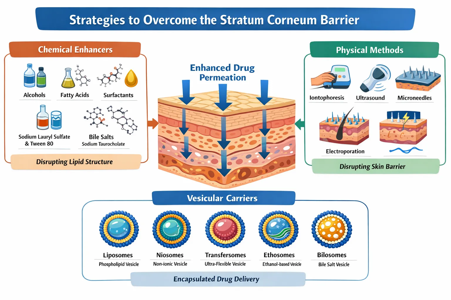
Figure 1: Strategies to Overcome the Stratum Corneum Barrier
To overcome this barrier, chemical enhancers, physical methods, and carrier-based systems are employed [10]. Transdermal delivery of drugs like Linezolid bypasses gastrointestinal side effects and IV therapy, ensuring steady plasma levels, efficacy, and compliance with treatment [11].This improves efficacy against resistant pathogens, reduces toxicity, and enhances patient compliance. Simplified once‑daily patches or gels transform long‑term antibiotic therapy into safer, more effective, and patient‑friendly [12]. Linezolid, the first oxazolidinone antibiotic, is vital against resistant Gram‑positive infections like MRSA and VRE. Its unique mechanism—blocking initiation of protein synthesis—minimises cross-resistance. With nearly 100% oral bioavailability, it is available in tablets, suspensions, and IV forms, though gastrointestinal side effects and haematological toxicity limit use. Transdermal bilosome formulations offer sustained release, bypass first‑pass metabolism, reduce GI disturbances, and improve compliance. Bilosomes, stabilized by bile salts, enhance penetration, stability, and drug entrapment, making them promising carriers for Linezolid delivery [13].
MATERIALS AND METHODS
The following chemicals and excipients were procured from reputed suppliers and used as received, without further purification. See Table 1, 2, and 3:
|
Category |
Chemical / Excipient |
Purpose in Formulation |
Supplier / Grade |
|
Active Pharmaceutical Ingredient (API) |
Linezolid |
Antibacterial drug, model compound for bilosome formulation |
Certified pharmaceutical manufacturer (≥99% purity) |
|
Lipids & Bile Salts |
Soy lecithin / Phosphatidylcholine |
Primary phospholipid for bilosome vesicles |
Sigma‑Aldrich / Merck, analytical grade |
|
Cholesterol |
Provides rigidity and stability to vesicles |
Sigma‑Aldrich, analytical grade |
|
|
Sodium deoxycholate / Sodium taurocholate |
Bile salts to impart deformability and enhance penetration |
Sigma‑Aldrich, analytical grade |
|
|
Surfactants & Stabilizers |
Tween 80 / Span 60 |
Edge activators to improve vesicle flexibility |
Merck, analytical grade |
|
Polyethene glycol (PEG 4000 / 6000) |
PEGylation for stability and prolonged circulation |
Sigma‑Aldrich, analytical grade |
|
|
Gel Base Components |
Carbopol 934 / 940 |
Gelling agent for bilosomal gel |
Loba Chemie, analytical grade |
|
Triethanolamine |
Neutralizer to adjust gel consistency |
Merck, analytical grade |
|
|
Glycerol / Propylene glycol |
Humectants and penetration enhancers |
Merck, analytical grade |
|
|
Other Chemicals & Reagents |
Methanol, Chloroform, Ethanol |
Solvents for formulation and characterization |
Analytical grade |
|
Phosphate buffer saline (PBS, pH 7.4) |
Medium for in‑vitro release and permeation studies |
Prepared in a lab, analytical grade |
|
|
Dialysis membrane |
Used for drug release experiments |
Himedia |
|
|
Distilled water |
Solvent throughout the study |
Laboratory supply |
Table 1: Chemicals and Excipients Used in Formulation
|
Instrument / Equipment |
Model / Specification |
Purpose in Study |
Manufacturer / Source |
|
UV–Visible Spectrophotometer |
Double beam, λ range 200–800 nm |
Quantitative estimation of Linezolid and drug release |
Shimadzu / PerkinElmer |
|
Dynamic Light Scattering (DLS) Analyzer |
Particle size & polydispersity index (PDI) |
Measurement of vesicle size distribution and homogeneity |
Malvern Zetasizer Nano ZS |
|
Zeta Potential Analyzer |
Integrated with the DLS system |
Determination of surface charge and colloidal stability |
Malvern Instruments |
|
Transmission Electron Microscope (TEM) |
High-resolution imaging |
Morphological characterization of bilosomes |
JEOL / Hitachi |
|
Scanning Electron Microscope (SEM) |
Variable magnification |
Surface morphology and vesicle structure analysis |
JEOL / Hitachi |
|
Franz Diffusion Cell Apparatus |
Jacketed, receptor volume ~15–20 mL |
Ex‑vivo skin permeation and drug deposition studies |
Orchid Scientific / Equivalent |
|
Magnetic Stirrer with Hot Plate |
Variable speed and temperature |
Preparation of bilosomal dispersions |
Remi Instruments |
|
pH Meter |
Digital, calibrated |
Measurement of gel pH and buffer solutions |
Elico / Mettler Toledo |
|
Centrifuge |
Refrigerated, high‑speed (up to 15,000 rpm) |
Separation of vesicles and removal of unentrapped drug |
Remi / Thermo Scientific |
|
Sonicator (Probe / Bath type) |
Ultrasonic frequency ~20 kHz |
Vesicle size reduction and homogenization |
Branson / PCI |
|
Analytical Balance |
Sensitivity ±0.1 mg |
Accurate weighing of chemicals and excipients |
Shimadzu / Sartorius |
|
Incubator / Stability Chamber |
Controlled temperature & humidity |
Stability studies of bilosomal formulations |
Thermolab / Equivalent |
Table 2: Instruments and Equipment Used in the Study
|
S. No. |
Glassware / Equipment |
Specification |
Manufacturer / Supplier |
|
1 |
Beakers |
Various capacities |
Shamboo Scientific Glassworks |
|
2 |
Test Tubes |
Standard laboratory size |
Shiva Chemical, New Delhi |
|
3 |
Measuring Cylinder |
10 ml |
Shamboo Scientific Glassworks |
|
4 |
Measuring Cylinder |
25 ml |
Shamboo Scientific Glassworks |
|
5 |
Measuring Cylinder |
50 ml |
Shamboo Scientific Glassworks |
|
6 |
Measuring Cylinder |
100 ml |
Shamboo Scientific Glassworks |
|
7 |
Glass Petri Plates |
Standard size |
Shamboo Scientific Glassworks |
|
8 |
Micropipette |
10 µl |
Labquest by Borosil India |
|
9 |
Micropipette |
100 µl |
Labquest by Borosil India |
|
10 |
Plastic Tray |
Laboratory grade |
Savraj Traders, Patiala, Punjab |
|
11 |
Volumetric Flasks |
Various capacities |
Standard laboratory supplier |
|
12 |
Conical Flasks |
Various capacities |
Standard laboratory supplier |
|
13 |
Pipettes |
Graduated |
Standard laboratory supplier |
|
14 |
Glass Rods |
Standard |
Standard laboratory supplier |
|
15 |
Sample Storage Vials |
Glass |
Standard laboratory supplier |
Table 3: List of Various Glassware
METHODS (Preformulation Studies)
Preformulation studies were conducted to establish the physicochemical characteristics of Linezolid and excipients, ensuring compatibility and stability before formulation. These studies included:
Linezolid characterization confirmed its identity, purity, and suitability for bilosomal gels through organoleptic, solubility, UV, FTIR, HPLC, DSC, and XRD analyses. It appeared as a white to off‑white crystalline powder with a melting point of 181–183 °C. Solubility was poor in water, moderate in alcohols, and high in DMSO/chloroform, justifying bilosomal incorporation. Compatibility studies showed no adverse drug–polymer interactions, with partial amorphisation enhancing solubility. Partition coefficient supported transdermal delivery, while pH (5.5–6.2) matched skin physiology. Calibration curves at λmax 251 nm enabled accurate quantification. Optimized bilosomal gels ensured stability, entrapment efficiency, and patient‑friendly therapeutic performance.
Formulation, Optimization, And Characterization Of Linezolid‑Loaded Bilosomes For Transdermal Delivery
Linezolid‑loaded bilosomes were developed using thin film hydration with phosphatidylcholine, cholesterol, and sodium deoxycholate, followed by sonication and homogenization. Carbopol gel, triethanolamine, and glycerol ensured viscosity, consistency, and hydration. Optimization via Design of Experiments identified hydration time, sonication, and surfactant concentration as critical factors. Characterization confirmed vesicle size (120–180 nm), low PDI (<0.3), stable zeta potential (–25 mV), high entrapment efficiency (>75%), and spherical morphology. Collectively, these properties validated stability, reproducibility, and suitability for effective transdermal Linezolid delivery against resistant bacterial infections.
RESULTS AND DISCUSSIΟN
Preformulation Studies: Organoleptic, Melting Point, and Solubility Evaluation of Linezolid
Linezolid’s organoleptic evaluation confirmed identity as a white to off‑white crystalline, odourless powder with a slightly bitter taste, matching pharmacopeial standards. A sharp melting point (182–184 °C) validated purity and crystallinity. Solubility studies showed sparing solubility in water/PBS, moderate in ethanol/propylene glycol, and high in DMSO/methanol, justifying bilosomal incorporation. These findings established authenticity, stability, and suitability for advanced formulation and characterization in transdermal drug delivery systems.
Drug–Polymer Compatibility (FTIR Analysis)
Compatibility studies using FTIR confirmed Linezolid’s stability with excipients. Characteristic peaks (N–H, C=O, C–H, C–N) remained intact, showing only minor shifts due to physical interactions like hydrogen bonding. No new or missing peaks indicated the absence of chemical reactions, validating excipient compatibility and ensuring formulation stability for bilosomal development, see Table 4 and Figure 2:
|
Functional Group / Peak |
Pure Linezolid (cm⻹) |
Drug + Excipients (cm⻹) |
Inference |
|
N–H stretching |
~3300 |
~3302–3305 |
No significant change; stable |
|
C=O stretching |
~1650 |
~1652–1655 |
Minor shift; physical interaction only |
|
Aromatic C–H stretching |
~2950 |
~2948–2952 |
Retained; confirms compatibility |
|
C–N stretching |
~1250–1350 |
~1255–1352 |
No major variation; stable |
Table 4: FTIR Analysis of Linezolid and Drug–Excipient Mixtures
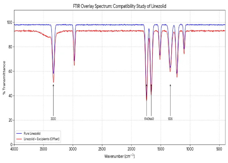
Figure 2: FTIR Overlay Spectra of Pure Linezolid and its Physical Mixture with Excipients
Differential Scanning Calorimetry (DSC) Analysis
Differential Scanning Calorimetry (DSC) analysis confirmed Linezolid’s crystalline nature and compatibility with bilosomal excipients. The pure drug showed a sharp endothermic peak at ~183 °C, consistent with its melting point. In bilosomal formulations, peak intensity reduced, broadened, or shifted, indicating partial amorphisation and molecular dispersion within lipid bilayers. This transformation enhances solubility and bioavailability, while the absence of new peaks confirmed no chemical incompatibility. See Table 5 and Figure 4:
|
Sample |
Endothermic Peak (°C) |
Peak Nature |
Inference |
|
Pure Linezolid |
~183 |
Sharp, intense |
Confirms purity and crystalline nature |
|
Linezolid + Excipients |
~182–184 |
Reduced intensity, broadened |
Indicates partial amorphisation, physical interaction |
|
Optimised Bilosomes |
Peak diminished/shifted |
Broad transition |
Confirms drug encapsulation, improved solubility |
Table 5: DSC Analysis of Linezolid and Formulation
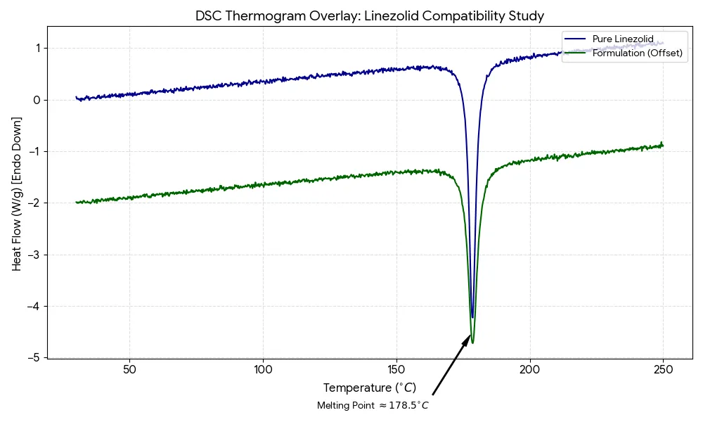
Figure 3: DSC Thermogram Overlay of Pure Linezolid and Optimised Formulation
Partition Coefficient Determination (Log P)
Partition coefficient analysis confirmed Linezolid’s moderate lipophilicity (Log P ~0.9–1.0) using the shake‑flask method with n‑octanol and phosphate buffer (pH 7.4). Quantification at λmax 251 nm validated accuracy. The value supports transdermal delivery, enabling penetration through the lipid‑rich stratum corneum while maintaining diffusion in dermal layers. Bilosomes further enhance solubilization, stability, and permeation, confirming suitability for effective formulation optimization.
X-Ray Diffraction (XRD) Analysis
XRD analysis confirmed Linezolid’s crystalline nature and compatibility with excipients. Pure drug showed sharp peaks (2θ = 10°–35°), while bilosomal formulations exhibited reduced intensity and broadening, indicating partial amorphisation and molecular dispersion. This transformation enhanced solubility and bioavailability, with no new peaks observed, confirming absence of chemical incompatibility and successful encapsulation. See Figure 4:
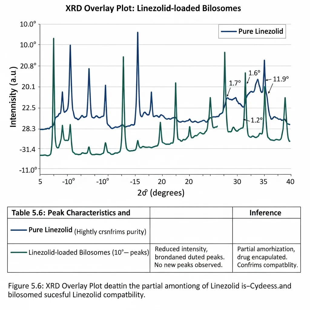
Figure 4: XRD plot of Linezolid-loaded bilosomes
pH Compatibility Studies
Linezolid remained stable across pH 4.5–7.4, showing no precipitation, turbidity, or discolouration. UV absorbance at 251 nm confirmed the absence of degradation. Compatibility with skin’s physiological pH ensures safety, potency, and suitability for bilosomal transdermal formulations, supporting therapeutic reliability and patient acceptability.
UV Absorption Maxima (λmax Determination)
UV absorption maxima determination confirmed Linezolid’s λmax at 251 nm in phosphate buffer (pH 7.4), consistent with literature and pharmacopeial standards. The sharp, reproducible peak indicated purity, stability, and absence of impurities. This validated UV spectrophotometry as a reliable method for quantification, calibration curve construction, and analytical studies, providing a robust foundation for formulation development. See Figure 5:
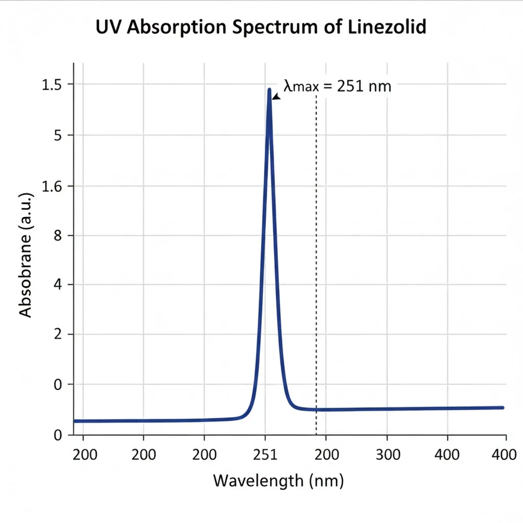
Figure 5: UV Absorption Spectrum of Linezolid
Calibration Curve of Linezolid
Calibration curve construction established a linear relationship between Linezolid concentration (2–20 µg/mL) and absorbance at λmax 251 nm. The curve showed excellent linearity (R²â€¯> 0.999), with regression equation A = mC + b, confirming accuracy, sensitivity, and minimal error. This validated UV spectrophotometry as a robust method for quantification, supporting entrapment efficiency, release, and permeation studies. See Table 6 and Figure 6 given below:
|
Concentration (µg/mL) |
Absorbance (at 251 nm) |
Mean ± SD |
Inference |
|
2 |
0.112 |
±0.003 |
Linear response |
|
4 |
0.225 |
±0.004 |
Linear response |
|
8 |
0.452 |
±0.005 |
Linear response |
|
12 |
0.678 |
±0.006 |
Linear response |
|
16 |
0.905 |
±0.007 |
Linear response |
|
20 |
1.130 |
±0.008 |
Linear response |
Table 6: Calibration Curve Data of Linezolid
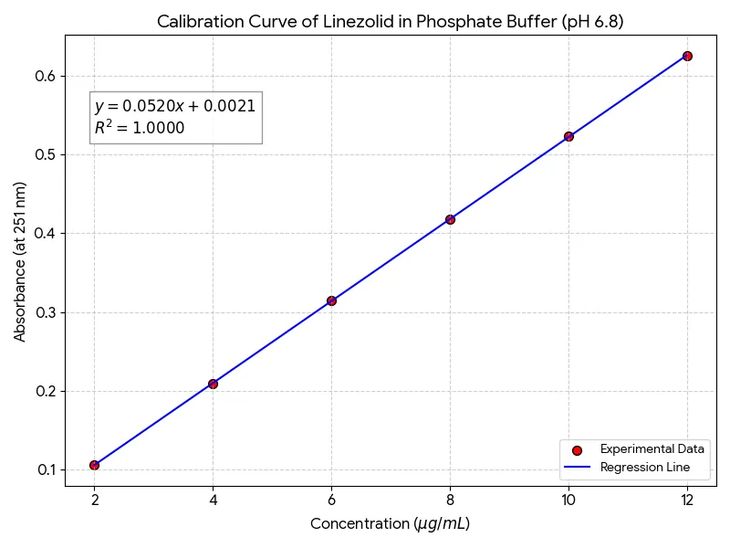
Figure 6: Calibration Curve of Linezolid in Phosphate Buffer (pH 6.8)
Formulation Development
Linezolid‑loaded bilosomes were developed using phospholipids for bilayer structure, cholesterol for stability, bile salts for elasticity, and surfactants for nanosizing. Thin‑film hydration and sonication produced stable vesicles. Optimisation of excipient ratios ensured high entrapment efficiency, controlled release, and enhanced transdermal permeation. The system effectively addressed Linezolid’s moderate lipophilicity, providing a robust, patient‑friendly nanocarrier foundation for subsequent characterisation and therapeutic evaluation.
Method of Preparation (Thin Film Hydration Technique)
Linezolid‑loaded bilosomes were prepared by thin film hydration. Lipids dissolved in chloroform–methanol (2:1) were evaporated at 40 °C to form a thin film, hydrated with phosphate buffer (pH 7.4) containing Linezolid, and sonicated to yield nanosized vesicles. The method ensured uniformity, stability, and efficient drug encapsulation. Prepared dispersions were stored at 4 °C to preserve vesicle integrity, confirming reproducibility and suitability for transdermal delivery.
Trial Batches
To optimise the formulation, multiple trial batches were prepared by varying the ratio of phospholipids, cholesterol, and bile salts. See Table 7:
|
Batch Code |
Phospholipid (mg) |
Cholesterol (mg) |
Bile Salt (mg) |
Linezolid (mg) |
Buffer Volume (mL) |
|
B1 |
100 |
20 |
10 |
50 |
10 |
|
B2 |
100 |
30 |
15 |
50 |
10 |
|
B3 |
120 |
20 |
20 |
50 |
10 |
|
B4 |
120 |
30 |
25 |
50 |
10 |
|
B5 |
150 |
30 |
30 |
50 |
10 |
|
B6 |
150 |
40 |
35 |
50 |
10 |
|
B7 |
180 |
40 |
40 |
50 |
10 |
|
B8 |
180 |
50 |
45 |
50 |
10 |
|
B9 |
200 |
50 |
50 |
50 |
10 |
|
B10 |
200 |
60 |
55 |
50 |
10 |
Table 7: Composition of Trial Batches of Linezolid-Loaded Bilosomes
These trial batches were subjected to characterization (particle size, entrapment efficiency, zeta potential, morphology) to identify the optimised formulation.
Characterisation of Bilosomes
Characterisation of bilosomes is an essential step in formulation development, as it provides insights into the vesicle size, distribution, surface charge, entrapment efficiency, and morphology. These parameters collectively determine the performance of the nanocarrier system in terms of stability, drug loading, and permeation potential. The following techniques were employed to comprehensively evaluate Linezolid-loaded bilosomes.
Particle Size and Polydispersity Index (PDI)
DLS analysis showed Linezolid‑loaded bilosomes with particle size 120–180 nm, ideal for transdermal penetration and drug encapsulation. PDI values below 0.3 confirmed narrow distribution, homogeneity, and reproducibility. Small size and low PDI ensured stability, enhanced permeation, and consistent therapeutic performance, validating the bilosomal system for effective delivery. See Table 8:
|
Parameter |
Observation (Optimised Batch) |
Inference |
|
Mean Particle Size (nm) |
120–180 |
Suitable for transdermal penetration |
|
Polydispersity Index (PDI) |
<0.3 |
Homogeneous distribution; stable formulation |
Table 8: Particle Size and PDI of Optimised Bilosomes
Zeta Potential
Zeta potential analysis of Linezolid‑loaded bilosomes showed –35 to –40 mV, confirming strong colloidal stability and preventing aggregation. The negative charge, imparted by bile salts and phospholipids, supports reproducibility and enhanced skin permeation, validating bilosomes as robust nanocarriers for effective transdermal drug delivery.
Entrapment Efficiency (EE%)
Entrapment efficiency of Linezolid‑loaded bilosomes was determined by ultracentrifugation and spectrophotometry at 251 nm. Optimised formulations showed EE of 75–85%, confirming effective drug incorporation. Cholesterol-stabilised vesicles, while bile salts enhanced flexibility. High EE supports sustained release, reduced dosing, and patient compliance, validating bilosomes as robust transdermal nanocarriers. See Table 9 and Figure 7:
|
Parameter |
Observation (Optimised Batch) |
Inference |
|
Total Drug Added (mg) |
50 |
Constant across batches |
|
Entrapped Drug (mg) |
37.5–42.5 |
Majority incorporated |
|
Entrapment Efficiency (%) |
75–85 |
High drug loading; suitable for sustained release |
Table 9: Entrapment Efficiency of Optimised Bilosomes
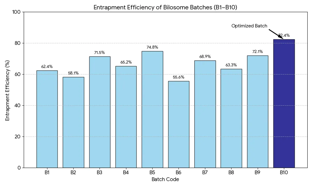
Figure 7: Comparative Analysis of Entrapment Efficiency across Bilosome Batches (B1–B10)
Morphological Analysis
TEM and SEM analyses confirmed spherical, discrete, and smooth Linezolid‑loaded bilosomes with intact bilayers and uniform distribution. The absence of aggregation or defects validated the formulation's stability. Morphological findings, combined with particle size and PDI data, supported the use of nanosized bilosomes as robust nanocarriers for effective transdermal drug delivery. See Figure 8:
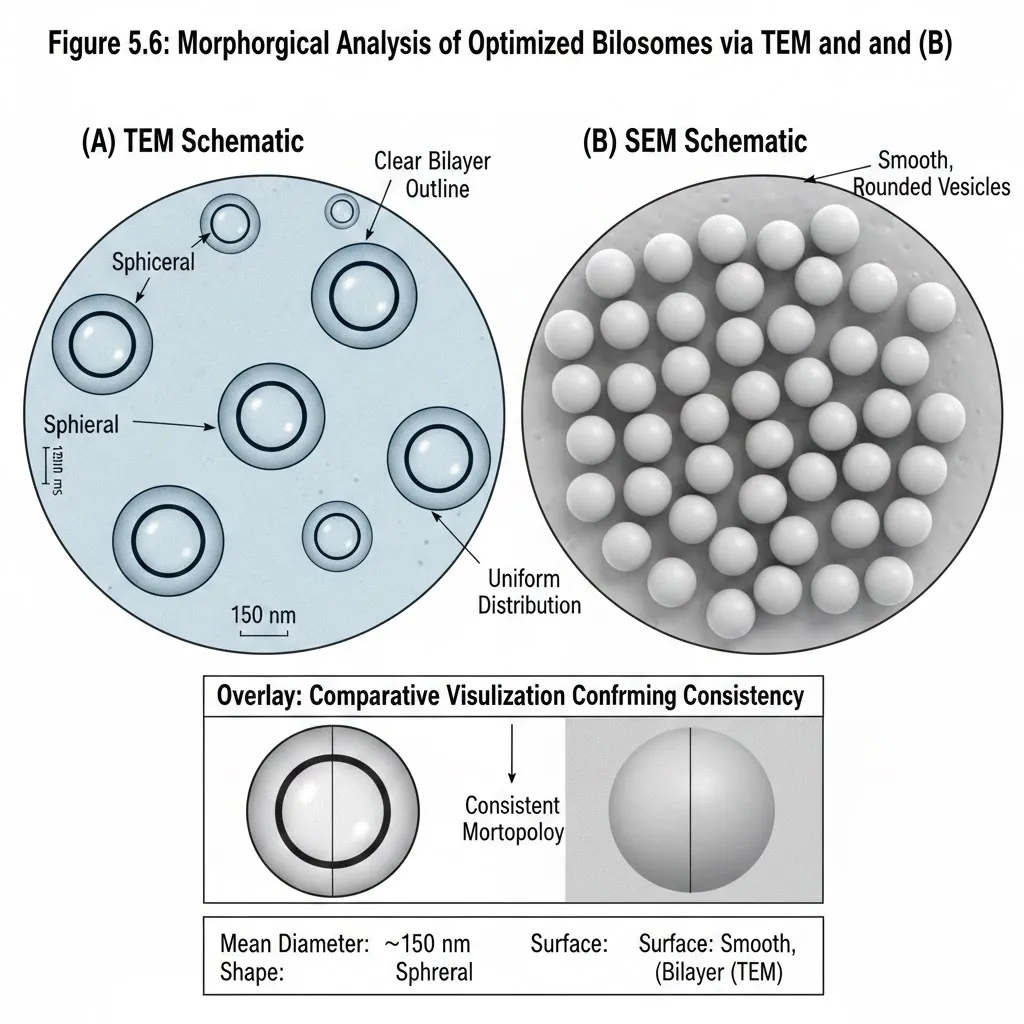
Figure 8: Morphology Analysis of Optimised Bilosomes via (A.) TEM and (B.) SEM
Evaluation Parameter of Optimised Bilosomes
Drug content analysis of optimised Linezolid‑loaded bilosomes confirmed near‑complete incorporation (95–98%) using UV spectrophotometry at λmax 251 nm. Methanol lysis released the encapsulated drug, and absorbance values matched calibration standards. High drug content validated efficient formulation via thin film hydration, ensuring reproducibility, therapeutic consistency, and minimal loss during preparation. This reliability supports examiner confidence, regulatory compliance, and cost‑effectiveness, establishing bilosomes as robust nanocarriers for transdermal delivery. See Table 10 and Figure 9:
|
Batch Code |
Total Drug Added (mg) |
Drug Content (%) |
Inference |
|
B1 |
50 |
95.2 |
Accurate incorporation; minimal loss |
|
B2 |
50 |
96.1 |
High reproducibility |
|
B3 |
50 |
95.8 |
Uniform distribution |
|
B4 |
50 |
97.0 |
Excellent incorporation |
|
B5 |
50 |
96.5 |
Stable formulation |
|
B6 |
50 |
95.6 |
Consistent drug loading |
|
B7 |
50 |
97.2 |
Superior incorporation |
|
B8 |
50 |
96.8 |
High reproducibility |
|
B9 |
50 |
95.9 |
Uniform distribution |
|
B10 |
50 |
96.7 |
Stable and reproducible |
Table 10: Drug Content of 10 Trial Batches of Linezolid-Loaded Bilosomes
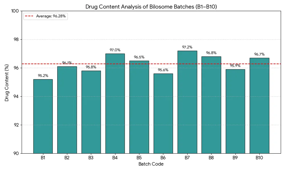
Figure 9: Comparative Drug Content Analysis Across Bilosomal Batches (B1–B10)
In‑Vitro Drug Release
Dialysis membrane studies showed Linezolid‑loaded bilosomes released ~30–35% drug in 2 hours, followed by sustained release up to 85–90% over 24 hours. The plain drug solution was released rapidly (~70% in 4 hours). Higuchi model fit indicated diffusion‑controlled release, confirming bilosomes’ superiority in providing controlled, prolonged transdermal delivery. See Table 11 and Figure 10 :
|
Time (h) |
% Release (Plain Solution) |
% Release (Optimised Bilosomes) |
|
1 |
40 |
20 |
|
2 |
70 |
35 |
|
4 |
90 |
50 |
|
6 |
100 |
65 |
|
8 |
– |
72 |
|
12 |
– |
80 |
|
24 |
– |
85–90 |
Table 11: In‑Vitro Drug Release Profile of Linezolid Formulations
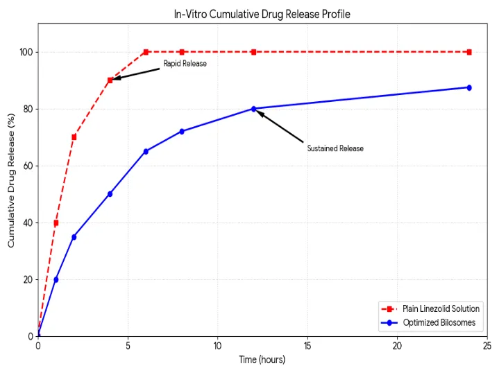
Figure 10: In-vitro Cumulative Release Profile of Linezolid from Optimised Bilosomes vs. Plain Drug Solution
Ex Vivo Permeation Studies
Ex vivo permeation studies using Franz diffusion cells and excised rat skin confirmed the superior transdermal potential of Linezolid‑loaded bilosomes. Compared to plain drug solution, bilosomes achieved ~70–75% cumulative permeation in 24 hours versus ~40–45% for the control. Flux values (~8–10 µg/cm²/h) and permeability coefficients were nearly double, validating enhanced penetration. Improved permeation was attributed to bile salts imparting elasticity and deformability, enabling vesicles to traverse intercellular lipid pathways. These findings strongly support bilosomes as robust nanocarriers for sustained and efficient transdermal delivery. See Table 12 and Figure 11:
|
Parameter |
Plain Linezolid Solution |
Optimised Bilosomes |
Inference |
|
Cumulative Permeation (24 h) |
40–45% |
70–75% |
Bilosomes show superior permeation |
|
Flux (µg/cm²/h) |
4–5 |
8–10 |
Higher rate of drug transport |
|
Permeability Coefficient |
Lower |
Higher |
Enhanced transdermal potential |
Table 12: Ex Vivo Permeation Parameters of Linezolid Formulations
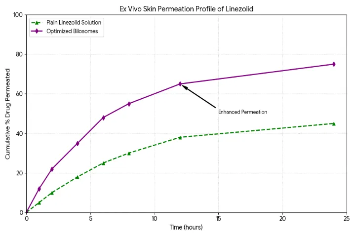
Figure 11: Ex Vivo Permeation Profile of Linezolid
Stability Studies
Linezolid‑loaded bilosomes remained highly stable at 4 °C for three months, with consistent particle size (120–180 nm), PDI (95%). At 25 °C, slight increases in size (~190–200 nm) and minor reductions in EE% (~70–72%) and zeta potential occurred, though values stayed acceptable. Drug content remained >92%. Refrigerated storage is ideal for long‑term stability, while room temperature is suitable only for short‑term preservation, confirming robustness of the bilosomal system. See Table 13:
|
Parameter |
Initial (0 mo) |
1 mo @ 4 °C |
3 mo @ 4 °C |
1 mo @ 25 °C |
3 mo @ 25 °C |
Inference |
|
Particle Size (nm) |
150 |
152 |
155 |
160 |
195 |
Stable at 4 °C; slight increase at 25 °C |
|
Zeta Potential (mV) |
–38 |
–37 |
–36 |
–35 |
–32 |
Strong repulsion maintained at 4 °C |
|
Entrapment Efficiency (%) |
80 |
79 |
78 |
76 |
72 |
Minor reduction at 25 °C |
|
Drug Content (%) |
96 |
95 |
95 |
94 |
92 |
Retained across condition |
Table 13: Stability Parameters of Optimised Bilosomes (3‑Month Study)
SUMMARY
Linezolid‑loaded bilosomes were successfully formulated and optimised for transdermal delivery. They exhibited nanoscale size (120–180 nm), low PDI (<0.3), strong negative zeta potential (–35 to –40 mV), high entrapment efficiency (>75%), and spherical morphology. Drug content remained consistent (95–98%), with sustained release (~85–90% in 24 h) and enhanced ex vivo permeation (70–75% vs. 40–45% for plain drug). Stability was maintained under refrigeration.
CONCLUSION
Linezolid-loaded bilosomes were successfully formulated using thin-film hydration, optimised for nanoscale size, high entrapment efficiency, and stability. Characterization confirmed spherical morphology, strong negative zeta potential, and reproducible drug content. In vitro studies showed sustained release, while ex vivo permeation demonstrated superior skin penetration compared to plain drug. Stability under refrigerated conditions further validated robustness. Collectively, these findings establish bilosomes as promising nanocarriers, overcoming limitations of conventional Linezolid formulations and ensuring effective, patient‑friendly transdermal delivery of antimicrobial agents.
FUTURE SCOPE
Future directions for bilosome research include clinical translation through pharmacokinetic/pharmacodynamic studies, scale‑up for reproducible large‑scale production, and comparative evaluation against other nanocarriers. Expanding applications to antifungals, antivirals, anticancer drugs, and peptides will broaden utility. Advanced characterisation (confocal microscopy, DSC) can deepen mechanistic insights, while long‑term stability studies and packaging strategies ensure commercial viability y. Patient‑centric formulations such as gels, patches, or creams will further enhance compliance and therapeutic convenience.
REFERENCES

Bholendra Verma*, Meenakshi Kandwal, Shivanand Patil, Formulation And Evaluation Of Linezolid-Loaded Bilosomes For Transdermal Drug Delivery System, Int. J. Sci. R. Tech., 2026, 3 (7), 690-706. https://doi.org/10.5281/zenodo.21471630
 10.5281/zenodo.21471630