

We use cookies to ensure our website works properly and to personalise your experience. Cookies policy
Department of Quality Assurance, Delight College of Pharmacy, Koregaon Bhima, Pune, Maharashtra, India¬-412216.
Neosomes are vesicular nanocarriers that have gained attention as potential systems for transdermal drug delivery. They may help improve drug penetration, retain drugs within the skin for prolonged release, and control the rate at which drugs enter systemic via the skin. Non-steroidal anti-inflammatory drugs (NSAIDs) are widely used to manage pain and inflammation; however, their oral administration may cause gastrointestinal side effects. Transdermal drug delivery offers an alternative way to improve drug delivery while reducing systemic adverse effects. Neosomes and liposomes are the vesicular carriers that enhance the drug penetration power through the skin and improves therapeutic efficacy. This review discusses the formulation, characteristics, and applications of neosomes for the transdermal delivery of NSAIDs, with a comparative overview of liposomes. It also describes their advantages, limitations, and future potential in developing effective and patient-friendly drug delivery systems.
Overview of Transdermal Drug Delivery –
Delivery of a drug via the skin is a highly effective approach to drug administration. The transdermal drug delivery system (TDDS) is an innovative approach in the pharmaceutical field for the administration of drugs because it has several advantages over other routes of drug administration. Conventional formulations of drug delivery systems require larger doses and larger regimens to achieve therapeutic effects, and long-term regimens may produce major side effects and ultimately low patient compliance. The oral route is generally considered the most suitable, convenient, and ideal route of drug administration because of the many advantages associated with it. However, there are several disadvantages associated with this route for drug delivery, including drug breakdown by enzymatic degradation in the gastrointestinal tract, gastrointestinal irritation, low absorption and low bioavailability, etc. Some drugs are also considered unsuitable via this route. In cases of drugs that are administered parenterally, trypanophobia is a common psychological problem associated with children and adults, as it also associated with risk of infection at site of administration. To overcome such challenges, TDDS is the ideal approach for such drugs [1].
Skin as a barrier to Drug Penetration
The skin acts as a protective barrier against external substances and prevents excessive water loss. It includes three main layers: the epidermis, dermis, and hypodermis. The stratum corneum, is the outermost layer of the epidermis, which act as the primary barrier to drug penetration. It consists of dead, keratin-rich cells embedded in a lipid matrix, which restricts the passage of many drugs, particularly those with unsuitable physicochemical properties [2].
Drug penetration through the skin depends on various factors such as molecular weight, lipid solubility, drug concentration, and skin hydration. These factors can limit the effectiveness of conventional transdermal formulations. These vesicular systems may enhance drug penetration by interacting with skin lipids and facilitating drug transport across the stratum corneum [2].
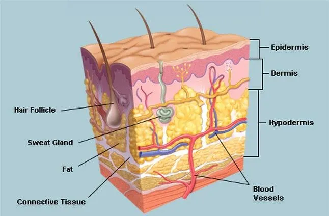
Figure 1: Skin as a Barrier to Drug Penetration
NSAIDs and their therapeutic importance –
Non-steroidal anti-inflammatory drugs (NSAIDs) are a class of medications widely used as analgesic, anti-inflammatory, and antipyretic. They primarily act by inhibiting cyclooxygenase (COX) enzymes, which reduces the synthesis of prostaglandins responsible for pain and inflammation. Common NSAIDs include Diclofenac, Ibuprofen, Ketoprofen, Piroxicam, and Indomethacin. Therefore, transdermal delivery of NSAIDs offers a promising alternative for localized pain and inflammation, potentially reducing gastrointestinal exposure. Neosomes and liposomes are being studied as vesicular carriers to improve skin penetration, increase drug retention, and provide the sustained drug release [3].
Need for Vesicular Drug Delivery System –
Vesicular drug delivery systems recived considerable attention in pharmaceutical research because of their potential to improve drug delivery, therapeutic efficacy, and patient compliance. Conventional transdermal formulations often face limitations due to the protective nature of the stratum corneum, which restricts the penetration of many drugs. Vesicular carriers, such as neosomes and liposomes, can encapsulate both hydrophilic and lipophilic drugs, protect drug molecules from degradation, and modify drug release [4].
In the transdermal delivery of non-steroidal anti-inflammatory drugs (NSAIDs), these systems are being investigated to enhance skin permeation, increase drug retention at the target site, and potentially reduce systemic exposure. Non-steroidal anti-inflammatory drugs (NSAIDs), such as diclofenac, ibuprofen, ketoprofen, and piroxicam, are widely used to manage pain and inflammation associated with musculoskeletal disorders. Although oral NSAIDs are effective, prolonged use may induce the risk of gastrointestinal adverse effects and other systemic complications. Among vesicular carriers, neosomes and liposomes are considered particularly important in novel drug delivery systems. Neosomes are generally composed of non-ionic surfactants and cholesterol, whereas liposomes primarily consist of phospholipid bilayers. Neosomes may offer advantages in terms of material cost and chemical stability in some formulations, while liposomes have well-established applications in drug delivery. Nevertheless, their performance depends on vesicle composition, particle size, drug entrapment efficiency, formulation stability, and skin permeability [4,2]
NSAIDs FOR TRANSDERMAL DELIVERY:
Non-steroidal anti-inflammatory drugs are drugs primarily used to relieve pain, reduce inflammation and manage conditions such as osteoarthritis, rheumatoid arthritis and other inflammatory disorders. NSAIDs act mainly by inhibiting COX enzymes, which are responsible for the synthesis of prostaglandins involved in pain, inflammation and fever.
Classification of NSAIDs –
1.Non-selective COX inhibitors
Salicylates: Aspirin
Propionic acid derivative: Ibuprofen, Ketoprofen
Enolic acid derivatives: Piroxicam, Tenoxicam
Acetic acid derivatives: Ketorolac
Pyrazolone derivatives: Phenylbutazone
2.Preferential COX-2 Inhibitors
Example—Diclofenac, Meloxicam
3.Selective COX 2 Inhibitors
Example—Celecoxib, Parecoxib
4.Analgesic antipyretic with poor anti- inflammatory action
Para amino phenol Derivative: Paracetamol
Pyrazolone Derivative: Propiphenazone
Benzoxazocine Derivative: Nefopam [10]
Mechanism of Action:
NSAIDs inhibit COX-1 and/or COX-2 enzymes, reducing the conversion of arachidonic acid into prostaglandins and related mediators. The reduction in prostaglandin synthesis decreases inflammation, pain sensitivity, and fever. The degree of COX-1 and COX-2 inhibition varies among the individual NSAIDs [5].
Challenges in convectional Transdermal delivery –
Conventional transdermal drug delivery systems (TDDS), such as transdermal patches, offer advantages including non-invasive administration, improved patient compliance, and avoidance of first-pass metabolism. However, their effectiveness is limited by several challenges:
1.Skin Barrier: The stratum corneum acts as the major barrier to drug penetration, restricting the permeation of many therapeutic agents.
2. Limited Drug Permeability: Conventional TDDS are mainly suitable for potent drugs with appropriate molecular size, lipophilicity, and physicochemical properties.
3. Limited Drug Loading: The amount of drug that can be delivered through the skin is often insufficient for drugs requiring high doses.
4. Slow Drug Onset: Drug absorption through the skin may be slow, making conventional patches unsuitable when rapid therapeutic action is required.
5. Skin Irritation and Sensitization: Adhesives, penetration enhancers, and prolonged patch contact may cause redness, itching, irritation, or allergic reactions.
6. Variability in Drug Absorption: Skin thickness, hydration, age, anatomical site, and individual skin conditions can influence drug permeation and lead to variable drug delivery.
7. Drug Stability Issues: Some drugs may undergo degradation during formulation, storage, or prolonged contact with the skin.
8. Adhesion Problems: Poor adhesion, sweating, movement, and environmental conditions may affect patch attachment and reduce delivery efficiency.
Addressing these limitations through advanced formulations and novel skin-permeation technologies may improve the efficacy, safety, and applicability of transdermal drug delivery systems [6].
Commonly Investigated NSAIDs:
Several non-steroidal anti-inflammatory drugs (NSAIDs) have been investigated for topical and transdermal delivery due to their analgesic and anti-inflammatory properties. Diclofenac, ibuprofen, ketoprofen, piroxicam, and indomethacin are important examples. Neosomes and liposomes have been explored as potential carriers to improve drug delivery across the skin, enhance local drug retention, and modify drug release [7].
| NSAIDs | Therapeutic use | Vascular Delivery Research |
|---|---|---|
| Diclofenac | Pain and Osteoarthritis | Skin Permeation and Retention |
| Ibuprofen | Pain and Inflammation | Drug release and Permeation |
| Ketoprofen | Musculoskeletal pain | Local Delivery and sustain release |
| Piroxicam | Arthritis and Inflammation | Solubilization and skin deposition |
| Indomethacin | Inflammatory disorders | Formulation optimization and skin delivery |
NEOSOMES:
Neosomes are microscopic, vesicular drug delivery systems composed mainly of non-ionic surfactants and cholesterol. They consist of one or more bilayers surrounding an aqueous core, enabling the encapsulation of both hydrophilic and lipophilic drugs. Hydrophilic drugs are generally entrapped within the aqueous compartment, whereas lipophilic drugs are incorporated into the bilayer membrane. The vesicular structure of niosomes can protect drugs from degradation, improve drug stability, and facilitate controlled drug release. In transdermal drug delivery, neosomes are investigated as carriers to enhance drug deposition and permeation across the skin barrier [8].
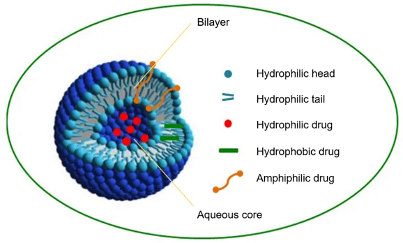
Figure 2: Structure of Neosomes
Composition–
Important two components use in neosomes preparation are cholesterol and non-ionic surfactants.
Cholesterol is a steroid derivative used to provide rigidity and proper shape, conformation.
Non-ionic Surfactants are generally used for the formulation.
Examples: Tweens (20, 40, 60, 80), Spans (Span 60, 40, 20, 80).
1.Non-ionic surfactants:
Non-ionic surfactants are an essential component of neosomes. To form neosomes, various types and their combinations are used to entrap various medications. Non-ionic surfactants are naturally amphiphilic, biodegradable, biocompatible, and non-immunogenic. The composition, concentration of additives, size, lamellarity, and surface charge of vesicles determine the properties of formulated neosomes. Non-ionic surfactants such as span (60, 40, 20, 85, and 80) and Tween (20, 40, 60, and 80) are used in the formation of neosomes [19].
2. Cholesterol:
It's acts as a crucial additive in the formulation of neosomes. It is not only required for the formation of neosomes, but it also influences many of their properties. It enhances the membrane's permeability, rigidity, entrapment efficiency, ease of rehydration of freeze-dried neosomes, stability, and storage period. Ifcholesterol is combined with low HLB surfactants, it increases vesicle stability, and if the HLB value is greater than 6, it aids in the creation of bilayer vesicles.
3. Charged molecule: Neosomes have some charged molecules that are added to increase stability by providing electric repulsion to prevent collisions. Diacetyl phosphate (DCP) and phosphotidic acid are examples of negatively charged compounds. Similarly, in neosomal preparations, stearyl amine and stearyl pyridinium chloride are well known charged compounds [9].
Types of Neosomes –
Neosomes are classified into different types based on their vesicle size, number of bilayers, and structural characteristics. Their size and structure influence drug encapsulation, release behaviour, stability, and potential applications in transdermal drug delivery. The major types of niosomes are described below [11]
1.Small Unilamellar Vesicles (SUVs):
Small uni-lamellar vesicles consist of a single surfactant bilayer surrounding an aqueous core. They generally have a small vesicle size and may be produced by methods such as sonication or extrusion. Their relatively high surface-area-to-volume ratio can influence drug release and interactions with the skin. They may be studied for topical delivery of NSAIDs when small vesicle size is desirable.
2.Large Unilamellar Vesicles (LUVs):
Large uni-lamellar vesicles contain a single bilayer surrounding a comparatively large aqueous compartment. They can carry hydrophilic drugs in the internal aqueous phase and lipophilic drugs within the bilayer. Their encapsulation capacity and release properties depend on the formulation and preparation method.
3.Multilamellar Vesicles (MLVs):
Multilamellar vesicles consist of several concentric bilayers separated by aqueous layers, resembling the layers of an onion. They are commonly formed using thin-film hydration method. Their multiple bilayers can accommodate drugs in different regions of the vesicle and may influence drug retention and release.
4.Oligolamellar Vesicles:
Oligolamellar vesicles consists small number of concentric bilayers. Their structure lies between unilamellar and highly multilamellar vesicles. Their drug-loading and release characteristics depend on the number of bilayers, vesicle composition, and preparation conditions.
5. Pro neosomes:
Proneosomes are dry or liquid-crystalline precursor systems that form neosomes upon hydration. They are designed to improve storage stability and reduce some of the handling problems associated with aqueous neosomal dispersions. Proneosomes may be useful for developing stable topical and transdermal formulations containing NSAIDs [11].
Methods of Preparation –
Neosomes can be prepared using different techniques depending on the nature of the drug, desired vesicle size, entrapment efficiency, and intended application. The preparation method significantly affects vesicle morphology, size distribution, drug loading, stability, and drug-release characteristics. For transdermal delivery of NSAIDs such as diclofenac, ibuprofen, ketoprofen, piroxicam, and indomethacin, selecting an appropriate preparation method is essential for obtaining an effective formulation [12]
1. Thin-Film Hydration Method:
The thin-film hydration method, also known as the hand-shaking method, is commonly used techniques for preparing neosomes.
Procedure:
1. The selected non-ionic surfactant and cholesterol are dissolved in a suitable volatile organic solvent.
2.The solvent is evaporated using a rotary evaporator to form a thin film on the inner wall of a round-bottom flask.
3. The dried film is hydrated with an aqueous phase containing the drug or a suitable buffer.
4. The flask is rotated or shaken at a suitable temperature to detach the film and form vesicles.
5. The resulting dispersion may be sonicated or extruded to reduce vesicle size and improve size uniformity.
Advantages: Simple, widely used, and suitable for preparing multilamellar vesicles.
Limitations: May produce heterogeneous vesicle sizes and involve organic solvents that require careful removal [9].
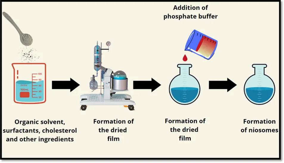
Figure 3: Thin- Film Hydration Method
2.Reverse-Phase Evaporation Method:
This method is used to prepare vesicles with potentially high entrapment of suitable hydrophilic drugs.
Procedure:
1.The surfactant and cholesterol are dissolved in an appropriate organic solvent.
2.An aqueous drug solution is added to form a water-in-oil emulsion.
3.The mixture is sonicated to improve emulsion uniformity.
4.The organic solvent is gradually removed under reduced pressure.
5.Vesicles form as the emulsion collapses and the solvent evaporates.
Advantages: May provide high entrapment efficiency for certain hydrophilic drugs.
Limitations: Uses organic solvents and may expose sensitive drugs to processing conditions that affect stability [11].
3. Ether Injection Method:
The ether injection method involves introducing a surfactant solution in an organic solvent into an aqueous phase.
Procedure:
1. The non-ionic surfactant and cholesterol are dissolved in diethyl ether or another suitable volatile solvent.
2.The organic solution is injected slowly into a heated aqueous phase, with continuous stirring.
3.Evaporation of the organic solvent promotes vesicle formation.
4. The dispersion is cooled and assessed for vesicle size and stability.
Advantages: Relatively straightforward and can produce small vesicles under suitable conditions.
Limitations: Requires careful handling of volatile, flammable solvents and control of solvent residues [9]
4. Sonication Method:
Sonication is generally used to reduce the size of previously formed neosomes rather than as a complete preparation technique on its own.
Procedure:
1. A neosomal dispersion is prepared using a suitable method, such as thin-film hydration.
2. The dispersion is exposed to ultrasonic energy using a bath sonicator or probe sonicator.
3. Sonication is conducted under controlled conditions to reduce vesicle size.
4. The resulting dispersion is evaluated for size distribution, drug leakage, and stability.
Advantages: Can reduce vesicle size and improve size uniformity.
Limitations: Excessive sonication may cause heating, drug degradation, or leakage of the encapsulated drug [9].
5. Microfluidization Method:
Microfluidization is a size-reduction and vesicle-processing technique in which a dispersion is forced through narrow channels under high pressure.
Procedure:
1.A preliminary niosomal dispersion is prepared.
2. The dispersion is passed through a microfluidizer under controlled pressure and processing conditions.
3. Repeated passes are used when necessary to obtain the desired vesicle size distribution.
4.The final formulation is evaluated for particle size, polydispersity, and stability.
Advantages: Can produce relatively uniform vesicles and offers potential for process scale-up.
Limitations: Requires specialized equipment and careful optimization of pressure and the processing cycles [12]
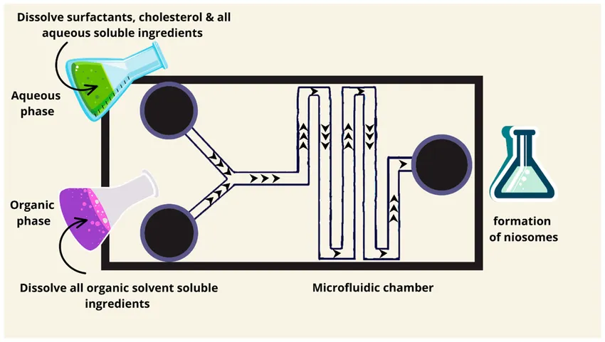
Figure 4: Micro Fluidization Method
6.Heating Method:
The heating method involves preparing neosomes by mixing surfactants, cholesterol, and an aqueous phase at an appropriate temperature.
Procedure:
1. The surfactant and cholesterol are mixed in the selected proportions.
2.An aqueous phase containing the drug or buffer is added.
3. The mixture is heated with continuous stirring until vesicle formation occurs.
4. The dispersion is cooled and characterized.
Advantages: May reduce or eliminate the need for certain organic solvents, depending on the formulation.
Limitations: Requires temperature optimization and may be unsuitable for heat-sensitive drugs [9]
7. Proneosome Method:
Proneosomes are dry or liquid-crystalline precursor formulations that form neosomes upon hydration. This approach is particularly relevant when improved storage and handling are desired.
Procedure:
1. A non-ionic surfactant and cholesterol are combined with a suitable carrier or solvent system.
2.The mixture is deposited onto a suitable carrier material or processed into a suitable precursor formulation.
3. The formulation is dried or otherwise processed to obtain proneosomes.
4. Upon hydration with an appropriate aqueous medium, the proneosomes form neosomal vesicles.
Advantages: May improve storage stability and reduce problems associated with long-term storage of aqueous vesicle dispersions.
Limitations: Hydration conditions, drug distribution, and conversion to vesicles must be optimized and verified. [13]
Evaluation and Characterization --:
Table 1: Evaluation Parameters
| Evaluation Parameters | Method / Instrument | Purpose |
|---|---|---|
| Vesicle size | Dynamic Light Scattering | To determine the average vesicle size |
| Polydispersity Index [PDI] | Dynamic Light Scattering | To assess vesicle size distribution |
| Zeta Potential | Zeta potential analyzer | To assess surface charge and colloidal stability |
| Morphology | TEM, SEM or AFM | To examine vesicle shape and surface structure |
| Entrapment Efficiency (EE%) | Centrifugation or ultrafiltration followed by UV / HPLC analysis | To determine the percentage of drug entrapped |
| Drug content | UV-visible Spectrophotometry or HPLC | To quantify drug present in the formulation |
| In-vitro drug release | Dialysis method or validated release apparatus | To determine the drug release profile |
| In-vitro skin permeation | Franz Diffusion cell | To evaluate drug permeation through the skin |
| Skin retention | Skin extraction followed by UV analysis | To determine drug deposited within skin layers |
| Surface morphology and structure | Microscopy techniques | To identify aggregation and structural changes |
| Skin irritation / Compatibility | Appropriate validated skin safety tests | To assess formulation tolerability |
NEOSOMES FOR ENHANCED TRANSDERMAL DELIVERY OF NSAIDS:
Mechanism of Skin Penetration–
Skin penetration is a critical factor in the transdermal delivery of non-steroidal anti-inflammatory drugs (NSAIDs). The stratum corneum, the outermost layer of the epidermis, acts as the principal barrier to drug permeation. Neosomes and liposomes may be used to improve the drug penetration by interacting with the skin barrier and influencing drug release, partitioning, and deposition within the skin. [2]
The main mechanisms involved in skin penetration are described below.
1.Interaction with Stratum Corneum Lipids:
Neosomes and liposomes can interact with the intercellular lipids of the stratum corneum. Depending on their composition, these interactions may alter lipid organization and facilitate drug partitioning into the skin. This can potentially improve the penetration of NSAIDs such as diclofenac, ibuprofen, and ketoprofen.
2. Vesicle Adsorption and Fusion:
Vesicles may adsorb onto the skin surface and interact with components of the stratum corneum. Under certain conditions, partial fusion or disruption of vesicle structures may occur, allowing the encapsulated drug to be released near the skin barrier. The extent of these processes depends on vesicle composition and skin conditions.
3.Drug Release and Partitioning:
After application, the drug may be released from the vesicular carrier and partition into the stratum corneum. The drug then moves through the skin according to its concentration gradient and physicochemical properties. 1Drug release and partitioning are influenced by the drug's solubility, vesicle composition, and formulation characteristics.
4. Penetration Through Skin Pathways:
Drugs can cross the skin through three principal pathways:
Intercellular pathway: Through the lipid matrix between corneocytes.
Transcellular pathway: Through corneocytes and the intervening lipid regions.
Appendageal pathway: Through hair follicles and sweat ducts.
For many drugs, the intercellular route is particularly important because of the lipid-rich structure of the stratum corneum.
6. Role of Formulation Components:
Non-ionic surfactants in neosomes and phospholipids in liposomes influence vesicle stability, drug release, and interactions with skin lipids. Cholesterol can modify membrane rigidity and permeability. Additional permeation enhancers, when included, may further affect drug transport, but their concentration must be optimized to avoid skin irritation. [2]
Drug Entrapment and Release–
Drug entrapment and release are important characteristics of neosomal drug delivery systems that influence their performance in transdermal delivery. In the transdermal delivery of non-steroidal anti-inflammatory drugs (NSAIDs), such as diclofenac, ibuprofen, ketoprofen, piroxicam, and indomethacin, effective drug entrapment and controlled release may improve drug availability at the site of application.
Drug Entrapment:
Entrapment efficiency refers to the percentage of the initially added drug that becomes associated with the niosomal vesicles. It depends on factors such as surfactant type, cholesterol concentration, drug solubility, drug-to-lipid or surfactant ratio, vesicle size, and preparation method. Lipophilic drugs are generally associated with the hydrophobic bilayer, whereas hydrophilic drugs may be entrapped within the aqueous compartment. Entrapment efficiency is commonly determined by separating free drug from vesicle-associated drug using a suitable technique, followed by drug quantification.
Higher entrapment efficiency may increase the amount of drug carried by the vesicles; however, it does not necessarily guarantee better skin penetration or therapeutic performance. [14]
Drug Release:
Drug release refers to the liberation of the encapsulated or vesicle-associated drug into the surrounding medium. Release may occur through diffusion across the vesicle membrane, desorption from the bilayer, or changes in vesicle structure. The release rate depends on vesicle composition, membrane rigidity, drug solubility, surfactant concentration, cholesterol content, and environmental conditions.
In transdermal NSAID delivery, neosomes may modify drug release and maintain drug availability near the skin surface. After release, the drug may partition into the stratum corneum and diffuse into deeper skin layers. An appropriate release profile may help improve local drug retention and prolong therapeutic action, depending on the formulation. [14]
Skin Permeation and Retention of NSAIDs from Neosomal Formulations –
Skin Permeation:
Skin permeation refers to the movement of a drug from the formulation through the different layers of the skin. In transdermal drug delivery, the stratum corneum is the main barrier that limits drug penetration. Neosomes are vesicular drug delivery systems that may improve the permeation of non-steroidal anti-inflammatory drugs (NSAIDs), such as diclofenac, ibuprofen, ketoprofen, and piroxicam.
Neosomes may enhance skin permeation through several mechanisms. Neosomes may also improve drug contact with the skin and modify the release and partitioning of the drug into skin layers.Skin permeation is commonly evaluated using a Franz diffusion cell with excised animal or human skin, where ethically approved and available. [15]
Skin Retention:
Skin retention refers to the amount of drug that remains within the skin after topical application. For NSAIDs, skin retention is particularly important because the therapeutic target may be located in local tissues beneath the skin.Neosomes may improve skin retention by promoting drug deposition in the stratum corneum and deeper skin layers.In some formulations, greater skin retention may be desirable even when the amount of drug reaching the systemic circulation is limited. [15,2]
LIPOSOMES:
Liposomes are tiny, spherical artificial vesicles made of one or more phospholipid bilayers that can encapsulate both water-soluble and fat-soluble active agents. Liposomes are spherical vesicular drug delivery systems composed mainly of one or more phospholipid bilayers surrounding an aqueous core. They are widely investigated as carriers for pharmaceutical agents because they can encapsulate both hydrophilic and lipophilic drugs. [16]
The phospholipid molecules contain a hydrophilic (water-loving) head and hydrophobic (water-repelling) tails. When phospholipids are dispersed in an aqueous medium, they can arrange themselves into bilayers, with the hydrophilic heads facing the surrounding water and the aqueous core. Their size, lamellarity, lipid composition, and surface properties influence drug loading, release, stability, and interaction with biological membranes. [17]
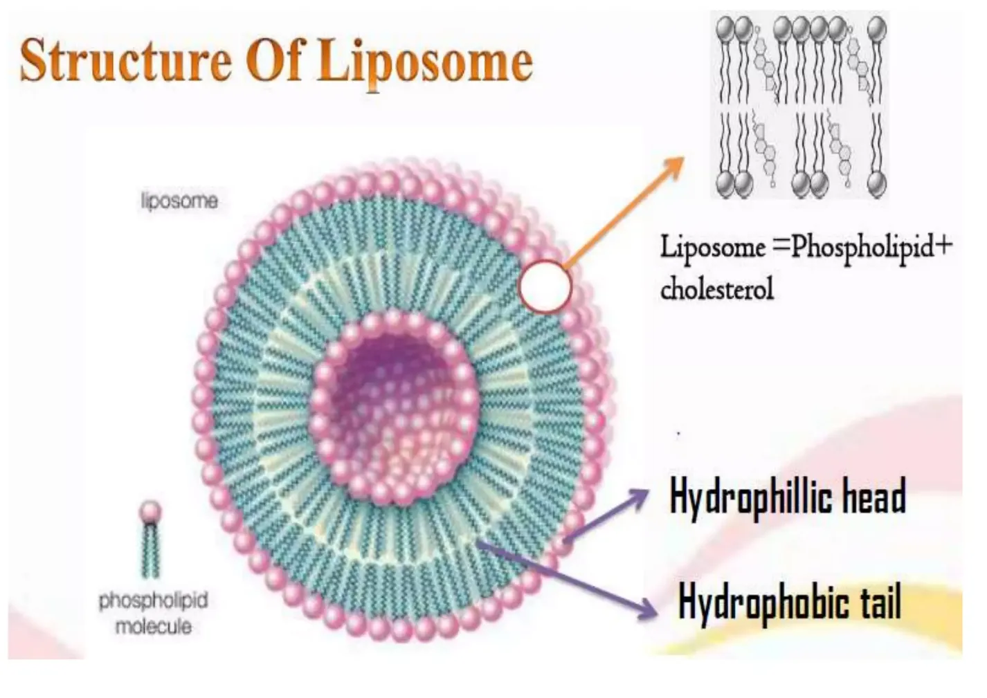
Figure 5: Structure of Liposomes
Composition of Liposomes –
Liposomes are mainly composed of phospholipids, cholesterol, and an aqueous phase. Depending on the intended drug delivery application, other components such as charge-inducing agents, surface-modifying agents, and antioxidants may also be incorporated. The composition of liposomes plays an important role in determining their vesicle formation, drug entrapment efficiency, stability, drug release, and skin permeation. [18]
1. Phospholipids:
Phospholipids are the major structural components of liposomes. They are amphiphilic molecules containing a hydrophilic head and hydrophobic fatty acid tails. In an aqueous environment, phospholipids arrange themselves into a bilayer, with the hydrophilic heads facing the aqueous medium and the hydrophobic tails directed toward the interior of the bilayer.
Commonly used phospholipids include phosphatidylcholine, phosphatidylethanolamine, and phosphatidylglycerol. The type and concentration of phospholipids influence vesicle size, membrane fluidity, drug loading, and release characteristics.
2. Cholesterol:
Cholesterol is commonly added to liposomal formulations to modify the physical properties of the phospholipid bilayer. It can regulate membrane fluidity, reduce excessive membrane permeability, and help minimize drug leakage under suitable formulation conditions.
The concentration of cholesterol affects the rigidity and stability of the bilayer. However, an excessive amount may reduce the space available for certain drugs within the membrane. Therefore, the cholesterol concentration must be optimized according to the drug and the intended delivery system.
3.Charge-Inducing Agents:
Charge-inducing agents may be incorporated to modify the surface charge of liposomes. Positively charged agents, such as stearyl amine, and negatively charged lipids, such as phosphatidylglycerol derivatives, can alter the electrostatic properties of the vesicle surface.
Surface charge may influence vesicle aggregation, physical stability, and interactions with biological membranes.
4. Aqueous Phase:
The aqueous phase is used to hydrate the phospholipids and form the internal aqueous compartment of liposomes. It generally consists of purified water, phosphate buffer, or another suitable aqueous medium.
Water-soluble drugs may be incorporated into the aqueous core during vesicle formation or by suitable loading techniques. The pH and composition of the aqueous phase can influence drug solubility, stability, and encapsulation efficiency.
5.Surface-Modifying Agents:
Surface-modifying agents may be added to alter the surface characteristics and biological behaviour of liposomes. For example, polyethylene glycol (PEG)-linked phospholipids can help reduce recognition and clearance by the body's immune system in certain systemic formulations.These agents are selected according to the intended application. They are not essential components of every liposomal formulation.
6. Antioxidants and Other Additives:
Phospholipids containing unsaturated fatty acids may be susceptible to oxidation during preparation and storage. Suitable antioxidants, such as alpha-tocopherol, may be incorporated when compatible with the formulation to help limit oxidative degradation.
Phospholipids form the bilayer, cholesterol helps regulate membrane properties, and the aqueous phase provides the internal compartment for hydrophilic drugs. In transdermal NSAID delivery, such as the delivery of diclofenac or ibuprofen, the composition should be optimized to achieve suitable drug loading, controlled release, skin permeation, skin retention, and skin compatibility. [18]
Types and Preparation Methods –
Types of liposomes-
Liposomes are classified according to their size, number of phospholipid bilayers (lamellarity), and structural characteristics. Different types of liposomes exhibit different drug-loading capacities, release profiles, and stability properties. The selection of an appropriate type depends on the physicochemical properties of the drug and the intended drug delivery route [18].
1.Classification based on Size and Lamellarity
1.Small Uni lamellar Vesicles (SUVs) –
SUVs are small liposomes consisting of a single phospholipid bilayer surrounding an aqueous core. They are generally prepared by reducing the size of larger vesicles using methods such as sonication or extrusion. Their small size may be useful for applications requiring relatively uniform vesicles.
2. Large Unilamellar Vesicles (LUVs) -
LUVs consist of a single phospholipid bilayer and have a relatively large aqueous core. They may provide greater aqueous volume for encapsulating hydrophilic drugs compared with smaller vesicles. They are commonly prepared using extrusion or other size-control techniques.
3. Multilamellar Vesicles (MLVs) -
MLVs contain multiple concentric phospholipid bilayers separated by aqueous layers. They are commonly formed during thin-film hydration. Their multilayer structure can accommodate lipophilic drugs within the bilayers and hydrophilic drugs in the aqueous compartments, although loading and release depend on the formulation.
4. Oligolamellar Vesicles -
Oligolamellar vesicles contain a small number of phospholipid bilayers. Their structure lies between single-bilayer and highly multilamellar systems. Their drug-loading and release properties depend on vesicle size and the number of bilayers.
2. Other Types of Liposomes
1. PEGylated liposomes:
Liposomes containing polyethylene glycol (PEG)-linked lipids on their surface. These are designed mainly to alter circulation time and biological interactions in systemic delivery.
2. Elastic or deformable liposomes:
Formulations containing membrane-softening agents or edge activators that increase bilayer flexibility. They are investigated for delivery across skin barriers.
3. Cationic liposomes:
Liposomes with a positively charged surface, often used in nucleic acid delivery and other applications requiring interaction with negatively charged materials.
4. Targeted liposomes:
Liposomes modified with ligands, antibodies, or other targeting components to promote interaction with selected cells or tissues[19].
Preparation Method of Liposomes-
Liposomes can be prepared using several techniques depending on the drug's properties, the required vesicle size, drug entrapment efficiency, and the intended application. The main steps generally involve lipid preparation, vesicle formation, size reduction when required, purification, and characterization [18]
1. Thin-Film Hydration Method:
This is one of the most commonly used laboratory methods for liposome preparation.
Procedure:
1. Phospholipids and cholesterol are dissolved in a suitable organic solvent.
2. The solvent is evaporated using a rotary evaporator to form a thin lipid film on the flask wall.
3. The dried lipid film is hydrated with an aqueous medium, with the hydrophilic drug included in the hydration medium when appropriate.
4. Gentle shaking or stirring promotes the formation of multilamellar vesicles.
5. Sonication or membrane extrusion may be used to reduce vesicle size and improve size uniformity.
6.The final formulation is purified and characterized.
Advantages: Simple, widely used, and suitable for different lipid compositions.
Limitations: May produce a heterogeneous vesicle population and often requires an additional size-reduction step. [20]
2. Reverse-Phase Evaporation Method:
This method is useful when relatively high entrapment of hydrophilic drugs is desired.
Procedure:
1.Lipids are dissolved in a suitable organic solvent.
2.An aqueous phase containing the hydrophilic drug is added.
3. The mixture is emulsified to form a water-in-oil system.
4. The organic solvent is gradually removed under reduced pressure.
5. Liposomes form as the emulsion breaks down and the lipid molecules reorganize around aqueous compartments.
6. The formulation is purified and characterized.
Advantages: Can provide a relatively large aqueous compartment and good entrapment of certain hydrophilic drugs.
Limitations: Requires organic solvents and careful processing to remove residual solvent. [17]
3. Ethanol Injection Method:
In this method, lipids are dissolved in ethanol and introduced into an aqueous phase.
Procedure:
1.Phospholipids and other lipid components are dissolved in ethanol.
2. The lipid solution is injected into an aqueous medium under controlled stirring.
3. Dilution of ethanol promotes the self-assembly of lipids into vesicles.
4. Residual ethanol is removed or reduced using a suitable method.
5. Vesicle size, drug entrapment, and formulation quality are evaluated.
Advantages: Relatively rapid and straightforward, with potential for producing small vesicles.
Limitations: Residual ethanol must be controlled, and vesicle characteristics depend on process parameters such as injection rate, lipid concentration, and stirring speed. [20]
4. Ether Injection Method:
In this method, lipids are dissolved in a suitable ether-based organic solvent and injected into a warm aqueous phase.
Procedure:
1. Lipids are dissolved in the organic solvent.
2. The solution is slowly injected into the aqueous phase at a controlled temperature.
3. The organic solvent is removed by evaporation.
4. Liposomes form as the lipids reorganize in the aqueous medium.
5. The resulting suspension is purified and characterized.
Advantages: Provides an alternative solvent-injection approach to vesicle formation.
Limitations: Requires careful control of solvent removal and temperature, and the use of volatile organic solvents requires appropriate laboratory safety measures.
5.Sonication Method:
Sonication is primarily a vesicle size-reduction technique, rather than a complete preparation method by itself.
Procedure:
1. A liposomal suspension is prepared, commonly by thin-film hydration.
2. The suspension is exposed to ultrasonic energy for a controlled period.
3. The energy disrupts larger vesicles and can produce smaller vesicles.
4. The formulation is checked for size distribution, drug leakage, and stability.
Advantages: Can reduce vesicle size and improve size uniformity.
Limitations: Excessive sonication may cause heating, lipid oxidation, or drug degradation. Temperature control is important.
6. Membrane Extrusion Method:
Extrusion is used to reduce and standardize the size of preformed liposomes.
Procedure:
1. A liposomal suspension is passed through a membrane with defined pore sizes.
2. The suspension may be passed through the membrane repeatedly.
3. The resulting vesicles generally have a narrower size distribution.
4. Particle size and polydispersity are measured to confirm the outcome.
Advantages: Offers useful control over vesicle size and size distribution.
Limitations: Requires suitable extrusion equipment and membranes, and may be less convenient for large-scale production without specialized systems [20].
7. Microfluidic Method:
Microfluidic methods use controlled mixing of lipid-containing solvent and an aqueous phase in small channels to produce liposomes.
Procedure:
1.Lipids are dissolved in a suitable solvent.
2. The lipid solution and aqueous phase are introduced into a microfluidic mixing system.
3. Controlled mixing and solvent dilution promote liposome formation.
4.The product is collected, and residual solvent is removed when necessary.
5.The liposomes are characterized for size, uniformity, and drug entrapment.
Advantages: Offers control over mixing conditions and may support reproducible production and scale-up.
Limitations: Requires specialized equipment, and optimization is needed for each lipid and drug formulations.
Mechanism of Skin Interaction –
Liposomes are vesicular drug delivery systems that have been investigated for improving the topical and transdermal delivery of non-steroidal anti-inflammatory drugs (NSAIDs). Their interaction with the skin depends on vesicle composition, size, lipid properties, formulation vehicle, and the physicochemical characteristics of the encapsulated drug.
1. Interaction with Stratum Corneum Lipids
The stratum corneum contains corneocytes surrounded by an organized lipid matrix. After topical application, liposomes may interact with these lipids through contact, adsorption, and lipid exchange.
Phospholipids present in liposomes may modify the organization or fluidity of skin lipids under certain conditions. This interaction may facilitate drug partitioning into the stratum corneum and improve its availability for subsequent penetration. [6,24]
2.Adsorption and Fusion with the Skin Surface
Liposomes may adsorb onto the skin surface and form close contact with the stratum corneum. Depending on their composition and the local conditions, interactions between liposomal membranes and skin lipids may occur. Partial fusion or lipid mixing has been proposed for some formulations.
3. Drug Release and Partitioning
Liposomes can act as reservoirs for drugs and release the encapsulated drug onto the skin surface or within superficial skin layers. Once released, the drug partitions between the formulation and the skin according to its physicochemical properties.
For example, diclofenac or ibuprofen incorporated into a liposomal formulation may be released from the vesicles and partition into the stratum corneum. The extent of penetration depends on factors such as drug solubility, ionization, lipid affinity, and the composition of the formulation. [25]
4. Penetration Through Skin Pathways
After release from the carrier or interaction with the skin barrier, the drug may enter the skin through three main pathways:
Intercellular pathway: Movement between corneocytes through the lipid matrix.
Transcellular pathway: Movement through corneocytes and the intervening lipid regions.
Appendageal pathway: Entry through hair follicles and sweat gland openings.
The relative contribution of these pathways depends on the drug and formulation. Detection of a drug in deeper skin layers does not, by itself, prove that intact liposomes crossed the stratum corneum.
5. Hydration and Occlusive Effects
A liposomal formulation can form a layer on the skin surface that may reduce water loss and increase hydration of the stratum corneum. Increased hydration can modify the barrier properties of the outer skin layer and may facilitate drug penetration.
The magnitude of this effect depends on the formulation vehicle, lipid composition, application conditions, and whether the skin is covered or occluded.
6. Role of Liposomal Composition
The interaction between liposomes and skin is strongly influenced by their composition.
1.Phospholipids: Form the bilayer and may interact with skin lipids.
2. Cholesterol: Modifies membrane rigidity, permeability, and drug retention.
3. Elasticity-enhancing components: In deformable liposomes, suitable edge activators may increase vesicle flexibility.
4. Topical vehicle: Influences spreading, contact time, drug release, and skin hydration.
COMPARATIVE EVALUATION OF NEOSOMES AND LIPOSOMES:
Table 2: Comparison of Neosomes and Liposomes
| Parameter | Neosomes | Liposomes |
|---|---|---|
| Composition and Stability | Mainly composed of non-ionic surfactants, often combined with cholesterol. They may offer good stability depending on the surfactant and formulation. | Mainly composed of phospholipids, often combined with cholesterol. Phospholipids oxidation and hydrolysis may affect stability. |
| Physical Stability | May undergo aggregation, fusion or drug leakage during storage. | May undergo aggregation, fusion or drug leakage during storage. |
| Chemical Stability | Stability depends on the type of surfactant, cholesterol content and storage conditions. | Stability depends on phospholipid composition, oxidation susceptibility, cholesterol content and storage conditions. |
| Drug Entrapment Efficiency | Can encapsulate suitable hydrophilic and lipophilic drugs. Entrapment depends on surfactant type, cholesterol concentration and drug properties. | Can encapsulate hydrophilic drugs in the aqueous core and lipophilic drugs within the phospholipid bilayer. Entrapment depends on lipid composition and drug properties. |
| Factors affecting Entrapment | Surfactant concentration, cholesterol ratio, vesicle size, preparation method and drug-surfactant interaction. | Phospholipid concentration, cholesterol ratio, vesicle size, preparation method and drug-lipid interaction. |
| Particle Size and Drug Release | Particle size and release rate can be adjusted by changing surfactant composition and preparation conditions. | Particle size and release rate can be adjusted by changing phospholipid composition and preparation conditions. |
| Drug Release Mechanism | Drug release may occur through diffusion, desorption, membrane disruption or vesicle leakage. | Drug release may occur through diffusion, de absorption membrane disruption or vesicle leakage. |
| Skin Permeation and Drug Retention | Surfactants may interact with stratum corneum lipids and influence drug penetration. The effect depends on the formulation. | Phospholipids may interact with skin lipids and support drug deposition or release near the skin surface. The effect depends on the formulation. |
| Skin Permeation Evaluation | Commonly evaluated using in-vitro or ex-vivo diffusion studies, such as Franz diffusion cells. | Commonly evaluated using in- vitro or ex-vivo diffusion studies, such as Franz diffusion cells. |
| Skin Drug Retention | May increase local drug deposition when the formulation is appropriately optimized. | May increase local drug deposition when the formulation is appropriately optimized. |
| Advantages | Non-ionic surfactants may be economical and offer flexibility in formulation design. Suitable systems may improve drug stabilization and controlled release. | Phospholipids have structural similarity to biological membranes and offer flexibility for incorporating drugs with different solubility characteristics. |
| Limitations | Surfactant selection is important because some surfactants may cause irritation. Vesicle leakage and instability can occur. | Phospholipids have structured similarity to biological membranes and offer flexibility for incorporating drugs with different solubility characteristics. |
| Cost consideration | May be less expensive when suitable non-ionic surfactants are cheaper than the required pharmaceutical grade phospholipids. | Cost may be higher when specialized or high purity phospholipids are required. |
COMPREHENSIVE STUDY OF TRANSDERMAL DELIVERY OF NSAIDs BASED NEOSOMES WITH LIPOSOMES:
Neosomes have emerged as promising alternatives to liposomes for the transdermal delivery of non-steroidal anti-inflammatory drugs (NSAIDs). Neosomes are composed of non-ionic surfactants and cholesterol, whereas liposomes primarily contain phospholipids. Neosomes may offer advantages such as lower formulation costs and improved chemical stability because their surfactants are generally less susceptible to oxidation than phospholipids. Both systems can improve drug incorporation, modify drug release, and enhance drug deposition within the skin; however, their performance depends on the formulation composition and the drug being delivered. [26,27]
Neosomes may offer several advantages over conventional liposomes for the transdermal delivery of NSAIDs. Their non-ionic surfactants are generally less susceptible to oxidative degradation than the phospholipids used in liposomes, potentially improving formulation stability and reducing preparation costs. Neosomes can also act as drug reservoirs, providing sustained drug release and potentially improving drug deposition within the skin. In a comparative study, Nasr et al. (2008) found that diclofenac-loaded neosomes showed better stability and superior anti-inflammatory activity than the corresponding liposomes under the investigated experimental conditions. These findings suggest that neosomes may be advantageous for certain topical NSAID formulations, although their superiority in skin permeation must be established for each drug and formulation. [26]
CONCLUSION:
Transdermal delivery of non-steroidal anti-inflammatory drugs (NSAIDs) represents a promising approach for the management of pain and inflammation, as it can reduce gastrointestinal exposure associated with oral administration and may improve patient convenience. However, the skin's outermost layer, the stratum corneum, presents a major barrier to drug permeation, while the poor aqueous solubility of several NSAIDs creates additional formulation challenges. Vesicular nanocarriers, particularly neosomes and liposomes, have therefore attracted considerable interest as potential systems for improving drug solubilization, skin penetration, and controlled drug release [21].
Neosomes, composed primarily of non-ionic surfactants and cholesterol, can accommodate both hydrophilic and lipophilic drugs. Their potential advantages include formulation flexibility, relatively good physical stability, and the possibility of sustained drug release. Nevertheless, their performance depends on several formulation variables, including surfactant type, cholesterol concentration, vesicle size, drug characteristics, and the composition of the final dosage form [6].
Compared with neosomes, liposomes are phospholipid-based vesicles with established applications in drug delivery and generally good biocompatibility. Nevertheless, neither carrier can be considered universally superior; their relative performance depends on the drug, vesicle composition, preparation method, and intended therapeutic outcome [22].
Neosomes represent a promising platform for the transdermal delivery of NSAIDs, with the potential to improve local drug deposition, regulate drug release, and enhance therapeutic performance. Future research should focus on systematic comparisons between niosomes and liposomes, long-term formulation stability, skin irritation and safety, reproducible manufacturing, and well-designed clinical studies. Although preclinicalfindings are encouraging, further evidence is required to establish their clinical effectiveness and demonstrate whether these systems provide meaningful advantages over conventional topical formulations [23].
REFERENCES

Pathan Yashfin N. Khan, Vishal S. Madankar, Sampat D. Navale, Neosomes, as Novel Vesicular Carrier for Enhanced Transdermal Drug Delivery of Nsaids: A Comprehensive Review Withliposomes, Int. J. Sci. R. Tech., 2026, 3 (11), 424-441. https://doi.org/10.5281/zenodo.24000132
 10.5281/zenodo.24000132