

We use cookies to ensure our website works properly and to personalise your experience. Cookies policy
Delight College of Pharmacy, Koregaon Bhima, Pune, Maharashtra, India-412216.
Psoriasis is a chronic, immune-mediated inflammatory skin disorder characterized by excessive keratinocyte proliferation, abnormal epidermal differentiation, and persistent inflammation. It presents as red, thickened plaques covered with silvery scales, often causing itching and discomfort. Conventional treatments, including topical corticosteroids, vitamin D analogues, and systemic medications, may be limited by poor skin penetration, adverse effects, frequent application, and reduced patient adherence. The stratum corneum acts as a major barrier to effective drug delivery. Transferosomes are flexible vesicular carriers composed of phospholipids and edge activators that can deform and penetrate narrow spaces within the skin barrier, enhancing drug permeation and targeted deposition. These properties make them promising systems for topical and transdermal delivery of anti-psoriatic drugs. Potential benefits include improved drug absorption, controlled release, enhanced therapeutic efficacy, reduced systemic exposure, and fewer adverse effects. This review examines the principles and mechanisms of transferosomal drug delivery, formulation techniques, and the influence of formulation components on vesicle performance. It also discusses characterization parameters, drug-loading strategies, advantages, and limitations in psoriasis management. Recent developments and potential clinical applications are explored, emphasizing skin permeation, therapeutic effectiveness, stability, safety, and formulation optimization. Overall, transferosomes represent a promising approach to psoriasis treatment, although further preclinical and clinical investigations are required to establish their long-term safety, stability, and therapeutic effectiveness in clinical practice.
Psoriasis is a chronic inflammatory skin condition that affects approximately 2–3% of the global population. It is characterized by red, scaly plaques that develop due to abnormal immune activity and excessive proliferation of keratinocytes. Conventional topical treatments, such as corticosteroids, vitamin D analogues, calcineurin inhibitors, and retinoids, are commonly used to manage the condition. However, their therapeutic effectiveness may be limited by inadequate drug penetration into the skin, potential adverse effects, and poor patient compliance due to the need for frequent application. The stratum corneum, which forms the outermost layer of the epidermis, acts as the main protective barrier against the entry of foreign substances. This barrier significantly limits the penetration of many therapeutic agents, particularly those with high molecular weights or hydrophilic properties, making it difficult to deliver sufficient drug concentrations to the affected skin layers. Therefore, the development of advanced drug delivery systems capable of overcoming this barrier is essential for improving the effectiveness of topical psoriasis treatment.[1-4]
Transferosomes were introduced in the early 1990s as advanced vesicular carriers designed to overcome skin penetration barriers. Unlike conventional liposomes, they contain edge activators, such as surfactants or bile salts, that provide high membrane flexibility and deformability. This allows them to pass through narrow intercellular spaces in the stratum corneum, even when these spaces are smaller than the vesicles themselves. Their ability to respond to skin hydration gradients further promotes deeper penetration and improved drug deposition in the epidermis and dermis, making them promising carriers for psoriasis treatment.[2,4,6]
Recent studies have shown that transferosomal formulations of anti-psoriatic agents, including methotrexate, cyclosporine A, betamethasone, and natural compounds such as 20-hydroxyecdysone, can improve skin permeation, provide sustained drug release, and enhance therapeutic efficacy compared with conventional formulations. This review examines transferosomes as a promising drug delivery approach for psoriasis management, focusing on their structure, formulation strategies, characterization, drug loading, and challenges in clinical translation.[4-8]
Psoriasis: Defination, Types & Mechanism:
1. Definition
Psoriass is a chronic inflammatory disorder involving the skin and sometimes joints, nails, and other organs. It results from interactions between genetic susceptibility, environmental triggers, innate immunity, and adaptive immunity.[9,10]
The most common form, plaque psoriasis / psoriasis vulgaris, accounts for roughly 85–90% of cases and presents as well-demarcated erythematous plaques with silvery-white scale.[10,11]
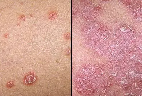
Figure 1: Skin Condition in Psoriasis
Table 1: Types of Psoriasis
| Type | Main clinical features |
|---|---|
| Plaque psoriasis (psoriasis vulgaris) | Raised, red, scaly plaques; most common form. [10,12] |
| Guttate psoriasis | Small, drop-like scaly papules, often after streptococcal infection. [12] |
| Inverse psoriasis | Smooth, red inflammation in skin folds such as axillae, groin, and under the breasts.[12] |
| Pustular psoriasis | Sterile pustules on red skin; may be localized or generalized.[12,13] |
| Erythrodermic psoriasis | Widespread redness, scaling, pain, and itching; potentially severe/systemic. [12] |
| Psoriatic arthritis | Inflammatory joint disease associated with skin psoriasis. [9] |
2. Mechanisms
1. Genetic predisposition
Psoriasis has a strong heritable component, particularly in early-onset disease. Risk variants affect skin-barrier function and immune regulation; HLA-C*06:02 is a well-known susceptibility allele.[10,14]
2. Environmental triggering
Triggers include skin trauma (Koebner phenomenon), infections, stress, smoking, obesity, and certain drugs. These events can initiate or worsen inflammatory signaling in genetically susceptible individuals.[10]
3. Innate immune activation
Keratinocytes and resident immune cells release antimicrobial peptides and inflammatory signals after injury or infection. Dendritic cells become activated and present antigens, promoting T-cell activation.[9]
4. Adaptive immune response
Activated dendritic cells drive differentiation of T cells, especially Th17 and Th1 cells. These cells infiltrate the skin and sustain chronic inflammation.[9]
5. Cytokine signaling: IL-23/IL-17 axis
The TNF-α–IL-23–Th17–IL-17 pathway is central to plaque psoriasis. IL-23 supports Th17 cells, while IL-17, IL-22, and TNF-α stimulate keratinocytes and amplify inflammation.[13]
6. Keratinocyte hyperproliferation
Inflammatory cytokines cause keratinocytes to proliferate excessively and differentiate abnormally. This produces the thickened epidermis, parakeratosis, and scaling characteristic of psoriatic plaques.[12]
7. Angiogenesis and reduced apoptosis
New blood-vessel formation and reduced apoptosis of keratinocytes help maintain the inflamed, thickened plaque.[12]
Simple pathway
Trigger → activation of dendritic cells and keratinocytes → T-cell activation → IL-23/IL-17/TNF-α signaling → keratinocyte proliferation and inflammation → scaly plaque formation.[12]
Transferosomes : defination types mechanisms:
1. Definition
A transferosome is a vesicular nanocarrier with at least one aqueous core enclosed by a lipid bilayer, modified by an edge activator to become ultra-flexible. Under non-occlusive conditions, it can deform and squeeze through skin pores much smaller than its own diameter.[15,16]
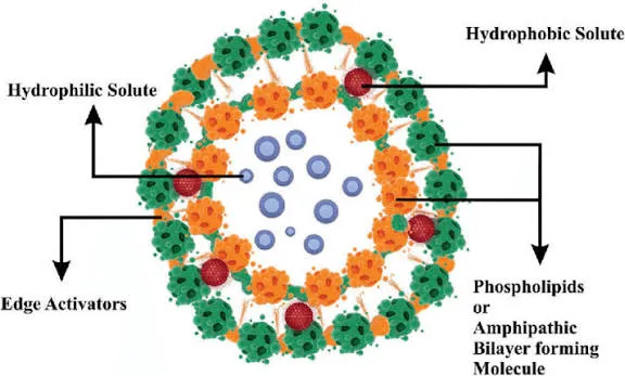
Figure 2: Struture of Transferosomes
Table 2: Composition of Transferosomes
| Component | Role |
|---|---|
| Phospholipids | Form the vesicle bilayer and encapsulate the drug. [18] |
| Edge activator | Usually a surfactant such as sodium cholate, sodium deoxycholate, Span, Tween, or bile salts; reduces interfacial tension and increases membrane elasticity. [19] |
| Water/aqueous phase | Forms the internal compartment and supports vesicle structure. [18] |
| Drug | May be hydrophilic, lipophilic, or macromolecular, depending on the formulation.[10] |
2. Types / related forms
Transferosomes are mainly classified by their composition and membrane-modifying agents:
3. Mechanism
1. Formation of elastic vesicles
The edge activator inserts into the phospholipid bilayer and destabilizes it locally. This lowers membrane rigidity and allows the vesicle to change shape without rupturing.[19,20]
2. Creation of an osmotic gradient
When applied to skin under non-occlusive conditions, water evaporates from the formulation surface. This creates a transepidermal hydration/osmotic gradient that drives transferosomes inward.[15]
3. Deformation and penetration
The vesicle follows the osmotic gradient and squeezes through narrow intercellular channels of the stratum corneum. It can pass through openings approximately one-tenth of its own diameter.[16]
4. Drug transport
Two mechanisms may operate:
5. Enhanced skin delivery
Their flexibility, lipid compatibility with skin, and ability to carry both hydrophilic and lipophilic drugs make them more effective than conventional liposomes for transdermal delivery.[17]
Advantages
Limitations
Drug Profile: Anti-Psoriatic Agents Suitable for Transferosomal Delivery
1. Small Molecule Drugs
Methotrexate (MTX): Methotrexate, an antifolate and antimetabolite drug commonly used to treat moderate-to-severe psoriasis, has limited skin permeability and may cause systemic toxicity when administered orally or parenterally. Transferosomal encapsulation has been reported to improve skin deposition by 3–5 times compared with conventional gels and provide sustained drug release for up to 24 hours, potentially reducing systemic absorption. Optimized methotrexate-loaded transferosomes also demonstrated a deformability index of 27.13, supporting their ability to penetrate narrow spaces within the stratum corneum.[1,7]
Cyclosporine A (CyA): Cyclosporine A (CyA) is a potent calcineurin inhibitor with immunosuppressive properties. Its high molecular weight (1202 Da) and lipophilicity limit its penetration through the skin. Ethosomal and transferosomal formulations can improve its skin delivery, with ethosomes often showing greater permeation due to ethanol-induced disruption of the stratum corneum. In contrast, transferosomes may promote localized drug deposition, making them a promising option for targeted psoriasis treatment with potentially reduced systemic exposure.[1]
Corticosteroids (Betamethasone, Clobetasol): Corticosteroids are potent anti-inflammatory agents widely used to treat psoriasis. Transferosomal delivery can enhance their skin penetration, including follicular delivery, and may help reduce local adverse effects such as skin atrophy. In preclinical psoriasis models, betamethasone dipropionate-loaded transferosomes showed improved tolerability and favorable safety outcomes, highlighting potential for topical psoriasis management.[4,8]
2. Natural Bioactive Compounds
20-Hydroxyecdysone (20-HE): 20-Hydroxyecdysone (20-HE) is a phytoecdysteroid with antioxidant, anti-inflammatory, and cytoprotective properties that may help reduce psoriatic lesions by modulating the IL-23/IL-17 inflammatory pathway. However, its poor skin permeability and limited physicochemical stability present challenges for effective delivery. Transferosomal formulations of 20-HE, alone or combined with resveratrol, have demonstrated high apparent encapsulation efficiency (~90%), actual encapsulation of 25–30%, particle sizes of 100–200 nm, and good stability over 30 days. These formulations also showed no observed cytotoxicity in human keratinocytes or zebrafish embryos, supporting their potential for further investigation in psoriasis treatment.[3]
Resveratrol: Resveratrol is a polyphenolic compound with antioxidant and anti-inflammatory properties that modulates NF-κB signaling and helps reduce excessive keratinocyte proliferation in psoriasis. However, its poor solubility and rapid degradation limit its therapeutic potential. Transferosomal encapsulation can improve its stability and enhance dermal delivery, making it a promising approach for psoriasis treatment.[3]
Berberine: Berberine is an isoquinoline alkaloid with anti-inflammatory and antiproliferative properties. Berberine-loaded transferosomes have shown improved skin penetration and enhanced therapeutic effects in preclinical psoriasis models, highlighting their potential for topical psoriasis treatment.[9]
3. Biologics and Peptides
Emerging research is investigating transferosomes for delivering biologic agents and peptides that target key inflammatory cytokines, including IL-17, IL-23, and TNF-α, involved in psoriasis. Although challenges related to drug stability and loading efficiency remain, transferosomes can accommodate both hydrophilic and lipophilic compounds, making them promising carriers for next-generation biologic therapies.[4,5]
Structural Basis and Formulation Design of Transferosomes
1. Defining Features
Transferosomes are phospholipid-based, ultra-deformable vesicles containing edge activators within their bilayer structure. Unlike conventional liposomes, they possess flexible membranes that allow them to change shape under stress while maintaining their structural integrity. This combination of flexibility and stability makes transferosomes promising carriers for transdermal drug delivery.[2,4]
2. Composition
Phospholipids: Phosphatidylcholine, commonly obtained from soy, egg, or hydrogenated sources, is the main component of transferosomes and forms their bilayer structure. It allows hydrophilic drugs to be incorporated into the aqueous core and lipophilic drugs into the lipid bilayer. The choice of phospholipid affects vesicle formation, membrane fluidity, drug permeability, and encapsulation efficiency. [2,4,17]
Edge Activators: Edge activators, such as Tween 80, Span 80, Tween 20, Span 20, and bile salts like sodium cholate and sodium deoxycholate, are incorporated into the phospholipid bilayer to increase membrane flexibility. Their concentration is important, as optimal levels improve vesicle deformability, whereas excessive amounts may destabilize vesicles and reduce drug entrapment efficiency.[2,4,17]
Cholesterol: Cholesterol is often incorporated into transferosomes (0–50 mol%) to regulate bilayer packing, control excessive permeability, and improve storage stability. However, higher concentrations may reduce membrane flexibility and vesicle deformability.[2,4]
Charge-Inducing Agents: Stearylamine and dicetyl phosphate are used to adjust the surface charge of transferosomes, producing positive and negative zeta potentials, respectively. This helps improve colloidal stability and influences interactions with the skin.[2,4]
Cryo/Lyoprotectants: Sucrose and trehalose are used as cryoprotectants to protect transferosomes during freeze-drying and improve their long-term stability.[2,4]
Table 3: Summarizes the Main Components and Their Functional Roles.[2,4,17]
| Component Class | Representative Examples | Main Functional Role | Impact on Performance |
|---|---|---|---|
| Phospholipids | SPC, EPC, DMPC | Bilayer framework, drug compartments | Influences vesicle formation, fluidity, encapsulation |
| Edge Activators | Tween 80, Span 80, sodium cholate | Membrane flexibility, deformability | Enhances skin penetration; excess causes instability |
| Cholesterol | Cholesterol | Bilayer stabilization | Improves integrity; high levels reduce deformability |
| Charge Agents | Stearylamine, dicetyl phosphate | Surface charge modulation | Affects zeta potential, aggregation, skin interaction |
| Cryoprotectants | Sucrose, trehalose | Lyophilization protection | Enhances storage stability |
3. Preparation Methods
Thin-Film Hydration: Phospholipids, edge activators, and the drug are dissolved in an organic solvent mixture, such as chloroform and methanol. The solvent is evaporated under reduced pressure to form a thin lipid film, which is then hydrated with an aqueous phase. Probe sonication or extrusion is subsequently performed to reduce vesicle size and obtain a uniform formulation.[3,4,17]
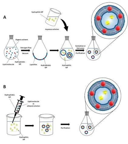
Figure 3: Thin-Film Hydration
Rotary Evaporation-Sonication: This method is similar to thin-film hydration, where rotary evaporation removes the organic solvent, followed by vortexing and sonication to produce uniform, nanosized transferosomal vesicles.[3,17]
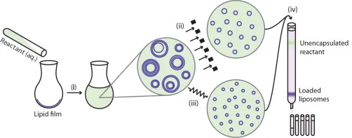
Figure 4: Rotary Evaporation-Sonication
Vortexing-Sonication: All formulation components are mixed in an aqueous buffer, followed by vortexing and sonication to form transferosomal vesicles. This method is suitable for heat-sensitive drugs due to its relatively mild processing conditions.[17]
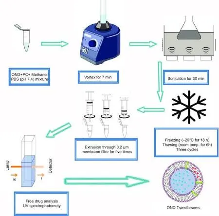
Figure 5: Vortexing-Sonication
Micellar Aggregation: The phospholipid and edge activator are mixed at a concentration above the critical micelle concentration, followed by dilution to form transferosomal vesicles.[4]
Optimization of phospholipid-to-edge-activator ratio, hydration temperature, sonication time, and aqueous phase composition is critical for achieving desired vesicle properties.[2,3,4]
Preparation methods
1. Thin-film hydration method[23]
This is the most widely used method for transferosomes.
Steps:
1. Dissolve phospholipid, edge activator, and drug in an organic solvent such as chloroform or methanol.
2. Evaporate the solvent under reduced pressure using a rotary evaporator to form a thin lipid film.
3. Vacuum-dry the film to remove residual solvent.
4. Hydrate the film with aqueous buffer or water above the lipid transition temperature, with gentle shaking or rotation.
5. Sonicate, extrude, or pass the suspension through a suitable membrane to reduce vesicle size.
Advantages: Good control over vesicle size, entrapment efficiency, and batch composition.
Limitations: Requires organic solvent removal, rotary evaporation, and careful control of hydration conditions.
2. Modified hand-shaking method[24]
A simplified version of thin-film hydration in which lipid, surfactant, and drug are dissolved, dried to a film, and hydrated by manual shaking or vortexing.
Advantages: Simple, inexpensive, and useful for preliminary screening.
Limitations: Poorer reproducibility and often larger, less uniform vesicles.
3. Reverse-phase evaporation method[23]
Phospholipid and drug are dissolved in an organic phase, mixed with an aqueous phase to form an emulsion, and the organic solvent is removed under reduced pressure. This produces vesicles with a relatively high aqueous-core volume.
Advantages: Suitable for hydrophilic drugs and can provide high encapsulation.
Limitations: Uses organic solvents; requires careful solvent removal and may produce heterogeneous vesicles.
4. Ethanol injection method[24]
Lipid, edge activator, and drug are dissolved in ethanol and rapidly injected into an aqueous medium under stirring. Ethanol diffuses out, allowing vesicles to self-assemble.
Advantages: Simple, rapid, and relatively easy to scale up.
Limitations: Residual ethanol must be removed; drug solubility in ethanol can limit use.
5. Supercritical fluid / supercritical anti-solvent method[24]
Lipids and drug are dissolved in supercritical carbon dioxide or an anti-solvent system, then rapidly depressurized or mixed with aqueous phase to form vesicles.
Advantages: Avoids or minimizes toxic organic solvents and can improve particle uniformity.
Limitations: Requires specialized equipment and higher cost.
Table 4: Key Formulation Variables
| Variable | Effect on transferosomes |
|---|---|
| Phospholipid concentration | Affects vesicle size, drug loading, and membrane strength.[24,25] |
| Lipid-to-surfactant ratio | Controls elasticity; excessive surfactant may destabilize vesicles. [25] |
| Lipid-to-drug ratio | Influences entrapment efficiency and drug-release behavior.[25] |
| Hydration medium and temperature | Affects vesicle formation, lamellarity, and encapsulation.[24,25] |
| Sonication/extrusion conditions | Determines particle size and polydispersity.[24] |
Evaluation Parameters
1. Vesicle size and size distribution
Measured by dynamic light scattering (DLS) or photon correlation spectroscopy (PCS). Typical transferosome sizes are about 50–300 nm, though the optimum depends on the drug and intended route.[24]
2. Polydispersity index (PDI)
PDI indicates uniformity of the vesicle population. A lower PDI means a more homogeneous formulation; values around 0.1–0.3 are generally considered desirable.[24]
3. Zeta potential
Zeta potential measures surface charge and predicts colloidal stability. Values greater than about ±25–30 mV generally indicate good electrostatic stabilization.[24]
4. Entrapment efficiency
Entrapment efficiency (EE%) shows how much drug is retained inside the vesicles. It is commonly calculated after separating free drug by ultracentrifugation, dialysis, or filtration. Reported EE% often ranges from about 70–95%, depending on formulation composition.[24]
5. Vesicle morphology
Examined by transmission electron microscopy (TEM), scanning electron microscopy (SEM), or cryo-TEM. This confirms vesicular structure, shape, lamellarity, and surface appearance.[26]
6. Deformability index
This is the most important transferosome-specific test. It measures the ability of vesicles to pass through a membrane or porous filter under pressure, reflecting their capacity to squeeze through skin pores.[24,26]
7. In vitro drug release
Performed using dialysis bags, Franz diffusion cells, or similar apparatus. It evaluates sustained or controlled release and is often expressed as cumulative percentage drug released over time.[24]
8. Ex vivo skin permeation
Conducted using Franz diffusion cells with excised human, porcine, rat, mouse, or other animal skin. Parameters include flux, permeation coefficient, lag time, and drug retained in skin layers.[23]
9. Drug content
Determines the actual amount of drug present in the final transferosomal formulation. It helps assess dose uniformity and preparation efficiency.[23]
10. Stability studies
Formulations are stored at different temperatures, commonly 4°C, 25°C, and sometimes 40°C, and monitored for size, PDI, zeta potential, drug content, leakage, and appearance over time.[24]
Practical evaluation checklist
Characterization of Transferosomes:
1. Particle Size and Polydispersity Index (PDI)
Dynamic light scattering (DLS) and nanoparticle tracking analysis (NTA) are used to measure vesicle size and size distribution. Transferosomes designed for transdermal delivery typically range from 100–300 nm, with a polydispersity index (PDI) below 0.3, indicating a relatively uniform vesicle population.[2,3]
2. Zeta Potential
Zeta potential measures the surface charge of transferosomes and helps assess their colloidal stability. Values above +30 mV or below −30 mV generally indicate better electrostatic stabilization against aggregation. Transferosomes commonly exhibit negative zeta potentials ranging from −25 to −50 mV, depending on the edge activator and charge-inducing agents used.[2,3]
3. Encapsulation Efficiency (EE%)
Encapsulation efficiency (EE%) is determined using HPLC or UV spectroscopy after separating free drug from vesicle-associated drug through size-exclusion chromatography (SEC) or dialysis. Apparent EE% may overestimate actual encapsulation because it includes surface-associated drug, whereas SEC-corrected measurements provide a more accurate estimate. Typical EE% values range from 25–90%, depending on the drug’s lipophilicity and formulation composition.[2,3]
4. Deformability Assessment
Extrusion Method: Vesicles are passed through polycarbonate membranes with pores smaller than their diameter to evaluate deformability. The deformability index is calculated using the vesicle size after extrusion and the membrane pore size. Lower values generally indicate greater vesicle deformability and flexibility.[4,7]
Atomic Force Microscopy (AFM): Microscopy techniques are used to observe changes in transferosome shape and structure under mechanical stress, helping assess their deformability and flexibility.[4]
Fluorescence Recovery After Photobleaching (FRAP): Fluorescence recovery after photobleaching (FRAP) measures the lateral movement of lipids within the bilayer, providing an indication of membrane fluidity.[4]
5. Morphology
Transmission electron microscopy (TEM) and cryo-TEM are used to examine vesicle morphology, lamellarity, and structural integrity. Transferosomes generally appear as spherical vesicles with a single or multiple lipid bilayers.[2,4]
6. Stability Studies
Transferosome stability is evaluated over 1–6 months at different temperatures (4°C, 25°C, and 40°C) by monitoring changes in particle size, PDI, zeta potential, encapsulation efficiency, and drug content. Lyophilized formulations containing cryoprotectants generally provide better long-term stability.[2,3,4]
7. In Vitro Drug Release
In vitro drug release is evaluated using dialysis or Franz diffusion cell methods in simulated skin fluid at pH 5.5 and 32°C. Transferosomal formulations often provide sustained drug release over 12–48 hours, following kinetic models such as Higuchi or Korsmeyer–Peppas.[4,7]
8. Skin Permeation and Deposition
Franz diffusion cells using excised human or animal skin, such as rat or pig skin, are used to measure drug permeation (µg/cm²/h) and skin retention (µg/cm²). Transferosomes generally achieve 2–5 times greater skin deposition than conventional liposomes or gels.[1,4,7]
Mechanism of Transdermal Penetration
Transferosomes enhance skin penetration through several mechanisms:
Advantages of Transferosomes in Psoriasis Therapy
Challenges and Limitations
Recent Advances and Future Perspectives
CONCLUSION
Transferosomes are a promising transdermal drug delivery system for psoriasis, designed to overcome the stratum corneum barrier through highly deformable vesicles. Their ability to improve skin penetration, sustain drug release, and deliver a wide range of anti-psoriatic agents offers potential advantages over conventional topical formulations. However, challenges related to large-scale production, storage stability, and regulatory approval remain. Advances in hybrid delivery systems, natural product encapsulation, and Quality by Design (QbD) approaches may further improve their effectiveness and reproducibility. Further research into their penetration mechanisms, clinical efficacy, and cost-effective manufacturing is essential for their successful translation into clinical practice.
REFERENCES

Suraj Jadhav, Vishal S. Madankar, Sampat D. Navale, Transferosomes for the Treatment of Psoriasis: A Novel Transdermal Drug Delivery Approach, Int. J. Sci. R. Tech., 2026, 3 (11), 353-364. https://doi.org/10.5281/zenodo.23975657
 10.5281/zenodo.23975657