

We use cookies to ensure our website works properly and to personalise your experience. Cookies policy
Kamla Institute of Pharmaceutical Sciences, Shri Shankaracharya Professional University, Bhilai, 490020, India
The present study aimed to computationally investigate a series of novel pyrrolidine sulfonyl hydrazone derivatives as potential COX-2 anti-inflammatory agents. Sixteen derivatives (P1-P16) were designed with ChemDraw and Chem3D, with minimized energies when done by the MM2 force field, and later docked to the COX-2 active site (PDB ID: 5F19) in the presence of ArgusLab v4.0.1. SwissADME, MolSoft, and ProTox-II were used to predict the pharmacokinetic, drug-likeness, and toxicity profiles, and Discovery Studio was used to visualize binding interactions. One of the study parts was to assess the binding affinity of docking the drug derivatives with the receptor with respect to the reference drug Celecoxib (-9.2924 kcal/mol). The most favorable docking scores were noted with compound P11 (-14.6191 kcal/mol) and followed by P10 (-14.0083 kcal/mol), P6 (-13.4422 kcal/mol), and P13 (-12.6843 kcal/mol), involving great interactions from some crucial COX-2 selectivity residues GLY526, TYR385, and VAL523. The ADMET analysis results showed that the gastrointestinal absorption was high, Lipinski drug likeness was good, and gastrointestinal total toxicity was low to moderate. The pyrrolidine sulfonyl hydrazone scaffold showed promising selective COX-2 inhibitory potential and will be synthesized for in vitro COX-2 and in vivo anti-inflammatory evaluation.
​
Inflammation is a highly coordinated biological defense response initiated by harmful stimuli such as microbial infection, chemical irritation, oxidative stress, and tissue injury. Acute inflammation is a fundamental problem underlying host defense and tissue repair, but unregulated or chronic inflammation plays a role in the pathogenesis of various chronic diseases, such as rheumatoid arthritis, osteoarthritis, cardiovascular diseases, neurodegenerative diseases, and cancer [1,2]. The inflammatory process is regulated by a complex network of cytokines, chemokines, lipid mediators, and signaling molecules that control cellular proliferation, tissue remodeling, and leukocyte recruitment as well as vascular permeability.
Prostaglandins are amongst the large group of mediators of inflammatory responses, which play a key role in the initiation and amplification of inflammatory signaling pathways. They are bioactive lipid mediators that are formed by the catalytic activity of enzymes of the cyclooxygenase (COX) family from arachidonic acid [3]. Three COX isoforms have been found (namely COX-1, COX-2, and COX-3), of which COX-1 and COX-2 are the two main forms that constitute the primary pharmacological target of nonsteroidal anti-inflammatory drugs (NSAIDs) [4]. COX-1 is expressed in most tissues constitutively, and it is involved in physiologic properties like renal blood flow, platelet aggregation, and gastric mucosal protection. COX-2, on the other hand, is an inducible COX that is avidly induced by pro-inflammatory cytokines, growth factors, endotoxins, and other inflammatory mediators during inflammatory conditions [5].
This selective inhibition of COX-2 is a therapeutic approach of great value in the control of pain and inflammatory process owing to its ability to block the overproduction of the vasodilating prostaglandin E2 (PGE2), which is responsible for edema, hyperalgesia, and inflammatory pain [6]. Although conventional NSAIDs block both COX-1 and COX-2, they also often suppress COX-1, leading to gastric ulceration, bleeding, and other side effects of COX-1 inhibition [7].
Hydrazone derivatives are an important class of bioactive compounds with a wide spectrum of pharmacologic activity, such as anti-inflammatory, antioxidant, antimicrobial, anticancer, etc., and are inhibitors of several enzymes. The hydrazone pharmacophore is a molecule with hydrogen bond donor and acceptor groups that can form extensive interactions with amino acid residues in enzyme active sites. The conformational rigidity, optimum physicochemical properties, and lipophilic properties of the pyrrolidine ring, along with the electrostatic and hydrogen-bonding interactions offered by the sulfonyl group, enhance the binding affinity. Consequently, the rational incorporation of pyrrolidine, sulfonamide, and hydrazone units is a promising preliminary structure for growing potent and selective COX-2 inhibitors.
The current study describes a computational investigation of a series of novel pyrrolidine sulfonyl hydrazone derivatives as selective COX-2 anti-inflammatory agents. The study aimed to assess their binding efficacy into the catalytic domain of COX-2 by molecular docking, evaluate their hydrogen-bond development interaction with the catalytic pocket, compare the docking score with a reference compound in the selective COX-2 inhibitor Celecoxib, and predict the ADMET properties using in silico methods. This integrated computational strategy offers a rational approach for serendipitously identifying new potent COX-2 binding pyrrolidine sulfonyl hydrazone derivatives that show favorable pharmacokinetic properties and emerging therapeutic feasibility for use in inflammatory diseases.
2. MATERIALS AND METHODS
2.1 Software and Online Databases
The designed compounds were built with ChemDraw Ultra 22.2.0 and Chem3D 22.2.0. Molecular docking was carried out in ArgusLab v.4.0.1, and SwissADME was used to analyze the pharmacokinetic properties and drug-likeness. The toxicological profiles of the synthesized derivatives were predicted using the ProTox-II online server.
2.2 Molecular Docking Simulation
2.2.1 Ligand Preparation
The chemical structures of the designed 1-((4-(2-(substituted benzylidene) hydrazineyl) phenyl) sulfonyl) pyrrolidine derivatives (P1-16) were drawn in ChemDraw Ultra 22.2.0 and converted to three-dimensional (3D) structures in Chem3D 22.2.0. The geometries thus obtained were energy minimized using the molecular mechanics MM2 force field to yield stable geometries that are appropriate for molecular docking. The optimized structures were then exported as Protein Data Bank (.pdb) files and subsequently used as input for the docking studies [8, 9].
1-((4-(2- (substituted benzylidene) hydrazineyl) phenyl) sulfonyl) pyrrolidine
|
Compound ID |
R |
Chemical Name |
|
1 |
4-H |
1-((4-(2-benzylidenehydrazineyl) phenyl) sulfonyl) pyrrolidine |
|
2 |
2-OH |
2-((2-(4-(pyrrolidin-1-ylsulfonyl) phenyl) hydrazineylidene) methyl) phenol |
|
3 |
3-Br |
1-((4-(2-(3-bromobenzylidene) hydrazineyl) phenyl) sulfonyl) pyrrolidine |
|
4 |
4-CH3 |
1-((4-(2-(4-methylbenzylidene) hydrazineyl) phenyl) sulfonyl) pyrrolidine |
|
5 |
3-OCH3 |
1-((4-(2-(3-methoxybenzylidene) hydrazineyl) phenyl) sulfonyl) pyrrolidine |
|
6 |
2-OH, 5-Cl |
3-chloro-2-((2-(4-(pyrrolidin-1-ylsulfonyl) phenyl) hydrazineylidene) methyl) phenol |
|
7 |
2-F |
1-((4-(2-(2-fluorobenzylidene) hydrazineyl) phenyl) sulfonyl) pyrrolidine |
|
8 |
4-NO2 |
1-((4-(2-(4-nitrobenzylidene) hydrazineyl) phenyl) sulfonyl) pyrrolidine |
|
9 |
3,4,5-(OCH3)3 |
1-((4-(2-(3,4,5-trimethoxybenzylidene) hydrazineyl) phenyl) sulfonyl) pyrrolidine |
|
10 |
4-Cl |
1-((4-(2-(4-chlorobenzylidene) hydrazineyl) phenyl) sulfonyl) pyrrolidine |
|
11 |
3,5(Cl)2 |
1-((4-(2-(3,5-dichlorobenzylidene) hydrazineyl) phenyl) sulfonyl) pyrrolidine |
|
12 |
4-CH(CH3)2 |
1-((4-(2-(4-isopropylbenzylidene) hydrazineyl) phenyl) sulfonyl) pyrrolidine |
|
13 |
5-NH2 |
3-((2-(4-(pyrrolidin-1-ylsulfonyl) phenyl) hydrazineylidene) methyl) aniline |
|
14 |
2,4-(OH)2 |
4-((2-(4-(pyrrolidin-1-ylsulfonyl) phenyl) hydrazineylidene) methyl) benzene-1,3-diol |
|
15 |
4-NH(CH3)2 |
N-isopropyl-4-((2-(4-(pyrrolidin-1-ylsulfonyl) phenyl) hydrazineylidene) methyl) aniline |
|
16 |
3-(OCH3),4(OH) |
2-methoxy-4-((2-(4-(pyrrolidin-1-ylsulfonyl) phenyl) hydrazineylidene) methyl) phenol |
2.2.2 Protein Preparation
The crystal structure of Aspirin acetylated human Cyclooxygenase-2 (PDB ID: 5F19, Resolution: 2.04 Å) was downloaded from the RCSB Protein Data Bank [10]. The preparation of the protein structure involved the removal of water molecules, co-crystallized ligands, and other heteroatoms. Moreover, hydrogen atoms were added and optimized to obtain a proper geometry for molecular docking studies.
2.2.3 Molecular Docking
Molecular docking was carried out using Argus Lab v.4.0.1. These prepared protein structures were subjected to full 3D ligand docking with the energy-minimized ligands into the active-site binding pocket formed by the co-crystallized ligand and the catalytic residues. The docking poses were ranked by binding energy, and the pose with the lowest binding energy was selected based on the lowest binding energy and important interactions at the receptor-ligand interface, such as hydrogen bonds, hydrophobic, and van der Waals interactions [11, 12].
2.2.4 Molecular visualization
The Discovery Studio Visualizer (BIOVIA) was used for visualization of the docked ligand-protein complexes. To understand the ligand orientation and the important intermolecular interactions in the active site of the target proteins, 3D binding poses and 2D interaction diagrams were generated [13].
2.3 In Silico ADMET and Drug Likeness
The pharmacokinetic and drug-likeness properties of the designed compounds (P1-16) were investigated on SwissADME, ProTox-II, and MolSoft online platforms. The chemical structures were drawn in ChemDraw Ultra 22.2.0 and converted into canonical SMILES, which were used as input for the in-silico analyses. SwissADME was used for the prediction of the physicochemical descriptors and the pharmacokinetic parameters: molecular weight, lipophilicity (LogP), water solubility, gastrointestinal absorption (GIT), blood-brain barrier (BBB) permeability, bioavailability, and synthetic accessibility (SA). ProTox-II [14-16] was used to predict toxicity profiles (LD50 Score). The drugs were assessed for their potential as drug candidates by their drug-likeness scores from the MuleSoft online database.
3. RESULTS AND DISCUSSION
3.1 Molecular Docking Simulation
Molecular docking is a pivotal computational technique in contemporary drug discovery that predicts the preferred orientation, binding affinity, and molecular interactions of a ligand within the active site of a target protein. The docking score and interaction energy calculations can help estimate the strength of the ligand–receptor association, giving insights into the structural factors involved in biological activity, selectivity, and inhibitory properties. Molecular docking in the field of anti-inflammatory drug design has special significance during drug search to dock the compounds that can selectively fit into the catalytic cavity and into the characteristic side pocket of cyclooxygenase-2 (COX-2) with a view to designing potent COX-2 inhibitors and optimizing them on rational grounds. The docking study yielded quantitative docking parameters such as Hydrogen bonds, binding energy, and residue-specific interaction profiles that were then analyzed to find the affinity, selectivity, and predicted Inhibitory activity of the designed derivatives with the COX-2 enzyme. The detailed docking results are shown in Table 1. The docking score for the reference ligand Celecoxib was found to be -9.2924 kcal/mol, and it involved 11 hydrogen-bond interactions with key active-site residues ARG44, ASN43, GLY63, LEU80, LYS79, LYS83, MET471, PHE64, THR62, and THR76. The residues are part of an important recognition network that is in the COX‑2 catalytic cavity, which is known to assist in stabilizing diaryl heterocyclic selective inhibitors. The interaction profile of Celecoxib also verified the reliability of the docking model that was used to assess the designed derivatives.
Among the designed compounds, the compound P11 showed the maximum docking score of −14.6191 kcal/mol, which is very high than Celecoxib. P11 made 26 H-bonds and presented a large complex of interactions with GLY526, LEU352, PHE198, PHE205, TYR385, VAL349, and VAL523. Concurrently, the interactions of TYR385 and GLY526, residues close to the catalytic channel, and VAL523, a characteristic residue of the COX-2 selectivity pocket, strongly indicate that the P11 side chain is located in a highly favorable position in the catalytic pocket. The derivatives P10 also showed excellent binding properties with a docking score of -14.0083 kcal/mol and 26 hydrogen bonds. Its interaction pattern included GLY526, LEU352, LEU384, PHE198, PHE205, PHE381, TYR385, VAL349, and VAL523. Four-LEU352, LEU 384, PHE381, VAL349, creating multiple hydrophobic contacts with LEU-352, LEU384, PHE381, VAL349, would lead to efficient accommodation within the elongated hydrophobic channel of COX‑2. In addition, the residue of VAL523. However, because the binding geometry of P10 indicates high COX‑2 selectivity as well as strong affinity, likely that selected variants will also have high COX‑2 selectivity.
Compound P6 had a docking score of -13.4422 kcal/mol with 25 hydrogen bonds. Both the canonical recognition residues (ARG44, ASN43, GLY63, THR62, THR76) and selectivity †associated residues (GLY526, TYR385, VAL523) were included in its interaction network. This complex interaction mode is a characteristic of the powerful and selective COX-2 inhibition and highlights the ability of P6 to interact with the entrance domain of an enzyme as well as with its selectivity pocket. Likewise, P3 had a good docking score of -13.0749 kcal/mol and 17 H-bonds. The number of hydrogen bonds was less than for P10 and P11, but P3 still had important contacts with other catalytic and hydrophobic residues inside the active site, indicating a stable binding conformation inside the active site. Other derivatives, such as P2 and P13, also showed an excellent affinity for COX‑2. The docking scores for P2 and P13 were -12.8706 kcal/mol and -12.6843 kcal/mol, respectively. The ability of P13 to form extensive hydrogen‑bonding could facilitate enthalpic stabilization of the ligand-protein complex. Importantly, both compounds bound to the same five residues (GLY526, LEU352, LEU384, TYR385, and VAL523), which suggests a similar binding mode to that of known selective COX‑2 inhibitors.
The interaction pattern was found to be consistent over most of the derivatives by detailed residue mapping. ARG44, ASN43, GLY63, LEU80, THR62, and THR76 were frequently found binding the ligand, forming a conserved recognition surface for the initial anchoring of the ligand. The most active derivatives (P6, P10, P11, and P13), by contrast, continued interacting with each other and with GLY526, TYR385, and VAL523, which are located in the catalytic/selectivity pocket of COX‑2. Mechanistically, the involvement of TYR385 is important since this residue is part of the catalytic radical transfer process, and VAL523 is part of the COX-2-specific side pocket in which this process occurs. These residues are therefore expected to find ligands that will be more. The 3D molecular docking interaction profiles of the most active derivative P14 and the reference selective COX-2 inhibitor Celecoxib are depicted in Figure 1, demonstrating their binding orientation and interactions with crucial residues of the COX-2 active site.
Overall, the docking study showed that the enzyme binding ability of the rationally designed derivatives is significantly higher predicted binding energy with the COX‑2 in comparison with Celecoxib. These compounds can form very stable complexes with the enzyme due to their high docking energies, large hydrogen-bonding networks, and direct interactions with the catalytic and selectivity residues.
|
Compound ID |
Docking Score (kcal/mol) |
No. of H-bonds |
Amino Acid Interactions |
|
Celecoxib |
-9.2924 |
11 |
ARG44, ASN43, GLY63, LEU80, LYS79, LYS83, LYS468, MET471, PHE64, THR62, THR76 |
|
P1 |
-12.3016 |
25 |
ARG44, ASN43, GLY63, GLY526, LEU80, LEU352, LEU384, LYS79, LYS83, MET471, PHE64, PHE205, PHE381, THR62, THR76, TYR385, VAL349 |
|
P2 |
-12.8706 |
25 |
ARG44, ASN43, GLY63, GLY526, LEU80, LEU352, LEU384, LYS79, PHE205, PHE381, THR62, THR76, TYR385, VAL349 |
|
P3 |
-13.0749 |
17 |
ARG44, ASN43, GLY63, LEU80, LEU359, LYS79, LYS83, MET471, PHE64, PHE357, THR62, THR76, TYR355, VAL349, VAL523 |
|
P4 |
-11.8763 |
15 |
ARG44, ASN43, GLY63, LEU80, LEU359, LYS79, MET471, PHE64, PHE357, THR62, THR76, TYR355, VAL349 |
|
P5 |
-11.2576 |
16 |
ARG44, ASN43, GLY63, LEU80, LEU359, LYS79, MET471, PHE64, PHE357, THR62, THR76, TYR355, VAL349, VAL523 |
|
P6 |
-13.4422 |
25 |
ARG44, ASN43, GLY63, GLY526, LEU80, LEU352, LEU384, LYS79, LYS83, MET471, PHE64, PHE205, PHE381, THR62, THR76, TYR385, VAL349, VAL523 |
|
P7 |
-11.2266 |
14 |
ARG44, ASN43, GLY63, LEU80, LEU391, LYS79, MET471, PHE64, PHE395, THR62, TYR404, VAL444, VAL447 |
|
P8 |
-10.4470 |
17 |
ARG44, ASN43, GLY63, LEU80, LEU359, LYS79, MET471, PHE64, PHE357, THR62, THR76, TYR355, VAL349 |
|
P9 |
-8.4563 |
12 |
ARG44, ASN43, GLY63, LEU80, PHE64, PHE96, THR62, VAL103 |
|
P10 |
-14.0083 |
26 |
ARG44, ASN43, GLY63, GLY526, LEU80, LEU352, LEU384, LYS79, LYS83, MET471, PHE64, PHE198, PHE205, PHE381, THR62, THR76, TYR385, VAL349, VAL523 |
|
P11 |
-14.6191 |
26 |
ARG44, ASN43, GLY63, GLY526, LEU80, LEU352, PHE64, PHE198, PHE205, THR76, TYR385, VAL349, VAL523 |
|
P12 |
-11.7381 |
16 |
ARG44, ASN43, GLY63, LEU80, THR62, THR76, TYR355, VAL349 |
|
P13 |
-12.6843 |
27 |
ARG44, ASN43, GLY63, GLY526, LEU80, LEU352, LEU384, LYS79, LYS83, MET471, PHE64, PHE198, THR76, TYR385, VAL349, VAL523 |
|
P14 |
-12.1443 |
25 |
ARG44, ASN43, GLY63, GLY526, LEU80, LEU352, MET471, PHE64, PHE205, PHE381, THR62, THR76, TYR385, VAL349, VAL523 |
|
P15 |
-9.7767 |
14 |
ARG44, ASN43, GLY63, LEU80, LEU359, LYS79, MET471, PHE64, PHE351, THR62, TYR115, VAL349 |
|
P16 |
-10.9383 |
18 |
ARG44, ASN43, GLY63, LEU80, LEU359, LYS79, MET471, PHE64, THR76, TYR355, VAL349 |
Table 1. Docking energies and residue interaction patterns of the designed derivatives with the COX‑2 binding site.
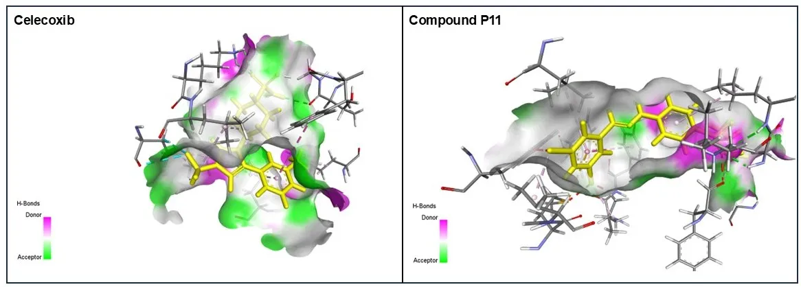
Figure 1. Three-dimensional (3D) molecular docking interaction visualization of the reference selective COX‑2 inhibitor Celecoxib and the most potent derivative P11 within the COX‑2 active site, illustrating ligand orientation, hydrogen-bond donor and acceptor regions, and interactions with key catalytic and selectivity-pocket residues.
3.2 In Silico ADMET and Drug Likeness
The physicochemical properties of the 1-((4-(2-(3-bromobenzylidene) hydrazineyl) phenyl) sulfonyl) pyrrolidine derivatives (P1-16) about their potential as anti-inflammatory agents targeted to COX-2 were evaluated (Table 2). The molecular weights of the compounds were between 329.42 g/mol and 455.31 g/mol, within the recommended range for orally active, small molecules. The number of hydrogen bond donors (1-3), hydrogen bond acceptors (4-7), and rotatable bonds (5-8) suggested a viable balance between flexibility and rigidity of the molecule that would lend itself to its binding in the COX-2 active site. The topological polar surface area ranged from 70.15 to 115.97 Å2, indicating good membrane permeability and intestinal absorption. The results of the physicochemical descriptors derived collectively indicate that the designed derivatives have characteristics associated with orally bioavailable anti-inflammatory drug candidates.
Based on in silico ADMET predictions, the profiles of these compounds are summarized in Table 3. All compounds exhibited high gastrointestinal (GIT) absorption, indicating their potential for efficient oral administration. Blood-brain barrier (BBB) permeability was predicted for P1, P3, P4, P6, P7, P10, P11, and P12, with the remaining derivatives having limited CNS penetration, a desirable characteristic for peripheral COX-2 inhibitors, as this could minimize the risk for central nervous system (CNS) side effects. The synthetic accessibility (SA) scores varied from 2.80 to 3.26, which indicated that all the molecules are easily synthesized. Furthermore, all the designed compounds did not violate Lipinski's rule of five, revealing their desirable oral drug likeness. Drug-likeness scores ranged from -0.33 to 0.61, and compounds P6, P9, P10, P12, P13, P15, and P16 showed positive scores, indicating good drug-like properties. The acute oral toxicity (LD50) estimates ranged from 800 to 2500 mg/kg body weight and are in the range of low to moderate toxicity, which gives an acceptable preliminary safety profile. Generally, the synthesized 1-((4-(2-(3-bromobenzylidene) hydrazineyl) phenyl) sulfonyl) pyrrolidine derivatives possessed good physicochemical properties, an appropriate pharmacokinetic profile, obeyed the Lipinski rule of 5, and exhibited a tolerable toxicity profile. Their findings validate their use as potential active COX-2 targeted anti-inflammatory agents, and further promote them for molecular docking, molecular dynamics simulations, and biological evaluation.
|
Compound ID |
MW (g/mol) |
nHA |
nAHA |
nRB |
nHBA |
nHBD |
TPSA (Å2) |
|
P1 |
329.42 |
23 |
12 |
5 |
4 |
1 |
70.15 |
|
P2 |
345.42 |
24 |
12 |
5 |
5 |
2 |
90.38 |
|
P3 |
408.31 |
24 |
12 |
5 |
4 |
1 |
70.15 |
|
P4 |
343.44 |
24 |
12 |
5 |
4 |
1 |
70.15 |
|
P5 |
359.44 |
25 |
12 |
6 |
5 |
1 |
79.38 |
|
P6 |
379.86 |
25 |
12 |
5 |
5 |
2 |
90.38 |
|
P7 |
347.41 |
24 |
12 |
5 |
5 |
1 |
70.15 |
|
P8 |
374.41 |
26 |
12 |
6 |
6 |
1 |
115.97 |
|
P9 |
419.49 |
29 |
12 |
8 |
7 |
1 |
97.84 |
|
P10 |
363.86 |
24 |
12 |
5 |
4 |
1 |
70.15 |
|
P11 |
398.31 |
25 |
12 |
5 |
4 |
1 |
70.15 |
|
P12 |
371.50 |
26 |
12 |
6 |
4 |
1 |
70.15 |
|
P13 |
344.43 |
24 |
12 |
5 |
4 |
2 |
96.17 |
|
P14 |
361.42 |
25 |
12 |
5 |
6 |
3 |
110.61 |
|
P15 |
386.51 |
27 |
12 |
7 |
4 |
2 |
82.18 |
|
P16 |
375.44 |
26 |
12 |
6 |
6 |
2 |
99.61 |
|
MW: Molecular weight, nHA: No. of heavy atom, nAHA: No. of Aromatic heavy atoms, nRB: No. of Rotatable bonds nHBA: No of H-bond acceptors, nHBD: No. of H-bond donor. |
|||||||
Table 2. Physicochemical characteristics of the designed 1-((4-(2-(3-bromobenzylidene) hydrazineyl) phenyl) sulfonyl) pyrrolidine derivatives.
|
Compound ID |
GIT (High/Low) |
BBB (Yes/No) |
SA |
LP |
Drug likeness |
LD50 (mg/kg) |
|
P1 |
High |
Yes |
2.80 |
0 |
0.09 |
2500 |
|
P2 |
High |
No |
2.87 |
0 |
-0.18 |
2500 |
|
P3 |
High |
Yes |
2.93 |
0 |
-0.15 |
2500 |
|
P4 |
High |
Yes |
2.91 |
0 |
-0.14 |
2500 |
|
P5 |
High |
No |
2.87 |
0 |
0.17 |
2500 |
|
P6 |
High |
No |
2.97 |
0 |
0.49 |
1000 |
|
P7 |
High |
Yes |
2.93 |
0 |
-0.33 |
2500 |
|
P8 |
High |
No |
2.98 |
0 |
-0.26 |
2500 |
|
P9 |
High |
No |
3.26 |
0 |
0.38 |
2000 |
|
P10 |
High |
Yes |
2.82 |
0 |
0.44 |
1000 |
|
P11 |
High |
Yes |
2.88 |
0 |
-0.03 |
800 |
|
P12 |
High |
Yes |
3.09 |
0 |
0.56 |
2500 |
|
P13 |
High |
No |
2.91 |
0 |
0.29 |
2500 |
|
P14 |
High |
No |
2.95 |
0 |
0.10 |
2500 |
|
P15 |
High |
No |
3.16 |
0 |
0.61 |
2500 |
|
P16 |
High |
No |
2.95 |
0 |
0.17 |
2000 |
|
GIT: Gastrointestinal tract, BBB: Blood-brain barrier, SA: Synthetic Accessibility, LP: Lipinski violations, LD50: Lethal dose. |
||||||
Table 3. Pharmacokinetics profile of the designed 1-((4-(2-(3-bromobenzylidene) hydrazineyl) phenyl) sulfonyl) pyrrolidine derivatives.
CONCLUSION
The present computational study revealed that the designed Pyrrolidine Sulfonyl Hydrazone derivatives have a good binding affinity to the active site of COX-2, wherein compounds P11, P10, P6, and P13 have better docking scores and have more interactions with the catalytic residues and selectivity pocket residues as compared to the reference drug Celecoxib. In silico pharmacokinetic and drug-likeness studies also showed acceptable oral bioavailability, good ADMET properties, and a low to moderate predicted toxicity profile. The results indicate that the pyrrolidine sulfonyl hydrazone structure is a viable structure for developing a selective COX-2 anti-inflammatory. Derivatives obtained as a future perspective will be synthesized and then fully tested both in vitro for COX-2 enzyme inhibition and in vivo for anti-inflammatory activity to confirm computational predictions and validate their therapeutic potential.
REFERENCES

Anju Daharia*, Pooja Verma, Mousmi Sahu, Structure-Based Molecular Docking And Pharmacokinetic Evaluation Of Pyrrolidine Sulfonyl Hydrazone Derivatives As Potential Selective COX-2 Inhibitors, Int. J. Sci. R. Tech., 2026, 3 (7), 981-990. https://doi.org/10.5281/zenodo.21641988
 10.5281/zenodo.21641988