

We use cookies to ensure our website works properly and to personalise your experience. Cookies policy
Department of Pharmacology, Sree Abirami College of Pharmacy, [Coimbatore, Tamilnadu], India
Background: Asthma is a chronic inflammatory airway disease in which histamine release, oxidative stress and pro-inflammatory cytokines drive bronchoconstriction and airway remodelling. Epiphyllum oxypetalum (DC.) Haw. (Cactaceae) is reported to have anti-inflammatory, antioxidant and analgesic properties, but its anti-asthmatic potential has not been evaluated. Objective: To evaluate the anti-asthmatic activity of the ethanolic leaf extract of E. oxypetalum against histamine-induced asthma in rats. Methods: Leaves were extracted with ethanol in a Soxhlet apparatus and screened for phytoconstituents. Thirty female Wistar rats were divided into five groups (n = 6): Group I, normal control; Group II, disease control (0.2% histamine aerosol); Group III, standard (ketotifen 2.4 mg/kg, p.o.); Group IV, E. oxypetalum 250 mg/kg, p.o.; Group V, E. oxypetalum 500 mg/kg, p.o., for 14 days. Body weight, pre-convulsive time, differential leukocyte count (DLC), serum SGOT and SGPT, lung tissue SOD, catalase, GSH and lipid peroxidation (LPO), TNF-?, wet lung weight and lung histopathology were evaluated. Data were analysed by one-way ANOVA with Dunnett's test. Results: The extract yield was 13.33% w/w, and the extract contained carbohydrates, alkaloids, flavonoids, saponins, tannins/phenolics, steroids and triterpenoids. Histamine exposure raised SGOT, SGPT, LPO, TNF-?, wet lung weight and inflammatory leukocyte counts and depleted SOD, catalase and GSH. Treatment with both doses of the extract significantly reversed these changes (P < 0.0001 versus disease control for the biochemical markers), with the 500 mg/kg dose generally giving the strongest effect, and prolonged pre-convulsive time. Histology showed less peribronchial smooth muscle hypertrophy, epithelial hyperplasia and inflammation, with intact blood vessels at 500 mg/kg. Conclusion: The ethanolic leaf extract of E. oxypetalum showed dose-dependent anti-inflammatory, antioxidant and bronchoprotective effects in a histamine-induced asthma model in rats and merits further investigation as a source of anti-asthmatic leads.
Asthma is a chronic inflammatory disorder of the airways characterised by wheezing, breathlessness and cough, with airway hyperresponsiveness, mucus hypersecretion and variable airflow obstruction [1,2]. Its clinical spectrum ranges from mild intermittent disease to severe persistent disease that can be fatal, and the pathogenesis involves acute and chronic inflammation, smooth muscle contraction, oedema and airway remodelling [2,3]. Current management is stepwise and relies on inhaled corticosteroids and long-acting β2-agonists, with leukotriene modifiers, oral corticosteroids and anti-IgE therapy added in severe disease [4]. Many patients nevertheless remain uncontrolled or respond poorly to usual therapy [5], and long-term use of these drugs carries adverse effects, which sustains interest in plant-derived alternatives. Oxidative stress and cytokines such as TNF-α are increasingly recognised as contributors to asthma severity [6,7].
Epiphyllum oxypetalum (DC.) Haw. (night-blooming cereus, "Nishagandhi", "Wijaya Kusuma") is an ornamental epiphytic cactus. Its leaf extracts contain phenolics, glycosides, saponins, steroids, terpenoids, tannins and resins and have shown antimicrobial activity [8]. Reported pharmacological activities include in vitro and in vivo anti-inflammatory activity [9], free-radical scavenging [10], analgesic activity [11], wound healing in diabetic mice [12] and anticancer activity in an NMU-induced colon cancer model [13]. Steroid-containing medicinal plants have also been discussed as potential antiasthmatic agents [14]. Despite this, no study has examined the effect of E. oxypetalum in an experimental asthma model. We therefore evaluated its ethanolic leaf extract against histamine-induced asthma in rats, using behavioural, haematological, biochemical, antioxidant, inflammatory and histopathological end-points. The pathophysiological sequence from mast cell degranulation to airway narrowing, and the study end-points aligned to each step, are summarised in Figure 1.
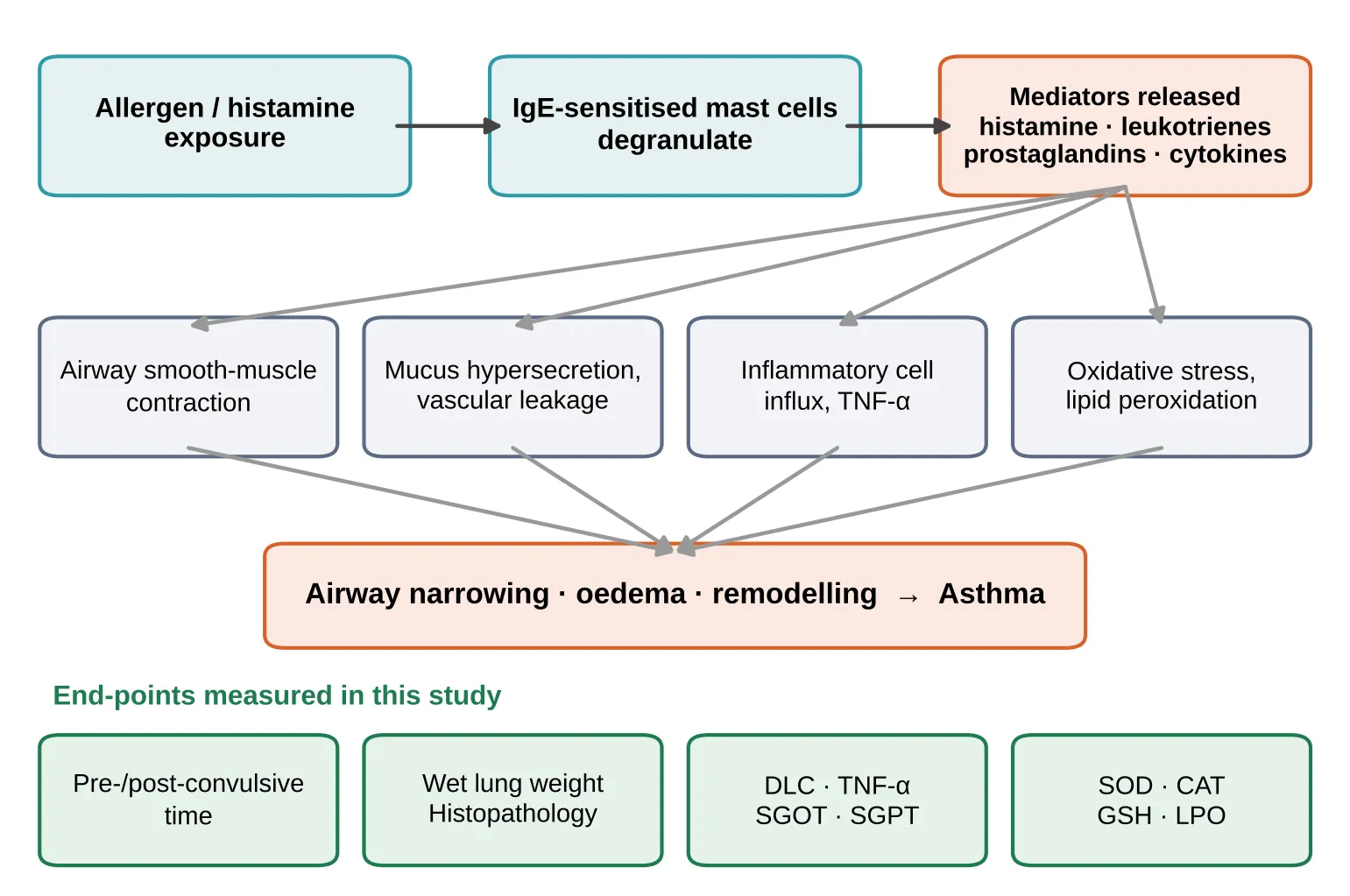
Figure 1: Pathophysiology of Histamine-Mediated Asthma and the End-Points Assessed in This Study. Allergen or Histamine Exposure Triggers Mast Cell Mediator Release, Leading to Bronchoconstriction, Mucus Hypersecretion, Inflammation and Oxidative Stress. DLC, Differential Leukocyte Count; SGOT/SGPT, Serum Aminotransferases; SOD, Superoxide Dismutase; CAT, Catalase; GSH, Reduced Glutathione; LPO, Lipid Peroxidation.
MATERIALS AND METHODS
2.1 Plant material and extraction
Leaves of E. oxypetalum were collected from a home garden in Bengaluru, Karnataka, and authenticated by the Botanical Survey of India, Southern Regional Centre, Coimbatore (Authentication No. BSI/SRC/5/23/2026-27/Tech./1271). The leaves were washed, shade-dried and coarsely powdered. The powder (105 g) was extracted with ethanol (400 mL) in a Soxhlet apparatus at 40–50 °C. The solvent was recovered by distillation and the extract was dried on a water bath at 60 °C. Percentage yield was calculated as (weight of extract / weight of plant material) × 100. The plant is shown in Figure 2.
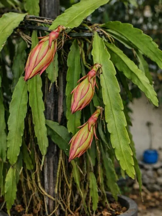
Figure 2: Epiphyllum Oxypetalum (DC.) Haw., Showing the Leaves (Flattened Phylloclades) and Flower Buds from Which the Extract Was Prepared.
2.2 Preliminary phytochemical screening
The extract was screened by standard qualitative tests for carbohydrates (Molisch, Benedict), alkaloids (Mayer, Wagner, Dragendorff), starch (iodine), glycosides (Keller-Kiliani), saponins (foam test), flavonoids (Shinoda, ammonia, lead acetate, ferric chloride), tannins and phenolics, steroids and triterpenoids (Salkowski, Hesse) and fats and fixed oils (saponification) [15,16].
2.3 Animals and ethical approval
Thirty healthy female albino Wistar rats (3 months old, 118–160 g) were obtained from the animal house of Sree Abirami College of Pharmacy (CPCSEA registration 2292/PO/Re/S/2024/CCSEA). They were housed under controlled temperature and humidity with a standard diet and water ad libitum, in accordance with CPCSEA guidelines. The protocol was approved by the Institutional Animal Ethics Committee (SACOP/Re/M.Pharm/03/2026).
2.4 Experimental design
Animals were randomly allocated to five groups of six (Table 1) and treated once daily for 14 days. The overall study workflow is shown in Figure 3.
Table 1: Experimental Design
| Group | Designation | Challenge | Treatment |
|---|---|---|---|
| I | Normal control | Saline (1 mL) | None |
| II | Disease control | 0.2% histamine aerosol | Vehicle |
| III | Standard | 0.2% histamine aerosol | Ketotifen 2.4 mg/kg, p.o. |
| IV | Test 1 (T-1) | 0.2% histamine aerosol | E. oxypetalum 250 mg/kg, p.o. |
| V | Test 2 (T-2) | 0.2% histamine aerosol | E. oxypetalum 500 mg/kg, p.o. |
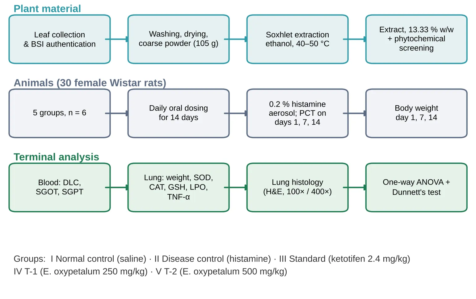
Figure 3: Study Workflow: Plant Processing and Extraction, Animal Grouping and Dosing, Histamine Challenge, and Terminal Analyses.
2.5 Induction of asthma and pre-convulsive time
Asthma was induced in Groups II–V by exposure to 0.2% histamine aerosol in a histamine chamber, with the nebulizer flow rate set at 0.2 mL/min [17]. Animals were observed for signs of asphyxia, and the time to onset of pre-convulsive dyspnoea (PCD/PCT) was recorded. Once dyspnoea appeared, rats were removed to fresh air and allowed to recover for 24 h before treatment. PCT was recorded on days 1, 7 and 14, and percentage protection was calculated as (1 − T1/T2) × 100, where T1 and T2 are mean PCT before and after drug administration. The histamine chamber used is shown in Figure 4.
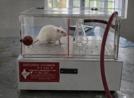
Figure 4: Histamine Chamber with a Rat During Exposure to 0.2% Histamine Aerosol.
2.6 Blood and serum analysis
Blood was collected by the retro-orbital route under light anaesthesia. Differential leukocyte count was determined on Leishman-stained smears (100× objective, 100–200 cells counted). For serum, blood was allowed to clot, centrifuged (2000 rpm, 10 min, 4–10 °C) and SGOT and SGPT were measured with commercial kits (ERBA) on a semi-auto analyser using the IFCC kinetic method (NADH oxidation at 340 nm).
2.7 Lung tissue parameters
After euthanasia, lungs were excised and weighed (wet lung weight). A 10% w/v homogenate was prepared in cold 0.05 M phosphate buffer and centrifuged at 4 °C. Catalase was measured by the decrease in absorbance of H2O2 at 240 nm. SOD was measured by inhibition of NBT reduction at 560 nm. GSH was measured with DTNB at 412 nm after TCA precipitation. LPO was measured as thiobarbituric acid-reactive substances at 532 nm. TNF-α was measured by ELISA (Krishgen Biosystems kit).
2.8 Histopathology
Lungs were fixed in 10% formalin for 24 h, embedded in paraffin, sectioned, stained with haematoxylin and eosin and examined by light microscopy [18].
2.9 Statistical analysis
Results are expressed as mean ± SEM (n = 6). Differences among groups were analysed by one-way ANOVA followed by Dunnett's multiple comparison test. P < 0.05 was considered significant.
RESULTS
3.1 Extract yield and phytochemistry
The ethanolic extract was a dark-brown powder with a yield of 13.33% w/w (14 g from 105 g). Preliminary screening showed carbohydrates, alkaloids, saponins, flavonoids, tannins/phenolics, steroids and triterpenoids; starch, cardiac glycosides and fats/fixed oils were not detected (Table 2).
Table 2: Preliminary Phytochemical Screening of the Ethanolic Extract
| Constituent | Test | Result |
|---|---|---|
| Carbohydrates | Molisch / Benedict / Dragendorff | − / + / + |
| Alkaloids | Mayer / Wagner | − / + |
| Starch | Iodine | − |
| Glycosides | Keller-Kiliani | − |
| Saponins | Foam | + |
| Flavonoids | Shinoda, ammonia, lead acetate, ferric chloride | + (all) |
| Tannins/phenolics | Lead acetate, potassium dichromate | + |
| Steroids and triterpenoids | Salkowski, Hesse | + |
| Fats and fixed oils | Saponification | − |
3.2 Body weight
All groups gained weight over 14 days (Table 3). Body weight did not decline in any group, indicating that the challenge and treatments were not accompanied by weight loss. Group means are plotted in Figure 5.
Table 3: Effect of E. oxypetalum on Body Weight (G, Mean ± SEM, N = 6)
| Group | Day 1 | Day 7 | Day 14 |
|---|---|---|---|
| Normal control | 118.17 ± 2.86 | 130.67 ± 3.21 | 143.33 ± 2.87 |
| Disease control | 122.33 ± 2.53 | 148.33 ± 3.94 | 170.17 ± 5.01 |
| Standard (ketotifen) | 125.17 ± 4.34 | 153.67 ± 4.26 | 172.00 ± 5.30 |
| T-1 (250 mg/kg) | 126.17 ± 7.07 | 158.33 ± 9.22 | 181.50 ± 12.11 |
| T-2 (500 mg/kg) | 125.50 ± 3.88 | 162.33 ± 8.11 | 185.67 ± 10.31 |
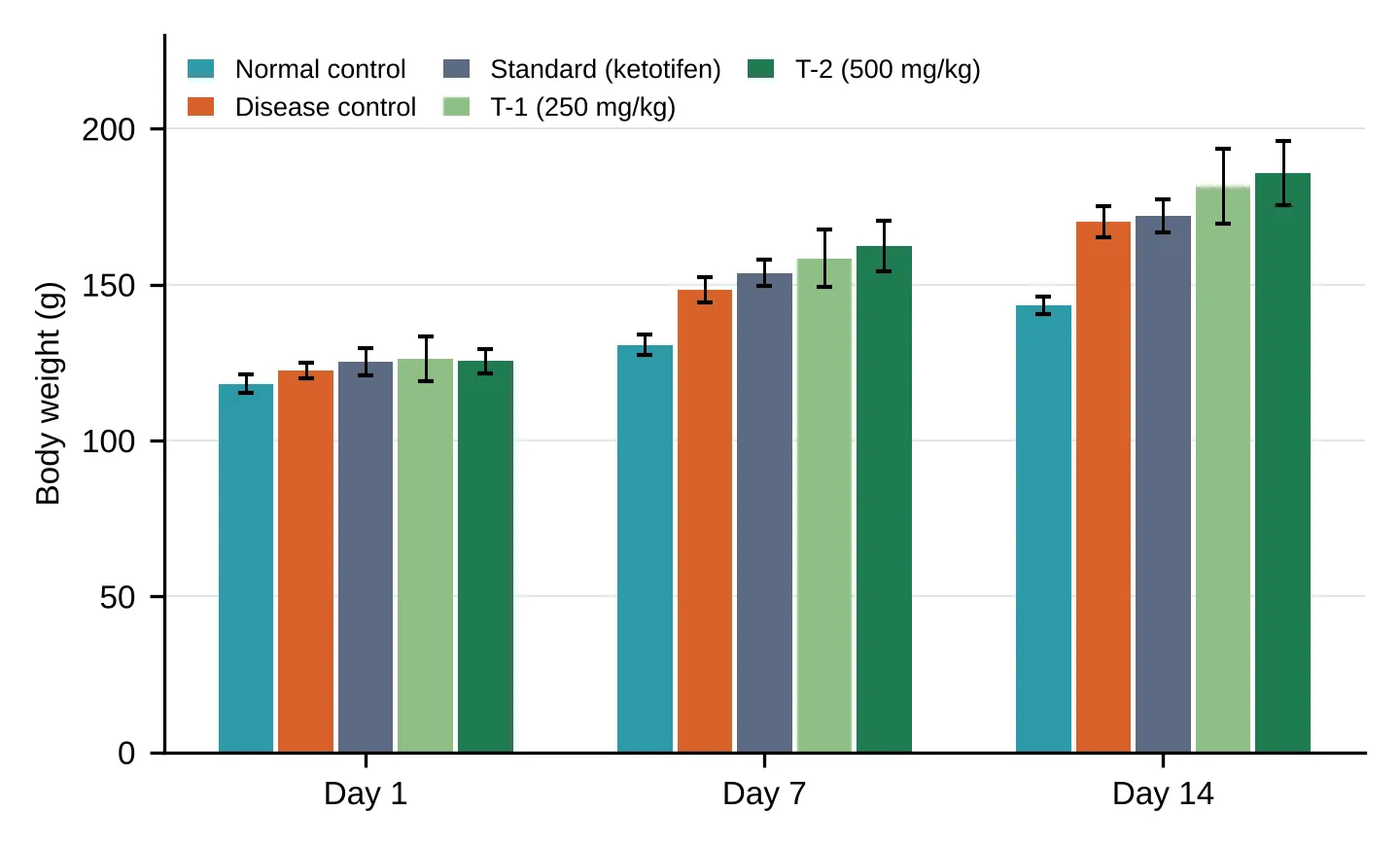
Figure 5: Effect of E. Oxypetalum on Body Weight on Days 1, 7 and 14. Bars Show Mean ± SEM (N = 6). All Groups Gained Weight; No Group Lost Weight.
3.3 Pre-convulsive time
In the disease control group, pre-convulsive time fell progressively (5.10 → 3.01 → 2.67 on days 1, 7 and 14), reflecting increasing airway sensitivity. Ketotifen and both extract doses maintained or increased PCT over the same period, and the 500 mg/kg dose and ketotifen produced the longest times on day 14 (Table 4). Post-convulsive time showed the same pattern, falling in the disease group (5.33 → 3.25 → 2.67) and remaining higher in the ketotifen and extract groups (Figure 6).
Table 4: Effect of E. oxypetalum on Pre-Convulsive Time (Mean ± SEM)
| Group | Day 1 | Day 7 | Day 14 |
|---|---|---|---|
| Normal control | – | – | – |
| Disease control | 5.100 ± 0.516 | 3.012 ± 0.154 | 2.667 ± 0.211 |
| Standard | 4.667 ± 0.256 | 5.331 ± 0.211 | 5.431 ± 0.145 |
| T-1 (250 mg/kg) | 5.667 ± 0.422 | 4.833 ± 0.401 | 4.500 ± 0.224 |
| T-2 (500 mg/kg) | 4.521 ± 0.342 | 5.167 ± 0.307 | 5.333 ± 0.211 |
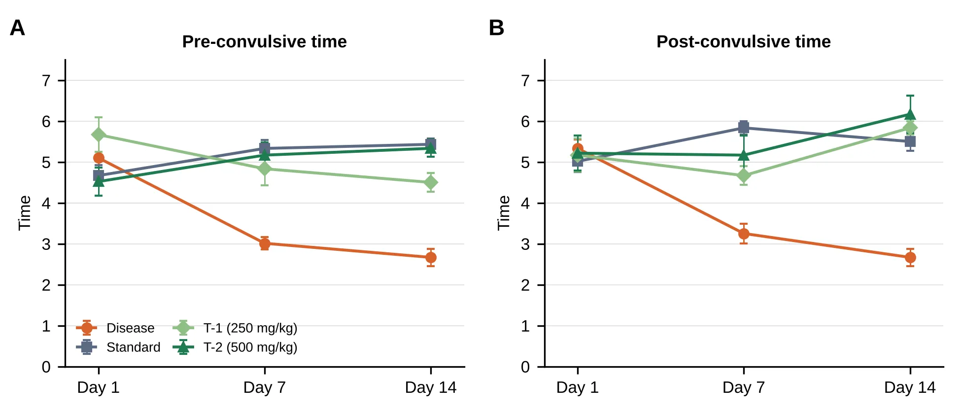
Figure 6: Effect of E. Oxypetalum on (a) Pre-Convulsive Time and (B) Post-Convulsive Time on Days 1, 7 and 14. Points Show Mean ± SEM (N = 6). the Normal Control Group Was Not Exposed to Histamine and Has No Values.
3.4 Serum enzymes, lung antioxidants, TNF-α and lung weight
Histamine challenge significantly raised SGOT, SGPT, LPO, TNF-α and wet lung weight and lowered SOD, catalase and GSH relative to normal control (Table 5). Treatment with ketotifen and with both doses of the extract significantly reversed these changes relative to disease control (P < 0.0001 for SGOT, SGPT, SOD, catalase, GSH, LPO and TNF-α; P < 0.0001 for wet lung weight). At 500 mg/kg, SGOT (148.0 IU/L), LPO (109.8) and TNF-α (355.2 pg/mL) approached normal-control values. For SGOT, LPO, catalase and wet lung weight the 500 mg/kg dose was closer to normal than ketotifen. Individual markers are plotted in Figures 7–10.
Table 5: Serum and Lung Tissue Parameters (Mean ± SEM, N = 6)
| Parameter | Normal | Disease | Standard | T-1 (250) | T-2 (500) |
|---|---|---|---|---|---|
| SGOT (IU/L) | 147.2 ± 1.96 | 226.1 ± 1.55 | 188.7 ± 1.23 | 157.6 ± 1.72 | 148.0 ± 1.26 |
| SGPT (IU/L) | 17.01 ± 1.08 | 38.33 ± 1.39 | 28.26 ± 1.52 | 19.46 ± 1.18 | 22.95 ± 1.28 |
| SOD | 0.605 ± 0.042 | 0.192 ± 0.028 | 0.608 ± 0.009 | 0.489 ± 0.027 | 0.539 ± 0.021 |
| Catalase | 17.76 ± 0.52 | 8.35 ± 0.52 | 11.46 ± 0.04 | 12.44 ± 0.25 | 15.12 ± 0.42 |
| GSH | 110.0 ± 3.03 | 29.74 ± 1.16 | 100.9 ± 1.51 | 81.17 ± 3.01 | 97.67 ± 1.98 |
| LPO | 111.4 ± 1.37 | 201.2 ± 4.10 | 170.1 ± 2.13 | 144.0 ± 1.68 | 109.8 ± 2.16 |
| TNF-α (pg/mL) | 353.1 ± 16.29 | 710.0 ± 2.73 | 325.3 ± 2.10 | 367.6 ± 1.77 | 355.2 ± 1.03 |
| Wet lung weight (g) | 1.398 ± 0.043 | 2.307 ± 0.115 | 2.050 ± 0.019 | 1.775 ± 0.023 | 1.573 ± 0.082 |
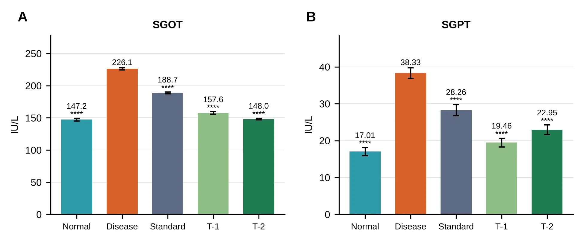
Figure 7: Effect of E. Oxypetalum on Serum (a) SGOT and (B) SGPT. Bars Show Mean ± SEM (N = 6). Group Codes: Normal, Normal Control; Disease, Histamine-Challenged Control; Standard, Ketotifen 2.4 mg/kg; T-1, E. Oxypetalum 250 mg/kg; T-2, E. Oxypetalum 500 mg/kg. ****P < 0.0001 Versus Disease Control (One-Way ANOVA, Dunnett's Test).
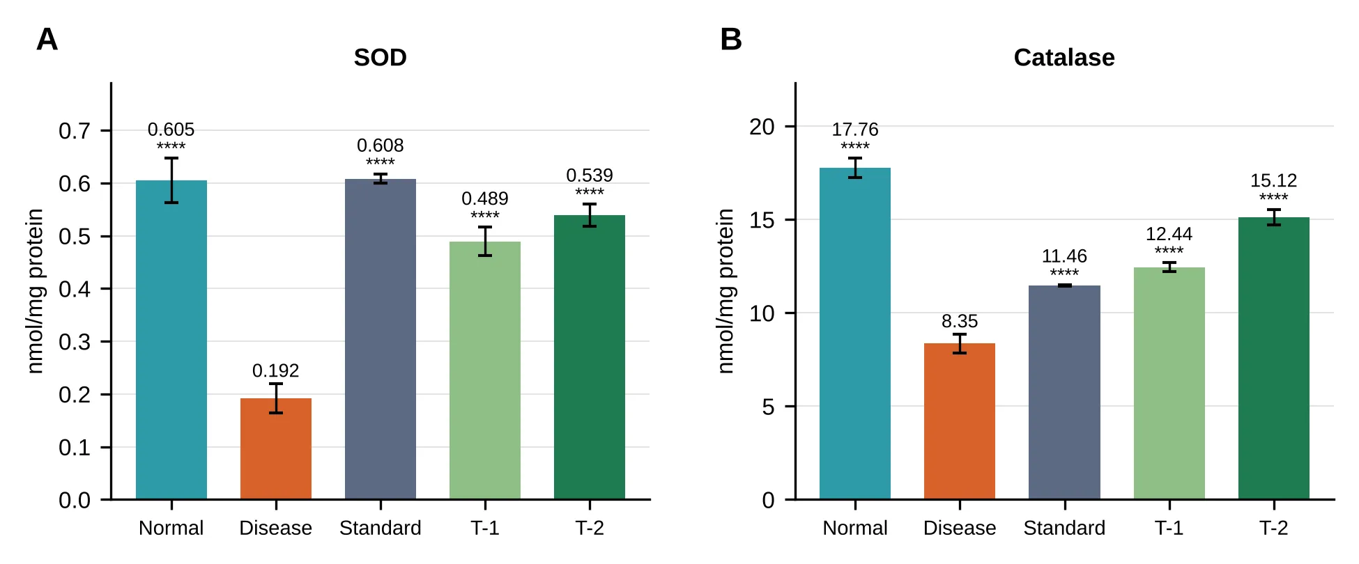
Figure 8: Effect of E. Oxypetalum on Lung Tissue (a) SOD and (B) Catalase. Bars Show Mean ± SEM (N = 6). Group Codes: Normal, Normal Control; Disease, Histamine-Challenged Control; Standard, Ketotifen 2.4 mg/kg; T-1, E. Oxypetalum 250 mg/kg; T-2, E. Oxypetalum 500 mg/kg. ****P < 0.0001 Versus Disease Control (One-Way ANOVA, Dunnett's Test).
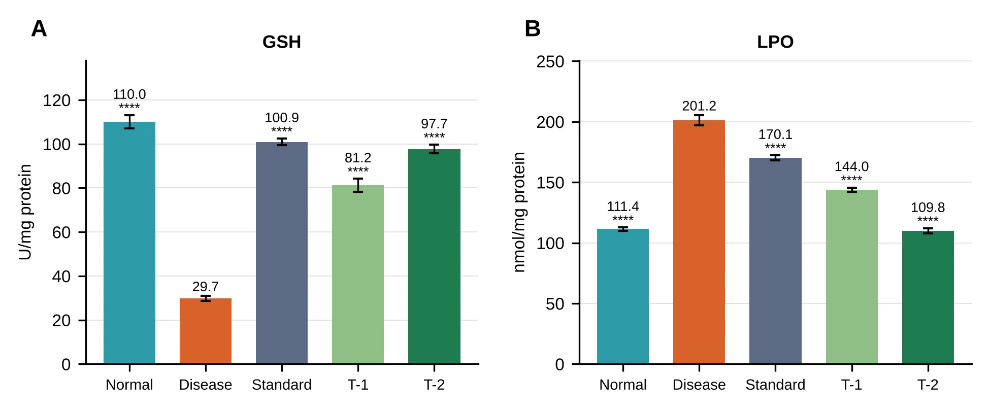
Figure 9: Effect of E. Oxypetalum on Lung Tissue (a) GSH and (B) Lipid Peroxidation (LPO). Bars Show Mean ± SEM (N = 6). Group Codes: Normal, Normal Control; Disease, Histamine-Challenged Control; Standard, Ketotifen 2.4 mg/kg; T-1, E. Oxypetalum 250 mg/kg; T-2, E. Oxypetalum 500 mg/kg. ****P < 0.0001 Versus Disease Control (One-Way ANOVA, Dunnett's Test).
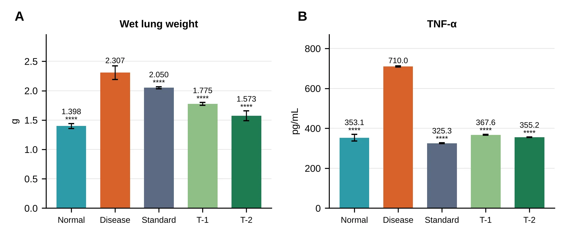
Figure 10: Effect of E. Oxypetalum on (a) Wet Lung Weight and (B) Lung Tnf-Α. Bars Show Mean ± SEM (N = 6). Group Codes: Normal, Normal Control; Disease, Histamine-Challenged Control; Standard, Ketotifen 2.4 mg/kg; T-1, E. Oxypetalum 250 mg/kg; T-2, E. Oxypetalum 500 mg/kg. ****P < 0.0001 Versus Disease Control (One-Way ANOVA, Dunnett's Test).
3.5 Differential leukocyte count
The disease control group showed a marked rise in neutrophils, eosinophils and monocytes compared with normal control (P < 0.0001). Ketotifen and both doses of the extract significantly lowered these counts (P < 0.0001 versus disease control), with a dose-related response for the extract (Table 6). The distribution of each cell type is plotted in Figure 11.
Table 6: Differential Leukocyte Count (%, Mean ± SEM)
| Group | Neutrophils | Eosinophils | Monocytes | Lymphocytes |
|---|---|---|---|---|
| Normal control | 65.50 ± 0.87 | 4.05 ± 0.66 | 4.79 ± 0.36 | 27.97 ± 0.75 |
| Disease control | 84.32 ± 0.55 | 10.57 ± 0.54 | 6.51 ± 0.36 | 46.10 ± 0.63 |
| Standard | 74.37 ± 0.56 | 5.17 ± 0.22 | 5.19 ± 0.19 | 34.80 ± 0.57 |
| T-1 (250 mg/kg) | 69.19 ± 0.50 | 5.11 ± 0.30 | 5.07 ± 0.41 | 30.65 ± 0.64 |
| T-2 (500 mg/kg) | 69.34 ± 0.67 | 5.18 ± 0.35 | 4.98 ± 0.24 | 29.24 ± 0.47 |
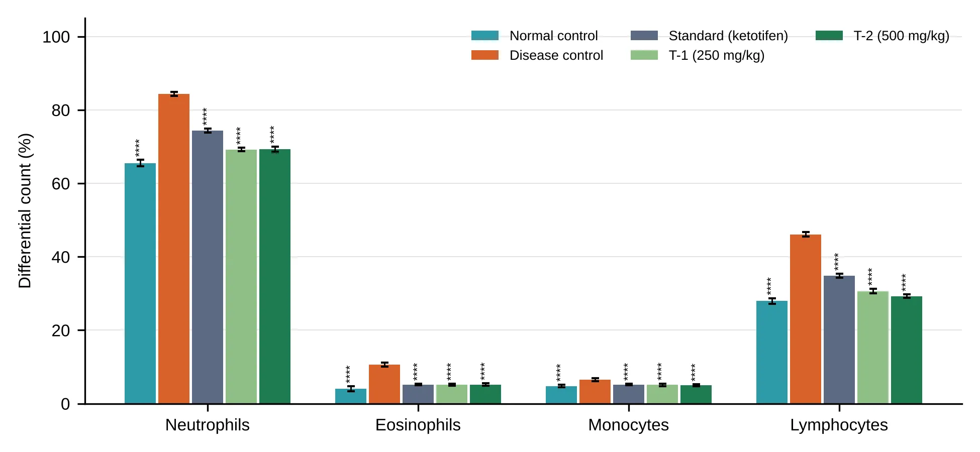
Figure 11: Effect of E. Oxypetalum on Differential Leukocyte Count. Bars Show Mean ± SEM (N = 6). ****P < 0.0001 Versus Disease Control. Group Colours: As in Figure 5.
3.6 Histopathology
Normal control lungs showed bronchioles lined by simple columnar epithelium, alveoli lined by cuboidal and flat cells, and interalveolar septa and vessels within normal limits. Disease control lungs showed hypertrophied peribronchial smooth muscle, epithelial hyperplasia with increased secretions, and moderate inflammation of septa and bronchioles with lymphocytes, plasma cells and eosinophils. Ketotifen-treated lungs showed focal smooth muscle hypertrophy, intact columnar epithelium, mild haemorrhage and mild inflammation, with intact vessels. The 250 mg/kg group showed hypertrophy at very few places, intact epithelium, mild haemorrhage and mild inflammation, with congested vessels. The 500 mg/kg group showed intact peribronchial smooth muscle and epithelium, only mild lymphocytic and plasmacytic inflammation, and intact blood vessels, the closest to normal histology among the treated groups. Representative sections are shown in Figure 12.
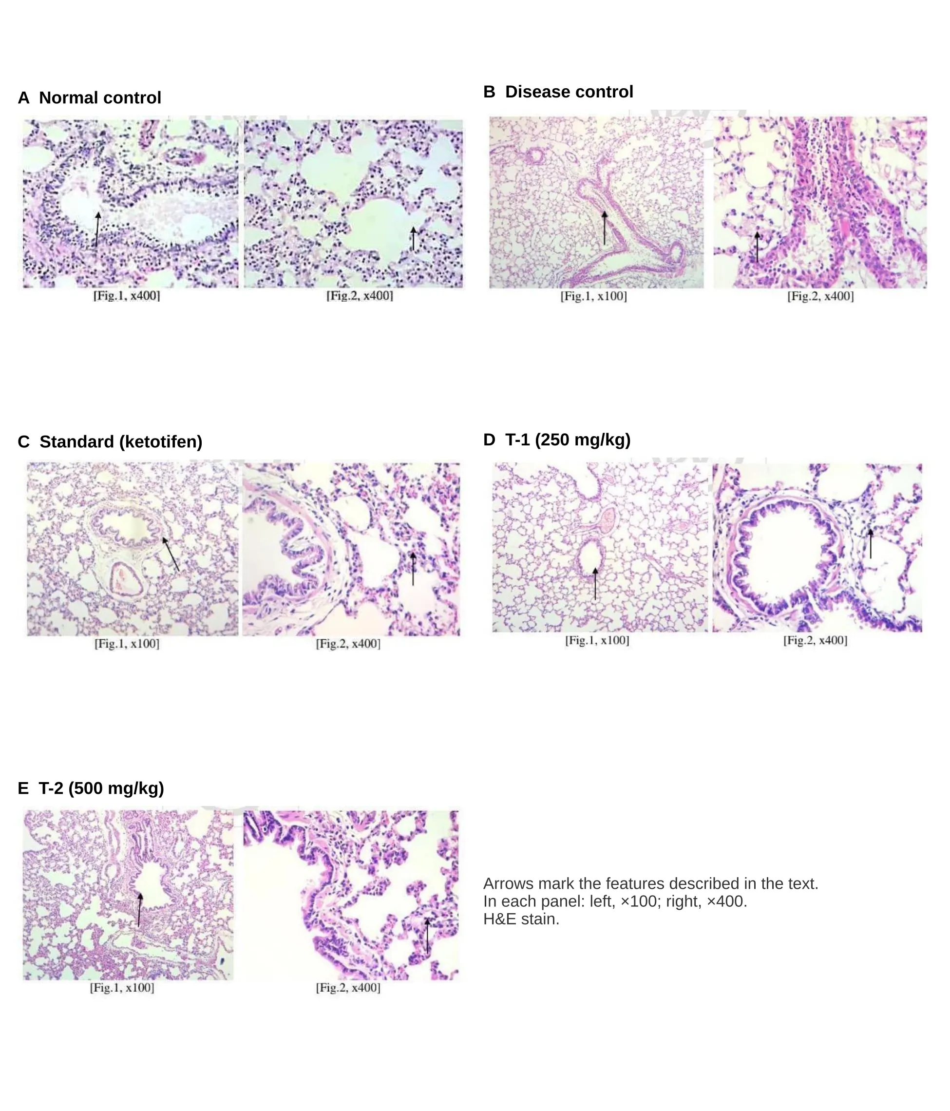
Figure 12. Haematoxylin and eosin-stained rat lung sections. (A) Normal control: bronchioles lined by simple columnar epithelium, septa and vessels within normal limits. (B) Disease control: hypertrophied peribronchial smooth muscle, epithelial hyperplasia, increased secretions and moderate inflammation. (C) Standard: intact columnar epithelium, mild haemorrhage and inflammation, vessels intact. (D) T-1: intact epithelium, mild haemorrhage and inflammation, congested vessels. (E) T-2: intact smooth muscle and epithelium, mild inflammation, intact vessels.
DISCUSSION
This is, to our knowledge, the first evaluation of E. oxypetalum in an experimental asthma model. Histamine aerosol produces bronchoconstriction, mediator release and airway inflammation and is a standard screen for antiasthmatic agents [17,19]. In our model it increased inflammatory leukocytes, TNF-α, lipid peroxidation and lung weight, depleted lung antioxidants and altered lung histology, and shortened pre-convulsive time on repeated exposure.
Prolonged pre-convulsive time in the treated groups indicates protection against histamine-induced bronchospasm, consistent with antihistaminic or bronchodilatory activity. The reduction in neutrophil, eosinophil and monocyte counts suggests suppression of allergic inflammation, possibly through mast cell stabilisation or reduced mediator release, as with the reference drug ketotifen. Because eosinophilic and neutrophilic inflammation underlies much of asthma pathology [3,6], this is a clinically relevant effect. The mechanism was not tested directly in this study.
TNF-α has been implicated in refractory asthma and airway inflammation [20,21]. The near-normalisation of lung TNF-α at both doses, together with lower wet lung weight, suggests a reduction in inflammation and pulmonary oedema, in line with the anti-inflammatory activity previously reported for the leaf extract [9].
Oxidative stress contributes to asthma pathogenesis [6]. Histamine challenge depleted SOD, catalase and GSH and raised LPO, indicating oxidant injury and membrane lipid peroxidation. The extract restored all four markers, and at 500 mg/kg LPO returned to normal-control levels. This agrees with the free-radical scavenging activity of the leaf extract in vitro [10] and may be attributable to its flavonoids and phenolic constituents, which are known antioxidants, although the active compounds were not isolated here. Saponins and steroidal or triterpenoid constituents, also detected, have been proposed as antiasthmatic principles in other plants [14]. Elevated SGOT and SGPT in the disease group are consistent with tissue injury and inflammation, and were reduced by treatment. These enzymes are not specific to the lung, so they are best read as indirect markers.
Histology supported the biochemical findings: the 500 mg/kg group retained intact epithelium, smooth muscle and vessels with only mild inflammation, comparable to or better than the ketotifen group in these qualitative descriptions.
Limitations. Only female rats and one extract were used, with n = 6 per group; lung function was not measured directly; histology was descriptive without scoring; the extract was not standardised to marker compounds; and mechanism-level work (IgE, mast cell counts, receptor studies) and acute toxicity data are needed. Histamine challenge models an acute bronchospasm rather than chronic allergic asthma.
CONCLUSION
The ethanolic leaf extract of Epiphyllum oxypetalum at 250 and 500 mg/kg significantly protected rats from histamine-induced asthma by prolonging pre-convulsive time, reducing inflammatory leukocytes, TNF-α, lipid peroxidation and lung weight, restoring lung antioxidant enzymes and improving lung histology, with the higher dose generally the most effective. These findings support further studies, including bioactivity-guided isolation, chronic allergen-induced models and toxicity evaluation.
Declarations
Ethics approval:
Approved by the Institutional Animal Ethics Committee, Sree Abirami College of Pharmacy (SACOP/Re/M.Pharm/03/2026); conducted per CPCSEA guidelines.
Conflict of interest:
[The authors declare no conflict of interest]
Funding:
[This research received no external funding]
Acknowledgements:[
I thanked your guide, the Principal and the Head of the Department of Pharmacology, Sree Abirami College of Pharmacy for the facilities and animal house, Dr. S. S. Hameed of the Botanical Survey of India, Coimbatore, for authenticating the plant, and the Institutional Animal Ethics Committee]
REFERENCES


Kalaimathivanan S., Dr. Srinivasan Ponmalai, Dr. Dhatchinamoorthi, Mohammad Fiaz, Evaluation of Anti-Asthmatic Activity of Epiphyllum Oxypetalum Leaf Extract Against Histamine-Induced Asthma in Rats, Int. J. Sci. R. Tech., 2026, 3 (10), 984-995. https://doi.org/10.5281/zenodo.23433071
 10.5281/zenodo.23433071