

We use cookies to ensure our website works properly and to personalise your experience. Cookies policy
Faculty of Pharmacy, Sree Balaji Medical College and Hospital, Bharath Institute of Higher Education and Research, Chennai 600044, Tamil Nadu, India.
Colorectal cancer (CRC) is one of the leading causes of cancer-related mortality worldwide, highlighting the need for the development of more effective and selective anticancer agents. Benzimidazole is a privileged heterocyclic scaffold with diverse biological activities, including significant anticancer potential. This review summarizes recent advances in benzimidazole derivatives evaluated for cytotoxic activity against human colorectal cancer HCT116 cells. The reported compounds exhibit anticancer effects through various mechanisms, including inhibition of cell proliferation, induction of apoptosis, cell cycle arrest, DNA damage, and modulation of key signaling pathways. Structural modifications of the benzimidazole nucleus have been shown to improve potency and selectivity, making these derivatives promising candidates for further drug development. Overall, benzimidazole-based compounds represent a valuable class of molecules for the discovery of novel therapeutic agents against colorectal cancer, although additional preclinical and clinical studies are required to establish their safety and efficacy.
The human colorectal carcinoma cell line HCT116 is one of the most extensively utilized in vitro experimental models in cancer biology and anticancer drug discovery. Since its establishment from the primary colon carcinoma of an adult male patient, HCT116 cells have become indispensable for evaluating the biological activity of newly synthesized compounds, particularly heterocyclic molecules such as benzimidazole derivatives. Their stable genetic profile, rapid proliferation rate, reproducibility, and molecular resemblance to human colorectal tumors make them one of the preferred models for investigating tumor biology, drug mechanisms, and therapeutic responses.
HCT116 cells closely mimic the molecular alterations observed in colorectal cancer patients, including constitutive activation of oncogenic signaling pathways, deregulated cell-cycle progression, enhanced proliferative capacity, and altered apoptotic mechanisms. Consequently, these cells provide an excellent platform for screening novel chemotherapeutic agents before progressing to animal studies and clinical investigations.
Origin and Characteristics of HCT116 Cells
HCT116 cells were established from the primary colon carcinoma of a 48-year-old male patient diagnosed with colorectal adenocarcinoma. The cells exhibit an epithelial morphology and grow as adherent monolayers under standard tissue culture conditions. They possess a doubling time of approximately 20–24 hours, allowing rapid experimental evaluation of cytotoxic compounds.
One of the major advantages of HCT116 cells is their high reproducibility under laboratory conditions. These cells can be cultured efficiently in McCoy’s 5A medium supplemented with fetal bovine serum and antibiotics while maintaining stable genetic characteristics over multiple passages.
Morphologically, HCT116 cells appear polygonal with distinct epithelial features. They exhibit strong proliferative potential, anchorage-dependent growth, and the ability to form colonies in soft agar assays. These characteristics make them highly suitable for evaluating antiproliferative and cytotoxic effects of newly synthesized benzimidazole derivatives.
Genetic Characteristics
The molecular profile of HCT116 cells resembles that of aggressive colorectal tumors, making them highly valuable in translational cancer research. The most important genetic characteristics include mutations in KRAS, activation of the Wnt/β-catenin signaling pathway, and defects in the DNA mismatch repair (MMR) system resulting in microsatellite instability (MSI).
HCT116 cells harbor an activating mutation in the KRAS oncogene, leading to constitutive stimulation of the MAPK signaling cascade. This mutation promotes uncontrolled cellular proliferation and contributes to resistance against several epidermal growth factor receptor (EGFR)-targeted therapies. Consequently, HCT116 cells are frequently employed to evaluate compounds capable of overcoming KRAS-mediated drug resistance.
Another hallmark feature is the mutation of β-catenin (CTNNB1), resulting in persistent activation of Wnt signaling. This pathway regulates genes associated with cellular proliferation, stemness, and tumor progression. Aberrant β-catenin signaling contributes significantly to colorectal carcinogenesis and serves as an important therapeutic target for novel benzimidazole derivatives.
HCT116 cells are also deficient in the MLH1 mismatch repair gene, leading to microsatellite instability (MSI). This defect causes accumulation of genetic mutations and genomic instability, closely resembling a subset of colorectal cancers observed clinically.
Despite these alterations, HCT116 cells retain wild-type TP53, allowing researchers to investigate apoptosis mediated through functional p53 signaling pathways. This characteristic distinguishes HCT116 cells from several other colorectal cancer cell lines that possess mutant TP53.
Advantages of HCT116 Cells in Drug Discovery
Several biological characteristics make HCT116 one of the preferred colorectal cancer models.
The cells exhibit rapid proliferation, enabling efficient evaluation of cytotoxic compounds within relatively short experimental periods. Their stable phenotype minimizes experimental variability and enhances reproducibility across laboratories.
The presence of clinically relevant genetic mutations allows accurate prediction of therapeutic responses against human colorectal tumors. HCT116 cells also demonstrate high sensitivity toward DNA-damaging agents, microtubule inhibitors, kinase inhibitors, and apoptosis-inducing compounds.
Furthermore, these cells are highly amenable to molecular biology techniques including RNA interference, CRISPR-Cas9 gene editing, quantitative PCR, Western blotting, transcriptomic analysis, proteomics, metabolomics, and fluorescence imaging. Such versatility facilitates comprehensive mechanistic investigations following treatment with newly synthesized benzimidazole derivatives.
Applications in Anticancer Research
HCT116 cells have become a standard experimental model for evaluating the anticancer potential of synthetic heterocyclic compounds. Medicinal chemists routinely employ these cells during early-stage drug discovery to identify lead molecules exhibiting potent cytotoxic activity.
Several biological endpoints can be investigated using HCT116 cells, including inhibition of cell proliferation, induction of apoptosis, disruption of mitochondrial membrane potential, generation of reactive oxygen species (ROS), inhibition of angiogenesis-related signaling, suppression of metastatic behavior, and modulation of oncogenic signaling pathways.
In addition to evaluating direct cytotoxicity, HCT116 cells are widely used to investigate multidrug resistance, synergistic drug combinations, nanoparticle-mediated drug delivery, photodynamic therapy, gene therapy, and targeted molecular therapies.
Cytotoxicity Assays
MTT Assay
The MTT assay remains the most widely used colorimetric method for evaluating cell viability following treatment with benzimidazole derivatives. Viable cells convert the yellow tetrazolium salt into purple formazan crystals through mitochondrial succinate dehydrogenase activity. Reduction in absorbance directly reflects decreased cell viability, allowing calculation of ICâ‚…â‚€ values.
Sulforhodamine B (SRB) Assay
The SRB assay measures total cellular protein content as an indicator of cell density and proliferation. Following treatment, cells are fixed with trichloroacetic acid and stained with sulforhodamine B dye. Absorbance is proportional to the number of viable cells and provides highly reproducible results.
CCK-8 Assay
The Cell Counting Kit-8 (CCK-8) assay utilizes a water-soluble tetrazolium salt that is converted into an orange-colored product by viable cells. Compared with MTT, CCK-8 is more sensitive, less toxic, and does not require crystal dissolution.
Live/Dead Cell Assay
Fluorescent dyes such as calcein-AM and propidium iodide distinguish viable from non-viable cells. Live cells fluoresce green, whereas dead cells exhibit red fluorescence due to compromised membrane integrity. cytotoxicity assays in a summary table1
|
Assay |
Principle |
Detection Method |
Outcome Measured |
Advantages |
Limitations |
References |
|
MTT Assay |
Mitochondrial succinate dehydrogenase in viable cells reduces yellow MTT to insoluble purple formazan crystals. |
Spectrophotometric measurement at 540–570 nm after dissolving formazan crystals. |
Cell viability, cytotoxicity, ICâ‚…â‚€ value. |
Simple, inexpensive, widely used, reproducible. |
Requires crystal solubilization; endpoint assay; affected by mitochondrial activity. |
Mosmann, 1983 [8] |
|
Sulforhodamine B (SRB) Assay |
Sulforhodamine B binds to basic amino acid residues of cellular proteins under acidic conditions. |
Spectrophotometric measurement at 564–565 nm. |
Cell density, cell proliferation, cytotoxicity. |
High sensitivity, suitable for high-throughput screening, stable endpoint. |
Does not directly measure metabolic activity; requires cell fixation. |
Skehan et al., 1990 [9] |
|
CCK-8 Assay |
Cellular dehydrogenases reduce WST-8 to a water-soluble orange formazan dye. |
Spectrophotometric measurement at 450 nm. |
Cell viability and proliferation. |
Higher sensitivity than MTT; no crystal dissolution; low cytotoxicity; rapid procedure. |
Slightly higher reagent cost than MTT. |
Tominaga et al., 1999 [10] |
|
Live/Dead Cell Assay |
Calcein-AM stains viable cells green, whereas propidium iodide (PI) or Ethidium Homodimer stains dead cells red. |
Fluorescence microscopy or flow cytometry. |
Live/dead cell discrimination, membrane integrity. |
Rapid, visual assessment; simultaneous detection of viable and dead cells. |
Requires fluorescence equipment; semi-quantitative unless analyzed by flow cytometry. |
Papadopoulos et al., 1994 [11] |
Table 1. Common Cytotoxicity Assays Used to Evaluate Benzimidazole Derivatives Against HCT116 Colorectal Cancer Cells
Apoptosis Detection
Apoptosis induction represents one of the principal mechanisms underlying the anticancer activity of benzimidazole derivatives.
Annexin V-FITC/Propidium Iodide staining enables discrimination between viable, early apoptotic, late apoptotic, and necrotic cells using flow cytometry.
Activation of caspase-3, caspase-8, and caspase-9 further confirms apoptotic cell death through intrinsic and extrinsic pathways.
DNA fragmentation can be evaluated using the TUNEL assay, whereas nuclear condensation is commonly visualized by DAPI or Hoechst 33342 staining.
Cell-Cycle Analysis
Many benzimidazole derivatives inhibit tumor growth by arresting cells at specific stages of the cell cycle. Flow cytometric analysis following propidium iodide staining enables quantification of cell populations in Gâ‚€/Gâ‚, S, and Gâ‚‚/M phases.
Numerous benzimidazole derivatives induce Gâ‚‚/M arrest by interfering with tubulin polymerization, whereas others produce Gâ‚€/Gâ‚ arrest through inhibition of cyclin-dependent kinases.
Another example was reported by Zhang et al. (2022), who synthesized fluorinated benzimidazole derivatives targeting the Wnt/β-catenin signaling pathway. Treatment of HCT116 cells resulted in significant downregulation of β-catenin, Cyclin D1, and c-Myc, producing pronounced Gâ‚€/Gâ‚ arrest and inhibition of colorectal cancer cell proliferation. These findings suggested that suppression of Wnt signaling effectively prevented progression into the DNA synthesis phase [15].
Overall, cell-cycle arrest represents one of the principal mechanisms through which benzimidazole derivatives exert cytotoxic effects against HCT116 colorectal cancer cells. Depending on their chemical structure and molecular target, these compounds may interfere with microtubule dynamics, resulting in Gâ‚‚/M arrest, or inhibit cyclin-dependent kinases and oncogenic signaling pathways, leading to Gâ‚€/Gâ‚ arrest. Such mechanistic diversity highlights the therapeutic potential of benzimidazole derivatives as multifunctional anticancer agents for colorectal cancer treatment [13-16].
Molecular Mechanisms Investigated in HCT116 Cells
Following exposure to benzimidazole derivatives, HCT116 cells are commonly analyzed for alterations in molecular signaling pathways.
Typical biomarkers include increased expression of Bax, cleaved caspase-3, cleaved PARP, and cytochrome c, accompanied by decreased expression of Bcl-2, Cyclin D1, CDK4, PI3K, Akt, mTOR, and β-catenin fig 1.
Reactive oxygen species generation, mitochondrial membrane depolarization, DNA damage, autophagy induction, and inhibition of angiogenic mediators such as VEGF have also been reported for several benzimidazole derivatives.
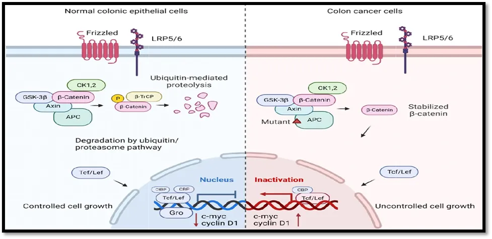
Fig 1: Molecular mechanisms of benzimidazole derivatives inducing cytotoxicity in HCT116 colorectal cancer cells."
Importance of HCT116 in Benzimidazole Research
The HCT116 cell line has become one of the most valuable experimental models for evaluating the cytotoxic potential of benzimidazole derivatives. Numerous studies have demonstrated that structural modification of the benzimidazole scaffold significantly enhances antiproliferative activity against HCT116 cells through multiple mechanisms, including apoptosis induction, inhibition of tubulin polymerization, suppression of PI3K/Akt signaling, ROS-mediated oxidative stress, and cell-cycle arrest. Because HCT116 cells possess clinically relevant genetic alterations and exhibit reproducible responses to chemotherapeutic agents, they provide an excellent platform for identifying promising lead compounds for colorectal cancer therapy. Consequently, continued utilization of HCT116 cells in medicinal chemistry and pharmacological research is expected to accelerate the development of next-generation benzimidazole-based anticancer agents with improved efficacy and safety profiles.
REFERENCES

D. Bharathi*, N. Deepa, M. D. Harish Ragul, D. Dilliganesh, A Review On Benzimidazole Derivatives As Cytotoxic Agents Against Human Colorectal Cancer Cells (HCT116), Int. J. Sci. R. Tech., 2026, 3 (8), 252-257. https://doi.org/10.5281/zenodo.21850358
 10.5281/zenodo.21850358