

We use cookies to ensure our website works properly and to personalise your experience. Cookies policy
Department of Electrical and Electronics Engineering, K.Ramakrishnan College of Technology, Tiruchirappalli, Tamil Nadu
Cardiovascular diseases are still one of the main causes of illness and death all over the world, which means that it is important to carry out continuous and reliable monitoring of the heart's electrical activity if an early evaluation of heart health is to be made. Electrocardiography (ECG) is one of the most commonly used non-invasive methods for recording the electrical activity of the heart and for detecting changes in heart rate and in the shape of the waveform. On the other hand, the conventional ECG monitoring equipment used in hospitals and diagnostic centres is generally rather expensive, bulky, and not very appropriate for use in portable or continuous monitoring situations. The paper describes the design and implementation of an ECG monitoring system that is low-cost, portable, and capable of real-time operation, the system making use of an Arduino UNO microcontroller and an AD8232 ECG sensor module. It obtains the cardiac electrical signals from the human body by means of disposable surface electrodes which are placed at suitable positions on the body. The weak biopotential signals picked up by the electrodes are fed into the AD8232 module, where they are conditioned by means of amplification and filtering in order to enhance the quality of the acquired ECG waveform and reduce the effect of noise, motion artifacts, and other undesirable signal components. The conditioned analog output from the AD8232 is then connected to the analog input of the Arduino UNO, where it is sampled and converted into a digital form for real-time monitoring. The ECG signal obtained can be displayed using a computer-based serial monitoring interface, allowing the characteristic changes in the cardiac waveform to be observed. The prototype suggested highlights simplicity, portability, affordability, and ease of implementation, and offers an effective means of carrying out real-time acquisition of cardiac signals. The system shows that it is possible to combine biomedical sensors with low-cost embedded technology for use in ECG monitoring. The platform could be improved by adding functions such as heart-rate estimation, automated extraction of ECG features, detection of abnormalities, wireless transmission of data, cloud-based monitoring, and classification of cardiac diseases using machine learning. Thus, the proposed system forms a practical basis for educational purposes in biomedical instrumentation, for initial cardiac monitoring, for portable healthcare applications, and for future remote patient monitoring systems.
The history of Electrocardiography (ECG) dates back to the late 18th century when Luigi Galvani discovered that living organisms produce internal electrical currents by experimenting with muscle contractions. In 1887, British physiologist Augustus D. Waller managed to record the world’s first ever human electrocardiogram using ground glass capillary electrometer with two electrodes attached to a subject’s legs. However, the ECG apparatuses used for the modern examination were pioneered by Dutch physiologist Willem Einthoven at the beginning of the 20th century. He presented the string galvanometer in 1901, an invention allowing to amplify cardiac biopotentials through a sensitive circuit without loss of details. Einthoven also introduced the universal P-Q-R-S-T wave nomenclature and laid a fundament for a three-lead system by establishing Einthoven’s Triangle.
During the 1930s and 1940s, the 12-lead ECG was developed by clinicians and researchers who wanted to perfect localized heart potential diagnostics. Leads V1-V6 were attached to the patient’s chest while three additional aVR, aVL and aVF augmented unipolar limb leads were invented by Emanuel Goldberger. All of them were integrated into the 12-Lead ECG Apparatus which was used as the clinical standard for decades. Then, in the middle of the last century, a new chapter in the history of ECG devices was opened by Norman Holter who introduced a 24-hour ECG monitoring method using a portable recorder. Such apparatuses were lightweight and easy to carry because of replacing vacuum tubes with miniature but powerful transistors. After the Analog ECG Signal is amplified and filtered, it is sent directly to the Analog input pin of the Arduino UNO microcontroller. The latter is the central processing unit of the whole system. Being a 10-bit ADC (Analog to Digital Converter), the microcontroller samples the incoming continuous voltage signal and transforms it into discrete bits. Then, an embedded algorithm tracks all prominent voltage variations to identify R-peaks of a cardiac Sine Waveform. It means that the microcontroller measures the R”-R intervals of a sinus rhythm and calculates the patient’s real-time Heart Rate (BPM) which is compared to the previously set thresholds: “NORMAL” or not. Fig. 1 shows the ECG Waveform.
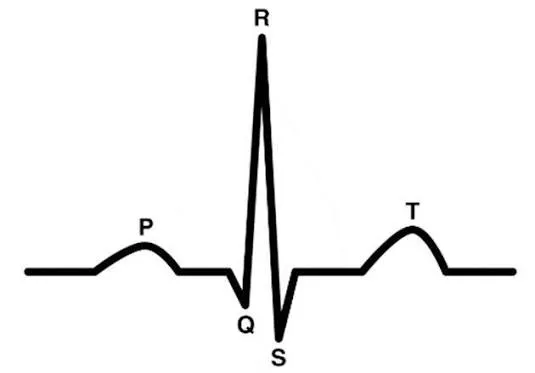
Figure 1: ECG Waveform
The main purpose of your proposed ECG system is to provide an affordable, compact, and real-time prototype for continuous cardiac monitoring without relying on bulky or expensive clinical equipment. Your system uses patient ECG electrodes connected to an AD8232 sensor module, which acts as an analog front-end to capture weak biopotential signals from the heart, amplify them, and filter out unwanted electromagnetic noise and motion artifacts to provide a clean analog signal, which is then fed into an Arduino UNO, which processes the incoming voltage and calculates the heart rate in beats per minute (BPM) and evaluates the operational status of the patient's heart rhythm.
To provide versatile data delivery, the system provides a dual-output interface for immediate reading and detailed waveform analysis. A local 16x2 LCD display powered by a regulated 5V power supply displays direct numeric updates of the calculated heart rate and status for quick point-of-care assessment. A USB serial connection is used to stream continuous real-time data to a laptop, where the Arduino IDE Serial Monitor and Plotter render the live P-Q-R-S-T ECG waveforms. In the end, your design demonstrates how one can combine integrated biomedical sensors and accessible microcontrollers to create an effective low-cost solution for vital signs tracking, educational instrumentation, and remote patient monitoring. Cardiovascular diseases remain one of the leading causes of global mortality, requiring continuous and early detection through electrocardiogram (ECG) monitoring. However, traditional clinical electrocardiographs are often expensive, bulky, and stationary, limiting their availability primarily to formal healthcare facilities and limiting access in low-resource or remote point-of-care environments.
Furthermore, the raw biopotential signals captured from skin surface electrodes are extremely weak and susceptible to high levels of electromagnetic noise from power lines and patient motion artifacts. Existing low-cost embedded monitoring solutions frequently present a major design trade-off, as they either display only numeric heart rates on a small screen, losing critical P-Q-R-S-T wave morphology required for diagnostic analysis, or rely entirely on an external computer for graphical plotting, which eliminates standalone field operation. There is then a need for a compact, low-cost, and dual-interface ECG monitoring system that utilizes an AD8232 analog front-end to filter noise and condition biopotential signals, an Arduino UNO to process digitized data, a 16x2 LCD display to provide immediate standalone numeric feedback (Heart Rate in BPM and Status), and a USB serial connection to stream real-time continuous ECG waveforms to a PC for detailed visual analysis.
This study describes the design and implementation of a portable, low-cost single-lead (Lead I) ECG monitoring system using an AD8232 sensor module and Arduino UNO microcontroller. The proposed design emphasizes acquiring biopotentials using surface electrodes, filtering baseline distortions, digitizing the signal, and calculating the real-time heart rate (BPM). The data are visualized locally using a 16 × 2 character LCD screen to provide textual status updates and a USB serial cable to view the live streaming of the P-Q-R-S-T waves on a PC. Advanced multi-lead (12-Lead) diagnostic capabilities, cloud connectivity, and medical device certification are beyond the scope of this project’s prototype.
LITERATURE SURVEY:
Pan and Tompkins et al. described a real-time QRS-detection algorithm for ECG signal. The main contribution was to enable accurate recognition of the QRS complex in order to determine the cardiac rhythm and heart rate. Their work introduced a series of signal-processing techniques including filtering, differentiation, squaring, moving-window integration, and adaptive thresholding. This study has made a significant contribution to the field of real-time ECG-signal processing and heart-rate detection.
Guedes et al. proposed a portable system based on an Arduino UNO and an AD8232 ECG sensor module for monitoring of the cardiac physiological signals. Their work was centered around acquiring the ECG signals from the electrodes and processing the signal using a low-cost embedded processing unit. The study demonstrated that Arduino-based hardware in combination with AD8232 ECG sensor could offer a viable option for ECG monitoring. A similar Arduino-based portable ECG monitor was described by Nissa et al.. The study concentrated on the use of an ECG sensor for acquiring and monitoring of the cardiac signals. The presented system emphasized on the portability of the system, low cost of implementation and real-time observation of the ECG signal. Their work showed that Arduino-based platforms could be viable for development of portable biomedical monitoring devices and form a baseline for developing portable ECG systems.
Hasini et al. presented a real-time ECG monitoring system based on an AD8232 sensor and Arduino UNO. Their system focused on acquiring of the ECG signals and processing the sensor output for monitoring the cardiac waveform in real time. The work demonstrated the possible integration of an AD8232 ECG sensor with microcontroller for biomedical signal monitoring. In addition, the study showed that low-cost embedded systems could be viable for ECG applications. Kamga et al. discussed the application of wearable ECG devices in clinical settings. The work described potential benefits of using wearable technologies for monitoring of the cardiac functions. The review suggested that portable technologies offer new opportunities for detection of the cardiac abnormalities outside the traditional hospital environment.
Bouzid et al. reviewed the state-of-the-art of remote and wearable ECG devices with diagnostic potential in adults. This review paper discussed the development of wearable ECG devices and their potential in remote cardiac monitoring. The authors highlighted the importance of continuous ECG acquisition and reliable signal quality for detection of cardiac abnormalities. Their review provided an insight into the current trends of using portable ECG devices in modern healthcare field. Alugubelli et al. reviewed wearable devices used in remote monitoring of heart rate and heart-rate variability. The work discussed the state-of-the-art of wearable-sensing technologies for continuous physiological monitoring. Their review demonstrated the potential of wearable devices for remote collection of cardiac information.
Ansari et al. presented an overview of deep-learning techniques for ECG arrhythmia-detection and classification. This work focused on different deep-learning algorithms in relation to ECG preprocessing, feature extraction and detection of the cardiac arrhythmias. The authors highlighted the potential of deep learning and its impact on the field of cardiac monitoring. However, they also identified potential limitations with regard to the quality and size of data sets; computational requirements; model generalization; and clinical validation. Hong et al. [9] performed a systematic review of deep-learning based ECG arrhythmia classification techniques. The review article discussed the use of convolutional neural networks and other deep-learning algorithms for detection of different types of cardiac arrhythmias. The manuscript demonstrated that deep-learning approaches could provide a reliable solution for automated analysis of the ECG recordings. However, it is necessary to ensure adequate dataset quality and sufficient computational resources.
Hannun et al. presented a deep neural network for detection and classification of the cardiac arrhythmias from ambulatory recordings. The study demonstrated that deep-learning approaches could offer exceptional performance in terms of automated analysis of the ECG recordings. In particular, the manuscript showed that training the deep neural network on large scale ECG data could offer a powerful tool for cardiac-rhythm analysis. This work provided an insight into the potential of artificial intelligence in healthcare-related ECG monitoring applications. Rajpurkar et al. proposed a convolutional neural network approach for automatic detection of the arrhythmias from ECG signals. The work demonstrated that with appropriate signal-preprocessing, deep-learning algorithms could be trained for automated recognition of various types of cardiac arrhythmias.
Acharya et al. investigated the potential of using deep learning techniques for automatic detection of the arrhythmias from ECG signals. This study concentrated on identification of the abnormal cardiac patterns automatically through computational analysis of the ECG recordings. Alfaras et al. proposed a fast machine-learning model for classification of the heartbeat from ECG. The study aimed at reducing the computational requirements of the ECG classification algorithm while ensuring acceptable levels of accuracy. The manuscript demonstrated that a relatively light-weighted machine-learning approach could be viable for real-time cardiac monitoring.
Han et al. investigated robustness of the deep-learning algorithms for ECG analysis against adversarial attacks. The work demonstrated that the deep-learning methods could offer high-performing ECG-analysis solutions. The manuscript discussed the importance of ensuring safe deployment of the deep learning algorithms for ECG monitoring. In this regard, appropriate validation procedures should be implemented to address the issues of adversarial attacks and model robustness. Ribeiro et al. discussed the use of the quantized deep neural networks for real-time ECG arrhythmia monitoring on low-power devices. The work aimed at reducing the computational requirements of the neural networks to ensure real-time ECG arrhythmia monitoring. The study demonstrated the possibility of implementing smart ECG monitoring systems on low-power embedded devices.
METHODOLOGY:
The proposed Electrocardiogram (ECG) monitoring system is an economical state-of-the-art biopotential biosensing instrumentation system intended for real-time acquisition, conditioning, processing, and graphic display of the cardiac bioelectric signals using an embedded system. The system is an affordable bedside solution that acquires heart beats, processes and stores them as numerical digitized electrical voltages for display of textual vital statistics and graphical electrocardiogram (ECG). Figure 1 below shows Real-Time ECG Monitoring Using Embedded System
The power supply design is the first level of this system’s schematic diagram, whose primary function is to provide a stable voltage to the entire active electronics circuitry. The system uses the standard 230V AC 50Hz commercial power supply as an input. A 5V DC Power Adapter or USB Power Supply is used to step down and regulate the power to 5V DC suitable for powering the sensitive on-board micro-controller circuitry and other low-power electronics. This power supply provides 5V DC power to the main microcontroller, which is the Arduino UNO microcontroller board (5V DC), 16x2 Character LCD Display (5V DC), and the required regulated supply voltage (3.3V-5V DC) for the AD8232 ECG Sensor Module. Fig. 2 illustrates Real-Time Cardiac Signal Monitoring and BPM Detection Using Embedded System
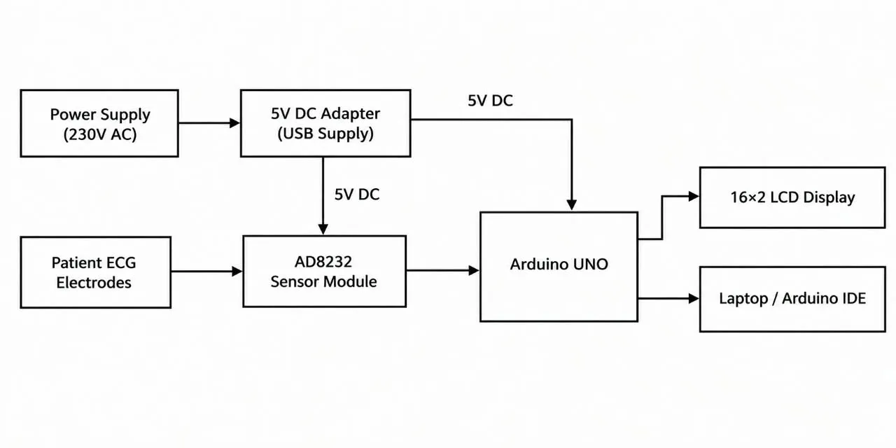
Figure 2: Real-Time Cardiac Signal Monitoring and BPM Detection Using Embedded System
The primary interface comprises conductive Patient ECG Electrodes positioned on the patient’s skin, which are connected to the ECG Sensor in a single-lead configuration to detect the cardiac vector. Raw biopotential signals from the human body are typically very weak (in the order of millivolts or microvolts) and consequently, they are extremely sensitive to external interference. The raw Analog ECG Signal is then passed to the AD8232 ECG Sensor Module, which is essentially an Analog Front-End (AFE) integrated circuit. The AD8232 houses an internal instrumentation amplifier and a number of active operational amplifiers (configured as high-pass and low-pass filters). The conditioned signal is then amplified (to counteract the attenuation of the weak bio-signal) and Common-Mode noise, movement artifacts, baseline wander and 50Hz power-line interference are rejected.
RESULTS AND DISCUSSION:
The given ECG waveform of output 1 over a 10-second period. The ECG has a regular cardiac pattern with visible R-peaks. The P-wave, QRS complex, and T-wave are present and are seen for each heart beat. The amplitudes of the R-peak appear almost the same throughout the waveform, signifying that the heartbeat is regular during the recording. The waveforms’ amplitude peaks at approximately 0.9-0.95 mV for the prominent R-peaks and is close to the baseline for other waves. The ECG pattern is regular and signifies that the system is able to record and display the cardiac electrical signal for further processing and displaying real-time BPM results. Figure 3 shows Normal ECG signal output 1
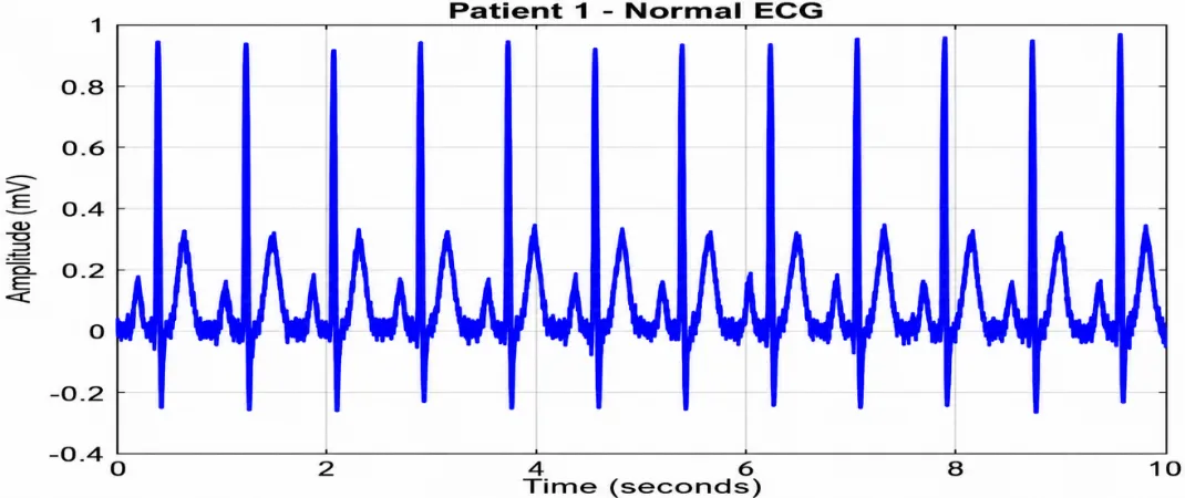
Figure 3: Normal ECG Signal Output 1
The ECG waveform of output 2 appears to be a regular cardiac pattern over the 10-second period under consideration. It is possible to notice prominent R-peaks that appear almost at the same interval in the ECG signal for this patient. The ECG waveform has all the typical features of the cardiac cycle, including P-wave, QRS complex, and T-wave. The amplitude of the R-peak is about 0.9 – 1 mV, while the other waves seem close to the baseline, which is characteristic of a normal heartbeat. Overall, the waveform observed from output 2 is normal, and it can be used to estimate the heart rate in beats per minute. In this case, the embedded system can process the signal to provide information about the heartbeat in real time. Figure 4: below presents a Normal ECG signal output 2.
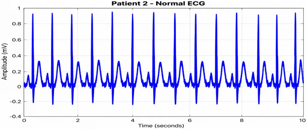
Figure 4: Normal ECG Signal Output 2
The ECG waveform of output 3 over a 10-second interval shows a regular and repeating cardiac pattern. The prominent R-peaks occur at nearly uniform intervals, indicating a relatively stable cardiac rhythm during the observation period. The waveform exhibits the characteristic P-wave, QRS complex, and T-wave components of the cardiac cycle. The R-peak amplitude reaches approximately 0.9–0.95 mV, while the signal remains close to the baseline between successive heartbeats. The clear and consistent waveform pattern demonstrates successful ECG signal acquisition and provides a suitable signal for BPM calculation and real-time cardiac monitoring. Figure 5 Normal ECG signal output 3
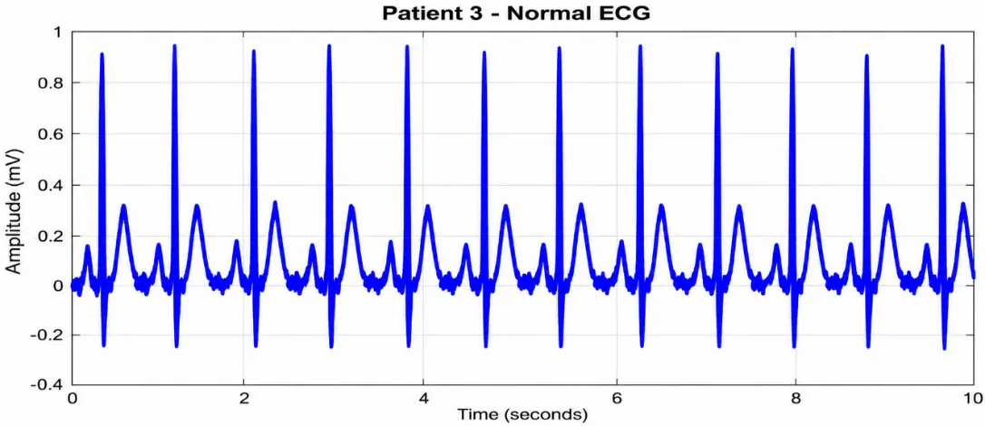
Figure 5: Normal ECG Signal Output 3
The ECG waveform of output 4 over a 10-second interval shows a noticeably higher frequency of cardiac cycles compared with the normal ECG recordings. The R-peaks occur at shorter and more frequent intervals, indicating an elevated heart rate during the observation period. The characteristic ECG waveform components are visible, with prominent R-peaks reaching approximately 0.9–0.95 mV. The increased number of R-peaks within the same 10-second duration demonstrates the higher cardiac rate. This result shows that the proposed system can capture variations in cardiac rhythm and provides an ECG signal suitable for BPM detection and real-time monitoring of elevated heart rate conditions. Figure 6 shows Abnormal ECG signal Output 4.
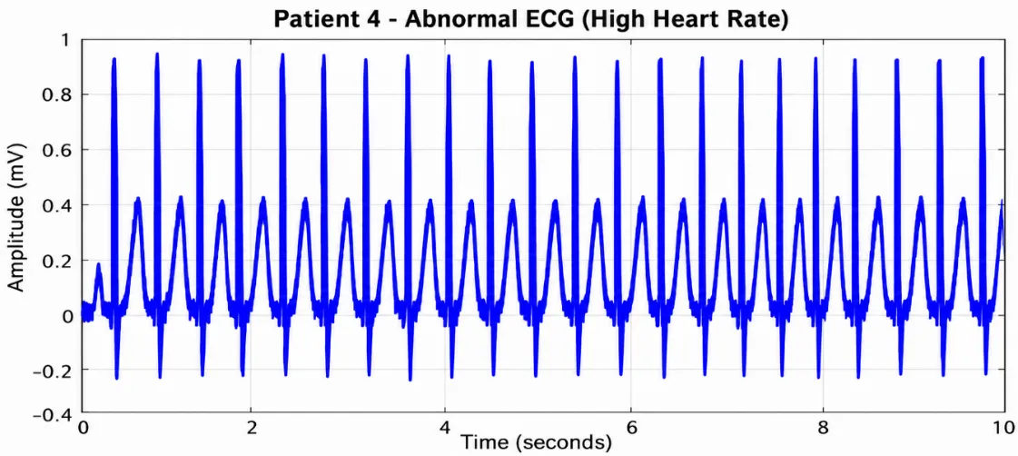
Figure 6: Abnormal ECG Signal Output 4
The ECG waveform of output 5 over a 10-second interval shows a lower frequency of cardiac cycles compared with the normal ECG recordings. The R-peaks are separated by relatively larger time intervals, indicating a reduced heart rate during the observation period. The characteristic P-wave, QRS complex, and T-wave components can be observed in the waveform. The prominent R-peaks reach approximately 0.9–1.0 mV, while the signal remains close to the baseline between successive cardiac cycles. The reduced number of R-peaks within the 10-second recording indicates a lower cardiac rate. This result demonstrates the capability of the proposed system to acquire ECG signals with different heart-rate conditions and provides a suitable basis for BPM detection and real-time cardiac monitoring.
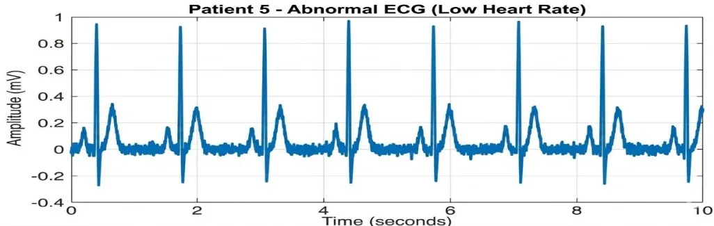
Figure 7: Abnormal ECG Signal Output 5
CONCLUSION:
The proposed Real-Time Cardiac Signal Monitoring and BPM Detection Using an Embedded System was a great success as a proof of concept of the viability of using low-cost embedded technology for real-time ECG monitoring. The design is built around the patient’s ECG electrodes, the AD8232 ECG sensor module, and the Arduino UNO which condition and process the given data. The ECG waves obtained from the two different patient readings are seen, with the variations in the R-peak being used to indicate different heart rates, such as normal, high, and low heart rates. The system can be used for real-time cardiac monitoring while also providing BPM detection and portable, low-cost cardiac surveillance. The developed prototype can be utilized in a multitude of different areas, such as biomedical instrumentation, educational purposes, introductory cardiac monitoring, and portable healthcare technology. The obtained results are a display of the potential of the given concept, with the AD8232 sensor and Arduino UNO microcontroller being an appropriate solution for low-cost and low-complexity ECG acquisition. While the given design is only a part of a larger system that would be used for monitoring, it can be integrated later with more complex technologies, such as wireless transmission and cloud-based data processing. Furthermore, the system can have additional features, such as advanced ECG analysis, arrhythmia detection, and the use of machine learning for cardiac abnormality classification to be implemented.
REFERENCES



Athi Meenatchi M., Haritha B., Visvesha R., Development of a Real-Time Embedded System for Cardiac Signal Monitoring and BPM Detection, Int. J. Sci. R. Tech., 2026, 3 (10), 570-577. https://doi.org/10.5281/zenodo.23257507
 10.5281/zenodo.23257507